Differential Impact of LPG-and PG-Deficient
Leishmania major
Mutants on the Immune
Response of Human Dendritic Cells
Michelle A. Favila1☯, Nicholas S. Geraci1☯, Asha Jayakumar1, Suzanne Hickerson2,
Janet Mostrom1, Salvatore J. Turco3, Stephen M. Beverley2, Mary Ann McDowell1*
1Eck Institute for Global Health, Department of Biological Sciences, University of Notre Dame, Notre Dame, Indiana, United States of America,2Molecular Microbiology Department, Washington University School of Medicine, St. Louis, Missouri, United States of America,3Department of Biochemistry, University of Kentucky College of Medicine, Lexington, Kentucky, United States of America
☯These authors contributed equally to this work.
Abstract
Background
Leishmania majorinfection induces robust interleukin-12 (IL12) production in human den-dritic cells (hDC), ultimately resulting in Th1-mediated immunity and clinical resolution. The surface ofLeishmaniaparasites is covered in a dense glycocalyx consisting of primarily lipo-phosphoglycan (LPG) and other lipo-phosphoglycan-containing molecules (PGs), making these glycoconjugates the likely pathogen-associated molecular patterns (PAMPS) responsible for IL12 induction.
Methodology/Principal Findings
Here we explored the role of parasite glycoconjugates on the hDC IL12 response by gener-atingL.majorFriedlin V1 mutants defective in LPG alone, (FV1lpg1-), or generally deficient for all PGs, (FV1lpg2-). Infection with metacyclic, infective stage,L.majoror purified LPG induced high levels ofIL12Bsubunit gene transcripts in hDCs, which was abrogated with FV1lpg1-infections. In contrast, hDC infections with FV1lpg2-displayed increasedIL12B expression, suggesting other PG-related/LPG2dependent molecules may act to dampen the immune response. Global transcriptional profiling comparing WT, FV1lpg1-, FV1 lpg2-infections revealed that FV1lpg1-mutants entered hDCs in a silent fashion as indicated by repression of gene expression. Transcription factor binding site analysis suggests that LPG recognition by hDCs induces IL-12 in a signaling cascade resulting in Nuclear FactorκB (NFκB) and Interferon Regulatory Factor (IRF) mediated transcription.
Conclusions/Significance
These data suggest thatL.majorLPG is a major PAMP recognized by hDC to induce IL12-mediated protective immunity and that there is a complex interplay between PG-baring Leishmaniasurface glycoconjugates that result in modulation of host cellular IL12.
a11111
OPEN ACCESS
Citation:Favila MA, Geraci NS, Jayakumar A, Hickerson S, Mostrom J, Turco SJ, et al. (2015) Differential Impact of LPG-and PG-Deficient Leishmania majorMutants on the Immune Response of Human Dendritic Cells. PLoS Negl Trop Dis 9(12): e0004238. doi:10.1371/journal.pntd.0004238
Editor:Christian R. Engwerda, Queensland Institute of Medical Research, AUSTRALIA
Received:June 1, 2015
Accepted:October 25, 2015
Published:December 2, 2015
Copyright:© 2015 Favila et al. This is an open access article distributed under the terms of the
Creative Commons Attribution License, which permits unrestricted use, distribution, and reproduction in any medium, provided the original author and source are credited.
Data Availability Statement:Complete array data generated in this study are accessible at the NCBI Gene Expression Omnibus database (http://www. ncbi.nlm.nih.gov/geo/, accession #GSE59766).
Funding:This work was supported by National Institutes of of Allergy and Infectious Diseases grants R01AI056242 (MAM) and NIH RO1AI31078 (SMB, SJT). MAF was a fellow of the
Author Summary
Leishmaniasis is a group of parasitic diseases caused by intracellular protozoa belonging to
the genusLeishmania, pathological manifestations ranging from self-healing cutaneous
forms to severe visceral infections that result in death. These clinical outcomes are dictated
by theLeishmaniaspecies initiating the infection and are influenced by early responses of
host immune cells, which ultimately initiate an IL12 mediated immune response in resolv-ing infections. Like the diseases themselves, the magnitude of IL12 induction in hDCs is Leishmania-species and strain specific, where species that elicit visceral disease do not
induce IL12, while most cutaneous disease-causingL.majorstrains induce robust IL12
responses and confer life-long immunity. The molecular mechanisms that mediate the ability of these innate immune cells to discriminate between pathogens remain elusive and
have been primarily investigated in murine model systems. Here we identifiedL.major
LPG as a major PAMP that induces IL12 in hDCs. Elucidation of this critical component
of human immunity toL.majorhas ramifications for leishmaniasis vaccine development.
Introduction
Leishmaniasis constitutes a group of vector-borne parasitic diseases that affects approximately
12 million people worldwide and results in diverse clinical pathologies [1]. The causative
intra-cellular protozoa belonging to the genusLeishmania, generally dictate disease outcome in a
dis-tinct species-specific manner. Visceral leishmaniasis may result from infection with Leishmania donovaniparasites that disseminate throughout the body, manifesting into fatal
systemic disease if left untreated. In contrast,Leishmania major, which is a causative agent for
cutaneous leishmaniasis, produces ulcerative lesions localized at the site of sand fly vector
inoc-ulation. In the majority ofL.majorpatients, lesions heal within several months, conferring
life-long acquired immunity [2]. Recovery of cutaneous leishmaniasis with a strong immune
response can be attributed to early cellular activities that occur following initial entry of the parasites into host cells.
Leishmaniaparasites have evolved mechanisms to survive within host cells and mediate infectivity in sand fly vectors through the interaction of their cellular surface coat molecules. TheLeishmaniasurface coat is densely packed with glycosylphosphatidylinositol (GPI)-anchored glycoconjugates, including lipophosphoglycan (LPG), proteophosphoglycans
(PPGs), glycosylinositolphospholipids (GIPLs), and glycoprotein 63 (GP63) [3–5]. Together
these molecules provide a protective barrier for parasites to persist within the host environment
[6]. LPG is one of the most intensely studiedLeishmaniasurface molecules, in both the sand
fly vector and vertebrate hosts, playing a distinct role in modulating host immune function [7]
and even vectorial capacity of various sand fly species [8]. LPG is polymorphic among
Leish-maniaspecies and developmentally regulated [6]. One dominant feature of LPG, the phospho-glycan repeating unit [Gal-Man-P] (PG), contains species-, strain-, and stage-specific
modifications usually on the Gal residues [9–13]. The number of PG repeat units almost
dou-bles during metacyclogenesis [14] and LPG is dramatically down regulated in the amastigote
stage [15]. Thus, the role of LPG in mammalian infections is limited to the initial period of
invasion and establishment of infection by metacyclic promastigotes.
Protective immunity to cutaneous leishmaniasis requires a robust IL12 driven type 1 helper T-cell (Th1) mediated response that produces high levels of interferon-gamma (IFNG), which ultimately promotes anti-microbicidal production of nitric oxide (NO) and reactive oxygen
species (ROS) that destroy invading pathogens [16,17]. Dendritic cells (DCs) and macrophages
design, data collection and analysis, decision to publish, or preparation of the manuscript.
are among the major cell sources of IL12, whose bioactive secretion is dependent upon the
covalent linkage between the p40 (IL12B) and p35 (IL12A) subunits [18]. The ability of
Leish-maniato selectively suppress IL12 production, as first established by using murine
macro-phages [19,20], occurs through the transcriptional inhibition of theIL12Bpromoter [21] and is
one immune evasion strategy employed by parasites to establish infection. Phagocytosis of Leishmaniaparasites by murine DCs induces IL12, driving the differentiation of Th1 cells to
elicit their effector function [22–27]. The precise role of different DC subsets during murine
infectionin vivois discordant depending on theLeishmaniastrain utilized, the infection route,
and the timing of analysis [28,29]. A role for DCs early in infection has been identifiedin vivo,
however, as DCs carryingLeishmaniaantigen produce IL12 within 8 hours following infection
[30]. The murine DC IL12 response can be altered depending on the biochemical composition
of the parasite surface, as evidenced by a study demonstrating that infection withL.major
LV39c5lpg2−, a mutant that lacks phosphoglycan (PG)-containing molecules and other
LPG2-dependent metabolites [31], induced IL12B in bone marrow derived mouse DCs
(BMDCs) co-stimulated with anti-CD40 and IFNG [32]. This effect along with the long-term
persistence of these parasites likely account for why vaccination with these LV39c5lpg2−
para-sites protects mice againstL.majorwild type (WT) challenge [33].
Remarkably, hDCs exhibit a dynamic range in IL12 production in response toLeishmania
infection that is largely dependent upon the nature of the infecting species or strain.L.
dono-vanifails to elicit IL12, whereas a general induction of IL12 is observed duringL.major
infec-tions [34]. However, IL12 production also varies acrossL.majorstrains. Strains LV39 and SD
do not induce IL12, whereas Friedlin V1 (FV1), IR173, IR176, and CC-1 strains elicit high
lev-els of IL12 [34,35]. These differences are not well-correlated with LPG structural
polymor-phisms, asL.majorLV39cl5 bears a highly poly-galactosylated LPG [36], whileL.majorSD
synthesizes an unsubstituted LPG similar to that ofL.donovani[37]. Several groups have
reported differences in lesion pathology followingin vivoinfection with these sameL.major
strains. For example,L.majorFV1 infected C57BL/6 mice develop lesions that eventually heal
over time, whereas mice infected withL.majorSD produce non-healing lesions [38]. BALB/C
IL4RA knockout mice are resistant toL.majorIR173 strain but susceptible toL.majorLV39
strain [39]. Moreover, whileL.majorFV1 strain infected BALB/C mice quickly develop lesions,
L.majorLV39c5, a clonal derivative of the LV39 strain, elicits slower lesion development. Hybrid crosses ofL.majorFV1 x LV39c5 segregate at a 1:1 ratio into“fast”or“slow”virulence
progeny [40]. These differential host responses to variant intra-species strains ofL.majorhave
important implications for the parasite strain-specific factors that could dictate disease persis-tence versus healing and induction of immunity.
In this study, we focus on elucidating whether parasite surface molecules are associated with
the robust cytokine response observed in hDCs using the‘high-IL12 inducing’L.majorFV1
strain. We generated parasite mutants lacking LPG alone, as done previously with the‘low-IL12
inducing’L.majorLV39c5, through inactivation of the LPG1 galactofuranosyl transferases
required for LPG core synthesis. Mutants generally lacking in all PG-containing structures were generated through inactivation of the Golgi GDP-mannose nucleotide sugar transporter gene,
LPG2[31]. This approach is powerful for probing the role of LPG as it allowed us to assess the
impact of LPG deficiency in the context of the parasite, rather than through exogenous and rela-tively artificial routes. A second advantage is that multiple mutants provided a means to dis-criminate between LPG effects and those of molecules that bear structures related to or shared with those found in LPG. Notably, the PG repeating units present on LPG also are abundant on secreted molecules, such as acid phosphatases and other PPGs, which can be anchored to the
parasite surface through glycosylphosphatidylinositol (GPI). Inactivation ofLPG1results in a
Our results demonstrate that hDC infection with the LPG-nullL.majorFV1lpg1−mutant
resulted in significantly diminishedIL12BmRNA, relative to FV1 WT parasites, indicating
that LPG is essential for stimulating host IL12 production. However, the PG-nullL.majorFV1
lpg2−mutant infected DCs exhibited an increase inIL12Bexpression, suggesting that PGs and/
or other LPG2-dependent metabolites may suppress IL12 induction. These results suggest that L.majorparasites balance stimulatory and inhibitory effects on the host cells to establish infection.
Materials and Methods
Ethics statement
The study protocol was approved by the University of Notre Dame Institutional Review Board in compliance with all applicable Federal regulations governing the protection of human sub-jects (Human Subsub-jects Assurance #M1262). The research was deemed exempt under exemp-tion #4. The samples were purchased from Central Indiana Regional Blood Center,
Indianapolis, IN and no identifying information was provided.
Dendritic cell generation and infection
Monocytes were isolated from healthy human donor buffy coats (Central Indiana Regional
Blood Center, Indianapolis, IN) by enriching for CD14+cells using a magnetic bead separator
(AutoMACs, Miltenyi Biotech systems, Germany). Monocytes from each donor were cultured
in 6-well plates at a concentration of 106cells/2ml of RPMI-complete media (10%
heat-inacti-vated FBS, 2mM l-glutamine 100U/ml, 1% penicillin/streptomycin) and supplemented with recombinant human IL4 (40U/ml, Peprotech, NJ) and granulocyte-macrophage colony-stimu-lating factor, GMSCF (1000U/ml, Peprotech, NJ) on days 0, 3, and 6 to allow differentiation into immature DCs. Cells were harvested, washed one day before infection to remove any residual cytokines, and assessed for DC marker CD1A to confirm a homogenous population of
immature DCs. All parasite strains were cultured at 26°C without CO2in M199 medium
con-taining 10% heat-inactivated FBS [42]. Metacyclic promastigotes were isolated according to
previously described methods [43] and opsonized by treatment with 5% human serum for 30
min at 37°C. DCs were then infected at a concentration of 10 parasites per 1 DC in
RPMI-com-plete media. As we previously demonstrated that the peak ofIL12Bexpression occurs at 8
hours postL.majorinfection [44] and to avoid the complication that mutant parasites might
be degraded at later time points as previously observed [31,41], samples were typically
har-vested at 8 hours post-infection. For kinetic analyses we focused on the early time points fol-lowing infection (2, 4, 8, or 24 hours). Cytospins were prepared at the conclusion of each experiment and Diff-quick stained (Fischer Scientific, Pittsburgh, PA) for visual analysis by light microscopy. Uninfected and infected DCs (100 total) were counted to calculate the infec-tion rate (% infected DCs) and the parasite indices (# parasites per 100 cells) for each infecinfec-tion sample. All parasite and human cell cultures tested negative for mycoplasma (PCR detection,
Takara) and tested below the limits of detection for endotoxin (<0.25U/ml) (Limulus
Amoe-boctye Assay, Endosafe, Charleston, NC).
Generation of
L
.
major
FV1
lpg1
−and
L
.
major
FV1
lpg2
−mutants and
complemented lines
Leishmania majorstrain Friedlin clone V1 (MHOM/IL/81/Friedlin) andL.donovanistrain 1S
Methods for electroporation of logarithmic phase promastigotes and plating on semisolid
media to obtain clonal lines were as described previously [46].
L.majorFV1lpg1−mutants were obtained by a gene disruption strategy, in which
autono-mous drug resistance cassettes were inserted within theLPG1coding region [41]. The methods
and constructs used were the same as in the prior study generating theL.majorLV39c5lpg1−
mutants [41]. In the first round, plasmid B2947 DNA was digested with restriction enzymes
XhoI and HindIII to yield theLPG1::HYGtargeting construct, conferring selective resistance
gene to hygromycin B (hygromycin phosphotransferase). 10μg of DNA was used for
electropo-ration and parasites were plated on semisolid medium containing 50μg/ml of hygromycin B.
Clonal parasite lines were obtained at typical frequencies and screened for the presence of the
expected heterozygousLPG1andLPG1::HYGinsertion by PCR (S1 Fig,S1 Table). Several
clones were inoculated into susceptible BALB/C mice (107stationary phase, footpad) and
recovered after 1 month; such mouse passaged lines are designated as‘M1’. These
heterozy-gotes underwent a second round of transfection; electroporating 10μg ofLPG1::PAC,
confer-ring a selective resistance gene to puromycin (puromycin acetyltransferase), derived from BamHI digestion of plasmid B2949, and followed by plating parasites on semisolid media
con-taining 50μg/ml hygromycin B and 30μg/ml puromycin. Clonal lines bearing disruptions in
bothLPG1alleles, and lacking unmodifiedLPG1(^LPG1::HYG/^LPG1::PAC), were identified
by PCR analysis and confirmed by Western blot analysis and agglutination tests. Several clones
were inoculated into susceptible BALB/C mice (107stationary phase, footpad) and recovered
after 1 month (M1). For simplicity, these lines are referred to as FV1lpg1−. To generate
com-plemented‘add back’lines, several FV1lpg1−clonal lines were electroporated with theLPG1
expression plasmid pSNBR-LPG1::NEO(B3340), conferring an episomal selective resistance
gene to the aminoglycoside antibiotic G418 via expression of the neomycin phosphotransferase
geneNEO, and clonal lines were recovered by plating on semisolid media containing 50μg/ml
HYG, 30μg puromycin, and 12μg/ml of G418. Successful transfection was established by PCR
tests and restoration of LPG expression by western blot, and agglutination tests. Formally, the
genotype of such lines is (^LPG1::HYG/^LPG1::PAC/+pSNBR-LPG1), which for simplicity is
referred to as FV1lpg1−/+LPG1. Sibling clonal lines displayed similar phenotypes and one
rep-resentative FV1lpg1−line (cl2.10, M1), and its complemented offspring (cl2.10 AB3, M1),
des-ignated FV1lpg1−/+LPG1were used in the experiments.
L.majorFV1lpg2−mutants were obtained by a gene replacement strategy; where the drug
resistance gene ORFS replaced theLPG2coding region. In the first round, plasmid B3950 was
digested with XhoI I, yielding theLPG2::HYGtargeting construct; 10μg was used for
electropo-ration and cells were plated on semisolid medium containing 50μg/ml of hygromycin B. Clonal
lines were obtained at typical frequencies and screened for the presence of the expected
hetero-zygousLPG2andLPG2::HYGinsertion by PCR (S2 Fig,S1 Table). Several clones were
inocu-lated into susceptible BALB/C mice (107stationary phase, footpad, M1). These heterozygotes
underwent a second round of transfection, electroporating 10μg ofLPG2::SAT, conferring a
selective resistance gene to nourseothricin (streptothricin acetyltransferase), derived from XhoI, HindIII digestion of plasmid B6598, followed by plating on semisolid media containing
50μg/ml hygromycin B and 100μg/ml nourseothricin. Clonal lines bearing disruptions in both
LPG2alleles and lacking unmodifiedLPG2(ΔLPG2::HYG/ΔLPG LPG2::SAT) were identified
by PCR analysis, and confirmed by Western blot analysis and agglutination tests. For
simplic-ity, these lines will be referred to as FV1lpg2−. To generate complemented
‘add back’lines,
sev-eral FV1lpg2−clonal lines were electroporated with theLPG2expression plasmid pXG-LPG2::
NEO(B4296) and clonal lines recovered by plating on semisolid media containing 50μg/ml
HYG, 100μg/ml SAT, and 15μg/ml of G418. Successful transfection was established by PCR
tests. Formally, the genotype of such lines is (ΔLPG2::HYG/ΔLPG LPG2::SAT/+pXG-LPG2),
which for simplicity is referred to as FV1lpg2−/+LPG2. Sibling clonal lines displayed similar
phenotypes and one representative FV1lpg2−line (cl6.1A, M1), and its complemented
off-spring (cl6.1A AB15, M1), designated FV1lpg2−/+LPG2 were used in the experiments.
Gene replacement plasmid generation
Plasmid B6598 was generated by a fusion PCR strategy. Briefly, the 5’LPG2flanking sequence,
3’LPG2flanking sequence,LPG2ORF, and selected drug marker,SATORF were amplified by
PCR and inserted into the pGEM-T-Easy vector by TA cloning according to manufacturer’s
instruction (Promega, Madison, WI) and transformed intoE.coli. Its structure was confirmed
by DNA sequencing. The primers used for constructing B6598 are provided inS1 Table.
Western blot
For Western blot analysis of PG-containing molecules, parasites were grown to logarithmic phase and harvested for cell lysate preparation in 4X Lamelli buffer (50 mM Tris-HCl pH 6.8, 2% SDS, 10% Glycerol, 1% 2-mercaptoethanol, 12.5 mM EDTA, and 0.02% Bromophenol
Blue). Samples were separated on 10–12% SDS-PAGE gels at a concentration of (3.5x10⁶cells/
well) and transferred onto methanol activated nitrocellulose membrane for 3 hrs at 60V, 4°C. Ponceau staining was performed to assure macromolecule transfer prior to blocking in 5% milk overnight. Membranes were stained with primary mouse monoclonal anti-sera WIC79.3 antibody (1:1000), recognizing galactosylated Gal-Man-P repeats on LPG, and detected using a goat anti-mouse HRP conjugated secondary antibody (1:5000) (Invitrogen, Carlsbad, CA). Membranes were developed using West-Pico detection solution assay (Thermo Scientific, Rockford, IL) and an X-ray film developer.
LPG purification
LPG was isolated from 109L.majorFV1 metacyclic promastigotes as previously described,
with minor modification [47,48]. Cellular membranes were disrupted by sonicating pelleted
cells suspended in a cold chlorform:methanol:water (1:2:0.8) solution, centrifuged (5000rpm, 10min, 4°C), and the top de-lipidated layer containing the majority of GIPLs and
phospholip-ids was removed. The remaining insoluble material was quick-dried under stream of N2and
further extracted with two rounds of 9% 1-butanol extraction to release LPG molecules into the top aqueous layer. Hydrophobic interaction chromatography was performed to purify LPG
molecules from theLeishmaniasurface coat. Briefly, LPG-containing butanol extracts were
pooled and added to a 20% Octyl-Sepharose column that was pre-equilibrated with (5% propo-nal, 1M ammonium acetate). A desalting gradient (5%-60%) was applied to the column to elute LPG fractions utilizing the fraction collector, (BioRad Fraction Collector, Model 2128). LPG was detected by thin layer chromatography (TLC) and quantified by phenol sulfuric assay. Sample fractions were spotted on silica containing TLC plate. Glycan determinants were visualized by spraying the plate with orcinol (0.5mg/ml in 95% ethanol), dried, and sprayed with 75% sulfuric acid. All LPG containing fractions were pooled and dried in speed-vacuum at room temperature. Lyophilized LPG was resuspended in water and quantified by a
colori-metric phenol-sulfuric assay [49]. Purification of LPG molecules was confirmed by a standard
Stains-All protocol. Briefly, 5–10μg of LPG was boiled in 2X Loading Dye and loaded onto 10%
SDS PAGE gel, running at 140V (room temperature). Gels were fixed in 25% 2-propanol and stained with stains-all solution (Fluka Analytical, Switzerland) containing 10% formamide, fol-lowed by destaining with 40% ethanol. Bands were visualized under white light, based on the
WIC79.3 western blot analysis was utilized to confirm LPG purification. Lyophilized purified
LPG was resuspended in serum free RPMI and a titration of LPG (0.5μg, 1μg, and 10μg) was
used for the hDC infection assay.
Quantitative real-time polymerase chain reactions
Relative levels of human gene transcripts were determined by qRT-PCR. Total RNA from
uninfected orLeishmania-infected DCs was isolated using an RNeasy kit (Qiagen, Valencia,
CA) and 1μg of RNA per infection sample was used to generate cDNA using SuperScript III
Synthesis (Invitrogen, Carlsbad, CA) according to manufacturer’s instructions. For analysis of
IL12B,IL12A,IRF1,IRF8,TNF,IL10,IL1B,SOCS3,TNFAIP3, andHPRT (hypoxanthine-gua-nine phosphoribosyltransferase) mRNA expressions, qRT-PCRs were conducted utilizing SYBR Green PCR Master Mix (Applied Biosystems by Life Technologies, Carlsbad, CA)
according to manufacturer’s protocol and detected with an ABI 7900HT Fast Real-Time PCR
System (Applied Biosystems by Life Technologies, Carlsbad, CA). All human primer sequences
were designed by Integrated Design Tools (IDT) and used at a concentration of 5μM per
reac-tion (S2 Table). For select analysis ofIL12B,IL12A, andGAPDH(glyceraldehydes
3-phospho-ate dehydrogenase) mRNA expressions, PCR reactions were setup employing TaqMan pre-developed assay kits (Life Technologies, Foster City, CA) and determined using an ABI 7500 Real Time PCR System (Applied Biosystems, Foster City, CA). For each gene, relative numbers
of mRNA copies were determined by theΔΔCTmethod [42].
Microarray expression profiling
Total RNA was isolated 8 hours post-infection from four additional donors’uninfected
mono-cyte-derived DCs and DCs infected withL.majorFV1 WT, FV1lpg1−, FV1lpg1−/+LPG1, FV1
lpg2−, and FV1lpg2−/+LPG2using RNeasy kits (Qiagen, Valencia, CA). RNA 6000 Nano kits
(Agilent Technologies, Santa Clara, CA) were used to determine total RNA integrity on a Bioa-nalyzer 2100 instrument (Agilent Technologies, Santa Clara, CA). 25ng of high quality RNA was converted to double stranded cDNA using a TransPlex Complete Whole Transcriptome Amplification kit (Sigma-Aldrich, Saint Louis, MO). RNA degradation, double stranded cDNA purification, and cDNA precipitation was conducted following NimbleGen Gene Expression
Array user’s guide protocols (Roche-NimbleGen, Madison, WI). A Nanodrop ND-2000
(Thermo Fisher Scientific, Waltham, MA) was used to determine total RNA and double stranded cDNA concentrations. Sample cDNAs were Cy3-labeled using NimbleGen Single Color Labeling Kit (Roche-NimbleGen, Madison, WI) per manufacturer's recommendations.
Labeled cDNAs were hybridized to 12-plex NimbleGenHomo sapiensExpression Arrays
(plat-form GPL16025), featuring 140,096 probes, representing 21,269 genes and transcripts, using Hybridization LS and Wash Buffer Kits (Roche-NimbleGen, Madison, WI) per manufacturer's recommendations. Image acquisition of arrays was performed using a NimbleGen MS 200 Microarray Scanner (Roche-NimbleGen, Madison, WI), at a 2 micron resolution. NimbleGen array image data were processed using NimbleScan version 2.5 (Roche-NimbleGen, Madison, WI) to extract intensity values for each gene. NimbleScan software automates the pre-process-ing of NimbleGen microarray image data, includpre-process-ing identifypre-process-ing the location of each probe, extraction of intensity data from the image, background correction, and obtaining expression summary values for each gene using a probe-level summarization robust multi-array average method (RMA). Probes with intensity values greater than twice that of background were
retained for downstream analysis. Log2normalized expression ratios for each gene were
calcu-lated between infected samples and paired uninfected samples. Z-scores were calcucalcu-lated
(log2(infected intensity value/inter-quartile mean of uninfected intensity values)Gi−average
(log2(infected intensity value/ inter-quartile mean of uninfected intensity values)Gi. . .Gn) /
stan-dard deviation(log2(infected intensity value/ inter-quartile mean of uninfected intensity
values)Gi. . .Gn). An absolute Z-score value of 1.96 may be inferred as significant (p
<0.05) [51].
Complete array data generated in this study are accessible at the NCBI Gene Expression Omni-bus database (accession GSE59766). Gene expression data of RMA normalized raw microarray probe hybridization fluorescence values, where at least one sample value was twice that of back-ground resulted in 12,911 genes.
Functional enrichment analyses
Genes that displayed significant differential expression from FV1 WT, FV1lpg1−, or FV1lpg2−
samples compared to uninfected samples on NimbleGen microarrays were fed into the Short
Time-series Expression Miner (STEM) program [52,53]. Briefly, log2ratio values for each of
four donors were loaded into the program as repeated data, where FV1 WT data represented a
“time point 1”, FV1lpg1−data represented“time point 2”, and FV1lpg2−data represented
“time point 3”. The datasets were clustered using the STEM clustering method with minimum
correlation values of 0.6. The genes from the resultant model expression profile containing IL12Bwere used for downstream enrichment analysis in the Web-based Gene Set Analysis
Toolkit (WEBGESTALT) [54,55] with a simple list of 233 official gene symbols as input.
KEGG Pathway enrichment was conducted on that list of genes with similar expression profiles
to that ofIL12Busing the following parameters: protein-coding EntrezGene database as a
refer-ence set and a hypergeometric test with Benjamini and Hochberg multiple test adjustments.
Pathways with an adjusted p-value<0.01 and a minimum of three genes found were
consid-ered significant. The same list of gene symbols was input to The Database for Annotation,
Visualization and Integrated Discovery (DAVID) v6.7 Functional Annotation Tool [56] and
transcription factor binding sites for each gene were identified using protein interaction enrich-ment. The annotations were cross-referenced to report the most common transcription factor
binding sites found in theIL12Bgene and genes withIL12B-like expression between DC
sam-ples infected withL.majorFV1 LPG mutants.
Statistical analysis
All statistical tests were performed using Graph Pad Prism version 5.0 (Graph Pad Software,
San Diego, CA). Statistical analysis was performed using Log2 transformedΔΔCTvalues using
a paired Student’s T-test. Differences were considered significant at p<0.05.
Results
Differential IL12 response in hDCs is
L
.
major
strain and developmental
stage specific
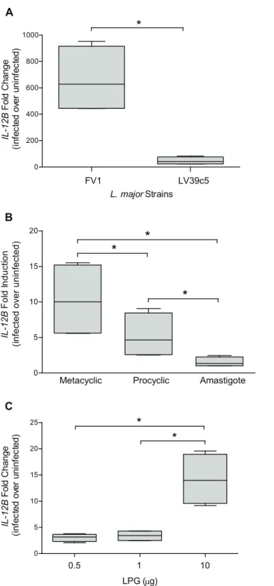
First, we confirmed that theIL12BmRNA expression in hDCs infected withL.majorstrains
FV1 WT was greater than DCs infected with LV39c5 WT. We demonstrated thatL.majorFV1
induced approximately 15-fold greater amounts ofIL12BthanL.majorLV39c5 (Fig 1A) at 8
hours post infection, the optimal time for peakIL12BmRNA expression followingL.major
infection [44]. These data support previous work that illustrated that the hDC IL12 response is
strain-specific, and also that infection withL.majorFV1 promotes a high induction of IL12
and that LV39c5 is similar to the LV39 strain tested previously [34]. We also confirmed that
the increasedIL12Bexpression observed duringL.majorFV1 WT infections were significantly
observed with the non-infective procyclic promastigote stage, and no response was elicited by
amastigotes (Fig 1B). These data are consistent with prior studies indicating that IL12
induc-tion depends on the life cycle stage ofLeishmaniaparasites [57,58].
Purified LPG induces
IL12B
expression
To determine whether the enhancedIL12Bproduction observed following infection withL.
majorFV1 metacyclic promastigotes was an LPG-dependent response, we first assessed the
role of purified LPG on the hDCIL12Bresponse. Human DCs were cultured in the presence of
varying amounts of purified metacyclicL.majorFV1 LPG for 8 hour and then assessed for
IL12Bexpression. At lower concentrations (0.5, 1μg), LPG induced a slight increase over
unin-fected samples, while at a higher concentration (10μg) a significant 15-fold induction ofIL12B
mRNA was observed (Fig 1C), indicating that LPG alone is capable of stimulating IL12B
pro-duction. Albeit to a lower level than what is observed withL.majorLPG, purifiedL.donovani
LPG induced a significant increase inIL-12Bexpression in 2 out of 3 donors (S5 Fig). Due to
variation amongst the human donors, however, this difference was not statistically significant.
Generation of
L
.
major
FV1 LPG- and PG-null mutants and
complemented lines
To probe the role of LPG and related PGs in host cell IL12 responses in the context of a
Leish-maniainfection, we generated parasites lacking LPG alone (FV1lpg1−) or all PGs (FV1lpg2−)
(Table 1). AsL.majorstrain FV1 is disomic for chromosomes 25 and 34 bearingLPG1and
LPG2respectively, two rounds of gene targeting were required to generate null mutants (S3A
and S3B Fig). PCR tests confirmed the loss ofLPG1(S1B Fig) andLPG2(S2B Fig) ORFs in the
FV1lpg1−and FV1lpg2−mutants, respectively. Similarly, PCR tests confirmed the generation
of the planned genetic alterations for theLPG1disruption (FV1lpg1−) (S1C Fig) and theLPG2
replacement (FV1lpg2−) (S2C Fig). Complemented
‘add back’lines were generated by
intro-ducing episomal constructs expressing theLPG1orLPG2genes into their respective null
mutants (S3A and S3B Fig, bottom), which were confirmed by PCR and drug sensitivity tests.
Western blot analysis with an anti-PG anti-sera (WIC79.3) showed that LPG expression alone
was lost in the FV1lpg1−mutant (S3C Fig, lane 6) and restored in the complemented FV1
lpg1−/+LPG1line (S3C Fig, lanes 4 and 5). Similarly, Western blot analysis with WIC79.3 generation and analyzed forIL12Bexpression by qRT-PCR. Fold change was calculated utilizing theΔΔCT
method and depicted as fold change over uninfected samples. Box plots display the median value (line), the interquartile range (box), and Tukey whiskers encompassing data within 1.5 fold of the interquartile range.
*Statistical significance as compared to uninfected control, (p<0.05). All values were significantly greater than uninfected.
doi:10.1371/journal.pntd.0004238.g001
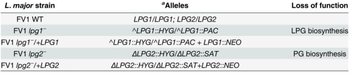
Table 1. Formal names forL.majorFV1 LPG and PG null mutants and add back lines.
L.majorstrain aAlleles Loss of function
FV1 WT LPG1/LPG1; LPG2/LPG2
FV1lpg1− ^LPG1::HYG/^LPG1::PAC LPG biosynthesis
FV1lpg1−/+LPG1 ^LPG1::HYG/^LPG1::PAC + LPG1::NEO
FV1lpg2− ΔLPG2::HYG/ΔLPG2::SAT PG biosynthesis
FV1lpg2−/+LPG2 ΔLPG2::HYG/ΔLPG2::SAT+LPG2::NEO
aA ^ denotes gene disruption and aΔ
denotes gene replacement
verified the absence of both PPGs and LPG in the FV1lpg2−mutant (S3C Fig, lane 2), and
their restoration in the complemented FV1lpg2−/+LPG2line (S3C Fig, lane 3).
L
.
major
FV1 LPG required for robust IL12 responses in hDCs
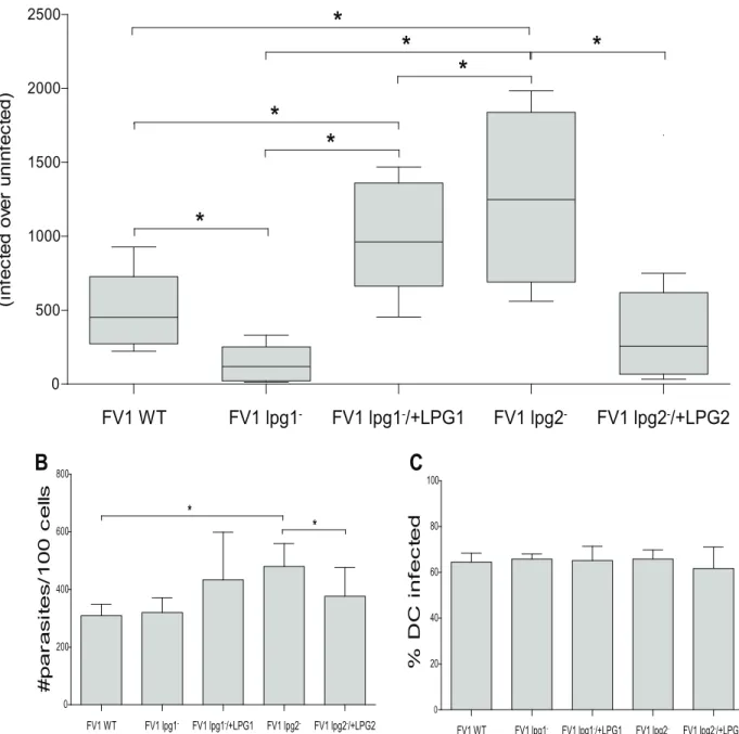
To explore the role of LPG on the IL12 response elicited fromL.majorinfected hDCs, we
quantified the relative amount ofIL12BmRNA in hDCs after 8 hours of infection with FV1
WT, FV1lpg1−, and FV1lpg1−/+LPG1parasites. Compared to FV1 WT, FV1lpg1−infected
hDCs displayed a substantial decrease in IL12 expression (3.2 fold;Fig 2A) that was restored to
levels approximately twice more than WT in the complemented FV1lpg1−/+LPG1line,
per-haps consistent with a slight elevation of LPG in this line (S3C Fig, lanes 4 and 5). Our results
indicate LPG plays a key role in IL12 induction in hDCs, consistent with the stimulatory effect
seen with purified LPG (Fig 1B).
Conversely, FV1lpg2−infected hDCs, relative to FV1 WT, displayed a significant increase
inIL12Bexpression, that returned to comparable FV1 WT levels in the complemented FV1 lpg2−/+LPG2line (Fig 2A). This observation was unexpected as FV1lpg2−lacks LPG as well as
other PGs, including PPGs (Fig 2C, lane 2). We considered the possibility that differences in
infectivity between the WT andlpg2−could contribute to this result asL.majorLv39c5lpg1−
andlpg2−mutants exhibit reduced survival in peritoneal macrophages [41,59]. While parasite
survival was slightly elevated in FV1lpg2−infections, a comparable fraction of DCs were
infected (Fig 2B and 2C), indicating, the differences observed in IL12 induction are likely not
related to parasite survival in hDC under the conditions tested.
Thus, our studies showed that LPG is associated with increased IL12 production when
tested biochemically (purified) or genetically (FV1lpg1−), while paradoxicallylpg2-which also
lacks LPG showed increased production. These data invoke the possibilityLPG2-dependent
molecules, such as phophoglycans including PPGs or other metabolites [60] may play a
sup-pressive role on IL12 production. Alternatively, the loss of allLPG2-dependent structures may
reveal another PAMP on the parasite surface that is able to induce IL12. Either scenario indi-cates a complex balance and interplay between parasite glycoconjugates and host cells.
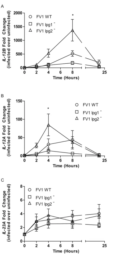
A kinetic analysis of these phenomena was conducted in DCs across four time points: 2, 4,
8, and 24 hours post-infection with FV1 WT and knockout mutants (Fig 3A). By 2 hours
post-infection, FV1lpg2−mutant infected hDCs induced slightly moreIL12Bcompared with FV1
WT infected DCs. Albeit at higher expression levels than FV1 WT, FV1lpg2−induced a similar
kineticIL12BmRNA response that declined by 24 hrs post infection. FV1lpg1−, on the other
hand, induced little to noIL12BmRNA (Fig 3A). Similarly, FV1lpg2−induced a quicker and
more robustIL12Aresponse compared to FV1lpg1−and FV1 WT infections (Fig 3B). There
were no differences between the WT and mutant strains for expression of the IL12 homolog IL23A(Fig 3C), suggesting that LPG and PGs regulate IL-12 production rather than IL-23.
Human DC
TNF
expression is reduced during
L
.
major
FV1
lpg1
−infections
In addition to IL12, DCs are strong producers of other Th1 proinflammatory cytokines. TNF,
for example, is significantly up-regulated inL.majorinfected hDCs [61]. We determined the
relative fold induction ofTNFin hDCs following infection with FV1lpg1−and FV1lpg2−
mutants. We demonstrated that FV1lpg1−induces significantly lessTNFmRNA compared to
WT or FV1lpg1−/+LPG1add back infections (Fig 4A), similar to the pattern ofIL12B
expres-sion (Fig 2A). Infection with FV1lpg2−, however, was not statistically different compared to
Down-regulation of
IL12B
in
L
.
major
FV1
lpg1
−infection is not
dependent upon IL10 induction
IL10 is generally implicated as a powerful inhibitor of IL12 production [62], and neutralizing
IL10 promotes the ability ofL.majorparasites to establish IL12 production [63]. Here we
Fig 2.L.majorFV1lpg1−and FV1lpg2−modulate theIL12Bresponse in hDCs.Human DCs (n = 9 donors) were infected withL.majorFV1 parasites:L.
majorFV1 (WT), LPG null mutant (FV1lpg1−), LPG add back (FV1lpg1−/+LPG1), PG null mutant (FV1lpg2−), or PG add back (FV1lpg2−/+LPG2). (A) At 8
hours post infection,IL12Bexpression was measured by qRT-PCR. Fold changes were calculated using theΔΔCTmethod and are represented as fold
change over uninfected samples. Box plots display the median value (line), the interquartile range (box), and Tukey whiskers encompassing data within 1.5 fold of the interquartile range. All values were significantly greater than uninfected. Aliquots from the infected hDC samples were prepared by Diff-Quick staining and visualized by light microscopy. (B) The parasite index (#parasites/100 cells) and (C) the percentage of infected cells (%DC infected) is displayed. Mean values of individual donors±SD are presented.*Statistical significance (p<0.05).
Fig 3. Kinetic analysis ofIL12andIL23expression modulation byL.majorLPG and PG null mutants. Human DCs (n = 3 donors) were infected withL.majorFV1 parasites:L.majorFV1 (WT), LPG null mutant (FV1lpg1−), LPG add back (FV1lpg1−/+LPG1), PG null mutant (FV1lpg2−), or PG add back (FV1
lpg2−/+LPG2). At 2, 4, 8, and 24 hours post infection,IL12B(A),IL12A(B), andIL23A(C) expression was
measured by qRT-PCR. Fold changes were calculated using theΔΔCTmethod and are represented as fold
change over uninfected samples. Mean values of individual donors±SD are presented. Statistical significance p<0.05, ANOVA with Bonferroni multiple comparisons test.
quantified theIL10mRNA levels in hDCs infected with our mutant parasites to determine
whether the failure of FV1lpg1−to elicit sustained host IL12 induction relative to FV1 WT is
due to the over-expression of IL10. TheIL10expression elicited from hDCs infected with FV1
lpg1−or FV1lpg2−did not differ from WT induced expression levels (Fig 4B), suggesting the
mechanism by which these mutant parasites modulateIL12Bexpression is not dependent
uponIL10.
Human microarray analysis reveals broader gene expression effects of
LPG and PPGs
To further assess the influence of LPG and PPGs on host immunological responses, we infected
additional DCs withL.majorFV1 WT, mutants, and complemented strains, collecting mRNA
at 8 hours post-infection. cDNA generated from these samples was hybridized to NimbleGen Homo sapiensExpression Microarrays. Expression of ten genes (IL12B,IL1B,IL8,TLR4,TLR2,
Fig 4. RelativeTNFandIL10levels inL.majorFriedlin V1 infected DCs.Human DCs were infected with
L.majorFV1 parasites:L.majorFV1(WT), LPG null mutant (FV1lpg1−), LPG add back (FV1lpg1−/+LPG1),
PG null mutant (FV1lpg2−), or PG add back (FV1lpg2−/+LPG2). At 8 hrs post infection, (A)TNF(n = 5
donors) and (B)IL10(n = 5 donors) expression was measured by qRT-PCR. Fold changes were calculated using theΔΔCTmethod and are represented as fold change over uninfected samples. Box plots display the
median value (line), the interquartile range (box), and Tukey whiskers encompassing data within 1.5 fold of the interquartile range; data outside this range are presented as individual data points (open circles).
*Statistical significance (p<0.05). All values were significantly greater than uninfected.
FKBP4,SOCS3,SMOX,FCGR1A, andTNFAIP3) correlated significantly using qRT-PCR (p<0.000001, Spearman correlation coefficient = 0.784), validating the array values (S4 Fig). Gene transcript expression values were transformed to Z-scores and those genes that were
sig-nificantly differentially expressed compared to uninfected cells (Z-score1.96) were retained
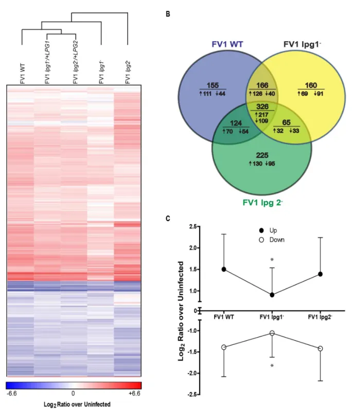
for downstream analysis. Hierarchical clustering of 730 genes that were expressed differently than FV1 WT infections in at least one mutant infection revealed that the complemented
strains clustered more closely to the WT strains than their respective mutant strains (Fig 5A).
Compared to uninfected cells, similar numbers of genes were regulated by infection with FV1
WT (771), FV1lpg1−(717) and FV1lpg2−(740) (Fig 5B). Infection with FV1 WT resulted in
more genes being up-regulated than either mutant strain (FV1 WT—524; FV1lpg1-—444; and
FV1lpg2−
—449). Notably, the magnitude of regulation (either up or down) was less during
infection with FV1lpg1−compared to either FV1 WT orlpg2−(Fig 5A and 5C), suggesting that
this strain enters hDC in a silent fashion.
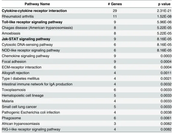
LPG regulates immune response and infectious disease pathways
To assess the pathways involved in the regulation of IL12 by LPG, we utilized STEM and
iden-tified 233 genes that exhibited expression patterns similar toIL12Bin response to infection
with FV1 WT, FV1lpg1−, and FV1lpg2−. Overalllpg2−resembled WT whilelpg1−differed (Fig
6). Pathway enrichment revealed 22 significantly enriched pathways, mostly belonging to the
immune response or infectious disease categories (Table 2). The most striking observation was
the enrichment of three pathways: Cytokine-Cytokine Receptor Interactions, JAK-STAT Sig-naling and Toll-like SigSig-naling, in which all the genes were down-regulated by infection with
FV1lpg1−compared to FV1 WT and FV1lpg2−(Fig 6). Although thelpg2-pathway genes did
not reflect any significance in this initial analysis compared to WT, future analysis of enriched pathways by criteria other than IL12 expression could reveal significant pathways enriched by lpg2- infection.
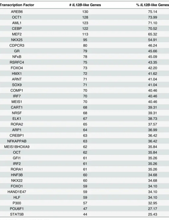
The most common transcription factor binding sites present in the promoters of genes
regu-lated similarly toIL12Bwere identified using the DAVID functional annotation tool [56]. Not
surprisingly, binding sites for transcription factor families known to regulateIL12Bwere
iden-tified, including, Octomer-binding transcription factor (OCT), Nuclear Factor Kappa B
(NFκB), Interferon Regulatory Factor (IRF), cAMP Response Element Binding protein
(CREB), and CCAAT/Enhancer Binding Protein families [64–68] (Table 3).
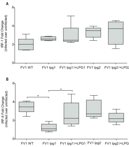
Human DC
IRF8
expression is reduced during
L
.
major
FV1
lpg1
−infection
Production of IL12 relies on the nuclear translocation and cooperative binding of IRF-1 and
IRF8 to IFNG-activated sequences (GAS) found within theIL12Bpromoter [18]. We
previ-ously demonstrated thatL.majorinfection of hDC results in the early activation of NFκB
tran-scription factors resulting in the trantran-scriptional induction and nuclear translocation of IRF-1
and IRF-8 and, ultimately, IL12 production [42]. To delineate the effect of FV1lpg1−and/or
FV1lpg2−on the upstream transcriptional features that regulateIL12Bexpression, we assessed
IRF1expression in hDCs and observed that infection with FV1 mutants up-regulatedIRF1, but
not significantly more compared to WT induced levels (Fig 7A). This result suggests that the
differentIL12Bresponses displayed during FV1lpg1−and FV1lpg2−DC infections are not
influenced by IRF1 expression.IRF8mRNA levels, however, were regulated by LPG. Infection
with FV1lpg1−resulted in a reduction ofIRF8that is restored following infection with the FV1
lpg1−add back strain (Fig 7B). Infection with FV1lpg2−did not significantly affectIRF8
Fig 5.Leishmania majorhuman host dendritic cells gene expression profiles.(A) Gene transcript expression heat map ofin vitroinfected monocyte-derived hDCs. The color scale is based on average log2ratios of RMA-normalized microarray gene probe set values for variably infected host cells over
uninfected cells. Only the genes that displayed significant differential expression by z-ratios, from both uninfected samples and between infections with FV1 WT and FV1lpg1−or FV1lpg2−mutants or their respective add back strains, were included in the map. Genes and sample types were clustered by city block
distance metric using average linkage in GENE-E. (B) Venn diagrams with the number of host DC genes significantly differentially expressed from uninfected samples in FV1 WT, FV1lpg1−, and FV1lpg2−mutants as quantified by microarrays. Values below the horizontal line indicate the number of genes from the
above total that were up- (") or down-regulated (#) compared to uninfected samples. (C) Total average log2ratios of up- and down-regulated genes
significantly differentially expressed from uninfected samples in FV1 WT, FV1lpg1−, and FV1lpg2−mutants as quantified by microarrays, plus or minus
standard deviation.*Significant difference of log2ratio values (p<0.05) between FV1lpg1−infected DCs compared to FV1 WT and FV1lpg2−infected DCs
by ANOVA with Bonferroni multiple comparisons test.
Fig 6. Enriched immunologically relevant pathways for genes expressed inIL12B-like patterns.Gene transcripts lists fromL.majorFV1 WT, FV1lpg1−, and FV1lpg2−mutantin vitroinfected monocyte-derived
hDCs microarray analysis, with log2ratio over uninfected value patterns between samples that clustered with
theIL12B, were analyzed to identify significantly enriched (Benjamini and Hochberg adjusted p<0.01) KEGG
Discussion
The major focus of this study was to investigate whether the enhanced IL12 immune response
observed inL.majorFV1 WT infected hDCs is dependent upon parasite LPG; as previous
studies have implicated LPG plays a major role in modulating immune function in murine cells [31,69,70], as well as in human mononuclear cells [71–73]. First, we showed that, for this
strain, infection with metacyclic promastigotes induces a highIL12Bresponse (Fig 1B),
com-pared to procyclic promastigotes and amastigotes, consistent with prior studies [57,58].
Addi-tionally, we demonstrated that purified LPG stimulates anIL12Bresponse in hDCs (Fig 1C).
Similar studies utilizing purifiedL.majorLPG from another strain have also highlighted the
stimulatory effect LPG has on IL12 in human PBMCs [72].
To assess the role of surface moleculesin situ, we employed genetic strategies to generate
parasite mutants devoid of LPG (FV1lpg1−) or PG molecules and other LPG2-dependent
metabolites (FV1lpg2−) in theL.majorstrain FV1 background (S3A Fig). Previous studies on
the‘low hDC IL12,L.majorstrain LV39c5 mutant parasites established several roles for LPG
and PGs in regulating immune function [31–33,41,60]. For example, LV39c5lpg2−induces
IL12 in mouse BMDCs co-stimulated with anti-CD40 or IFNG [32,33]. In the absence of
co-stimulation, however, there was no significant difference between IL12 elicited from LV39c5
WT or LV39c5lpg2−parasites. We observed a similar result in our hDC assay where there was
signaling pathway (C). All graphs display average log2ratio over uninfected values for genes present in the
expression datasets of FV1 WT, FV1lpg1−, and FV1lpg2−mutant infected samples which were members of
the corresponding enriched pathways.
doi:10.1371/journal.pntd.0004238.g006
Table 2. Significantly enriched pathways for genes regulated similarly toIL12Bby LPG.
Pathway Name # Genes p value
Cytokine-cytokine receptor interaction 29 2.31E-21
Rheumatoid arthritis 11 1.52E-08
Toll-like receptor signaling pathway 9 5.96E-06
Chagas disease (American trypanosomiasis) 8 5.22E-05
Amoebiasis 8 5.22E-05
Jak-STAT signaling pathway 9 8.16E-05
Cytosolic DNA-sensing pathway 6 8.16E-05
NOD-like receptor signaling pathway 6 8.16E-05
Chemokine signaling pathway 9 0.0003
Focal adhesion 9 0.0004
ECM-receptor interaction 6 0.0004
Allograft rejection 4 0.0011
Type I diabetes mellitus 4 0.0021
Intestinal immune network for IgA production 4 0.0032
Toxoplasmosis 6 0.0033
Hematopoietic cell lineage 5 0.0033
Malaria 4 0.0033
Small cell lung cancer 5 0.0033
Pathogenic Escherichia coli infection 4 0.0038
Phagosome 6 0.0061
African trypanosomiasis 3 0.0082
RIG-I-like receptor signaling pathway 4 0.0082
little difference in IL-12 induction between LV39c5 WT, LV39c5lpg2−, and LV39c5lpg2−
/+-LPG2infections (S6 Fig). Compared to FV1 WT, LV39c5 WT does not induce the same robust
levels ofIL12B(Fig 1A,S6 Fig).
Here, we generatedLPG1andLPG2knockout mutants in the‘high hDC IL12’L.majorFV1
background strain, in order to directly assess the parasite-derived molecular factors that
con-tribute to the robust hDC IL12 response elicited by this strain ofL.major. Our data
demon-strated that the FV1lpg1−mutant does not induce a high amount ofIL12Btranscript in hDCs
as compared to FV1 WT (Figs2Aand3A). Consistent with this observation, we showed that
application of purified LPG was able to induce significant IL12 expression (S5 Fig), with both
Table 3. Enriched Transcription Factor Binding sites inIL12B-like gene promoters.
Transcription Factor #IL12B-like Genes %IL12B-like Genes
AREB6 130 75.14
OCT1 128 73.99
AML1 123 71.10
CEBP 122 70.52
MEF2 113 65.32
NKX25 95 54.91
CDPCR3 80 46.24
GR 79 45.66
NFκB 78 45.09
RSRFC4 75 43.35
FOXO4 73 42.20
HMX1 72 41.62
ARNT 71 41.04
SOX9 71 41.04
COMP1 70 40.46
IRF7 70 40.46
MEIS1 70 40.46
CART1 68 39.31
NRSF 68 39.31
ELK1 67 38.73
RORA2 65 37.57
ARP1 64 36.99
CREBP1 63 36.42
NFKAPPAB 63 36.42
MEIS1BHOXA9 62 35.84
OCT 62 35.84
GFI1 61 35.26
IRF2 61 35.26
RORA1 61 35.26
HNF3B 60 34.68
NKX22 60 34.68
FOXO1 59 34.10
HAND1E47 59 34.10
HLF 59 34.10
P300 57 32.95
POU6F1 47 27.17
STAT5B 44 25.43
metacyclicL.majorLPG which bears abundant PG side chain modifications, andL.donovani LPG, which is unmodified.
In contrast, and somewhat surprisingly given its similar lack of LPG, FV1lpg2−up-regulates
theIL12Bresponse (Figs2Aand3A) relative to FV1 WT. While in macrophage and animal infections thelpg1-andlpg2- mutants are typically attenuated [41,59], in our studies the survival of the WT and two mutant parasites did not differ significantly in DC survival over the course of these studies (Fig 2B and 2C). One explanation for this finding is that inL.majorstrain FV1,
LPG and otherLPG2-dependent glycoconjugates play inverse roles in stimulating the IL12
response in human DCs. One candidate for such an inhibitory LPG2-dependent molecule are
the proteophosphoglycans (PPGs), which remain intact in thelpg1−mutant. Compared to LPG,
little is known about the function of PPGs on host cell immune response, with evidence
sup-porting roles as both an inhibitor or enhancer depending on the species and study [74–77].
PPGs vary structurally across species both in their PG and protein composition, and their large
size and tendency to form polymeric aggregates renders their study more challenging [78].
Fig 7. Thelpg1−mutant affects IL12 associated gene regulatorIRF8, and notIRF1.Human DCs were
infected withL.majorFV1 parasites:L.majorFV1(WT), LPG null mutant (FV1lpg1−), LPG add back (FV1
lpg1−/+LPG1), PG null mutant (FV1lpg2−), or PG add back (FV1lpg2−/+LPG2). At 8 hrs post infection, (A)
IRF8(n = 3 donors) and (B)IRF1(n = 5 donors) expression was measured by qRT-PCR. Fold changes were calculated using theΔΔCTmethod and are represented as fold change over uninfected samples. Box plots
display the median value (line), the interquartile range (box), and Tukey whiskers encompassing data within 1.5 fold of the interquartile range.*Statistical significance (p<0.05). All values were significantly greater than uninfected.
Clearly, the development of mutants lacking only PPGs would be beneficial for future studies to directly assess the role these molecules have on the host cell response. Interestingly, amastigotes
do not express significant amounts of the‘pro-IL12’LPG but do express high levels of PPG,
which may further contribute to their inability to stimulate IL12 expression in hDCs.
Impor-tantly, the LPG2-dependent effect was also observed in the‘low hDC IL12’LV39 line, where
ablation of LPG2 similarly resulted in increased IL12 production (S6 Fig)
Thus our data cause us to infer the presence of other LPG2-dependent PAMPs beyond LPG, with PPG as a possible candidate, and acting in an inhibitory fashion. The potential dom-inance of these inhibitory LPG2-dependent PAMPs provides an explanation for the
conun-drum that while allLeishmaniaspecies express LPG, despite that many do not induce IL12
[79]. Potentially, the strength of these suppressive LPG2-dependent PAMPs/processes may
vary in different species and/or strains.
As it has been established that IRF1 and IRF8 are up-regulated inL.majorinfected hDCs
and positively regulateIL12Bgene expression [42], we assessed whether FV1lpg1−or FV1
lpg2−affected the expression ofIRF1andIRF8. Interestingly, FV1lpg1−parasites caused a
sig-nificant decrease inIRF8expression compared to WT (Fig 5A), indicating that LPG may
influ-ence the induction ofIL12Bby targeting upstreamIL12Bassociated transcription factors that
mediate its expression. Although IRF1 and IRF8 are known to cooperatively regulateIL12B
gene transcription [42,80], we report that the FV1lpg1−mutant does not affectIRF1expression
compared to WT at 8 hours post-infection (Fig 4B). The distinct expression phenotypes
exhib-ited byIRF1andIRF8following infection with FV1lpg1−may be due to the difference in
regu-lation of these two transcription factors. IRF1 is ubiquitously expressed, whereas IRF8 is preferentially expressed in immune cells and in response to activating signals. Furthermore,
IRF1 and IRF8 can be differentially expressed in hDCs [81]. To bind target DNA sequences,
IRF8 must bind to another transcription factor, compared to other IRF family members that
can bind DNA sequences alone [82]. It is possible that infection with FV1lpg1−reduces the
amount of IRF8, which in turn inhibits the capacity of other transcription factors, such as
IRF1, to form heterodimeric complexes that bind theIL12Bpromoter. These data suggest that
LPG and not other PGs, enhance theIL12Bresponse by a common mechanism involvingIRF8.
Like IL12,L.majorinducesTNFin both human macrophages and DCs [61]. We therefore
evaluated the relationship between parasite derived PG-bearing molecules onTNFusing our
LPG and PG null mutants. Our results demonstrate that thelpg1−mutant exhibits a significant
decrease ofTNFexpression, similar to the reduction observed forIL12B(Fig 5A). Interestingly,
the promoter regions forIL12BandTNFhave similar transcription factor binding sequences,
namely NFκB and ETS sites; the latter containing ISRE sequences that promote gene
transcrip-tion upon IRF8 complex binding [83]. Therefore, it is possible that the reduction inTNF
expression observed during FV1lpg1−infection (Fig 5A) may also be IRF8-specific. A murine
study demonstrated that cholera toxin (CT) inhibits plasmacytoid dendritic cellular IL12 by
blocking the ability of IRF8 to bind to the ISRE sequence within theIL12Bpromoter, while
IRF1 phosphorylation and subsequent binding to its DNA target sequence remained unaf-fected [84]. It is feasible that a similar mechanism exists inL.majorinfected cells, whereby IRF8 is specifically targeted for induction downstream of parasite LPG binding, subsequently
leading to the induction ofIL12BandTNF. Altogether, our data indicates thatL.majorFV1
skews the hDC response in an LPG-dependent manner towards a Th1-like polarization charac-terized by an increase in IL12 and TNF production which may be regulated by a common
mechanism involving IRF8. A recent study demonstrated that macrophage induction ofIL12B
is controlled at the level of IRF8, which is specifically targeted for activation downstream of
TLR4 in concert with Notch signaling pathways [85]. Interestingly, TLR4 [86] and other TLRs
An alternative explanation for the lack of an IL12 signal observed in the FV1lpg1−
infec-tions may be a consequence of other functionally active PG-containing molecules, such as the
PPGs which remain intact in thelpg1−mutant. These PPGs could provide an inhibitory IL12
signal. This theory is supported by our results demonstrating that FV1lpg2−, which lacks both
LPG and PPGs, induces higher levels of IL12 compared to WT (Fig 2A), suggesting that some
PG-containing molecules actually inhibit IL12 responses. In addition, amastigotes, on which LPG expression is drastically down-regulated and high levels of other PG containing
glycocon-jugates are highly expressed [15], do not induce IL12 (Fig 1B). Compared to LPG, little is
known about the function of PPGs on host cell immune response. Previous work illustrating the ability of PPGs to induce complement activation by triggering the mannose binding protein
pathway [76] and their inability to elicit CD4+T-cell response in murine bone marrow derived
macrophages [74], concludes that PPGs may contribute to the chronic infections observed
dur-ingL.mexicanainfections. However, it has been demonstrated thatL.majorPPGs require
IFNG priming to induce TNF and NO production in murine macrophages [77]. In human
PBMCs, PPGs cause an induction of IL10 and to a lesser extent NO and IL12 [75]. Although
these studies provide conflicting implications for PPGs role as either inhibitor or enhancer of immune response, it is difficult to compare studies because the repertoire of PPGs structure
varies across species [78]. Additionally, the use of purified PPGs can be problematic because
the amount of purified PPGs added is often higher than what is biologically present during an actual infection, therefore the development of mutants lacking only PPGs would be beneficial for future studies to directly assess the role these molecules have on the host cell response. We
measuredIL-10mRNA levels in our mutant-infected DCs, because of the generally inhibitory
effects of IL-10 on IL12 [62]. However, IL10 expression exhibited between FV1lpg1−, FV1
lpg2−, and WT infected hDCs did not differ (Fig 5B), ruling out one theory that the decrease in
IL12Bexpression observed during FV1lpg1−could be consequence IL10 overproduction.
Another explanation for the induction of IL12 by FV1lpg2−, is the possibility that the absence
of all surface and secreted PGs reveals a molecular pattern or some other molecule that induces IL12.
Our microarray analyses of FV1WT, FV1lpg1−, and FV1lpg2−infected hDCs revealed that
FV1lpg1−enter hDC in a relatively silent fashion as indicated by the overall down-regulation
of significantly expressed transcripts, (Fig 6), and the overall reduction in genes belonging to
cytokine and TLR related gene pathways, (Fig 7). Altogether these data suggest that a lack of
LPG molecules results in silent entry and that LPG is a major pattern recognized by pattern recognition receptors on DCs. As with the IL12 response, the absence of all PGs appears to either release some sort of repression or reveals a molecular pattern that compensates for the lack of LPG, highlighting the complexity of DC pattern recognition receptor interactions in
controlling host responses toLeishmaniainfection. Future analyses focusing on FV1
lpg2-mutant infections may reveal pathways uniquely regulated by PGs.
This work adds to the growing set of genetically modified parasites (lpg1−,lpg2−in theL.
majorFV1 background) providing biologically relevant tools for assessing the role of para-site surface glycoconjugates on cellular function in human and mouse model systems, as well as, provides insight into the complex interplay of LPG and other PG molecules on the
cellu-lar immune response elicited followingL.majorinfections by global gene expression
analyses.
Supporting Information
S1 Fig. Confirmation ofL.majorFV1lpg1−mutant.(A) Schematic representation of WT
analysis for one representative FV1 WT, FV1lpg1−(cl 2.10) and FV1lpg1−/+LPG1(cl 2.10
AB3) is depicted. (B) Primers 1/2 (SMB1023/SMB1626) confirmedLPG1disruption: WT
(420bp), FV1lpg1−(3200bp) and FV1lpg1−/+LPG1(420bp & 3200bp). (C) Primers 3/11
(SMB4183/SMB2566), and 12/6 (SMB4185/SMB4184) established the integration of the 5’
flanking (3300bp) and 3’flanking (2600bp) sequences of theLPG1::HYGdisruption cassette,
respectively. Primers 3/7 (SMB4183/SMB2889), and 8/6 (SMB2888/SMB4184) established the
integration of the 5’flanking (3400bp) and 3’flanking (2800bp) sequences, of theLPG1::PAC
disruption cassette, respectively. Primers 9/10 (SMB2891/SMB2892) and 4/5 (SMB1568/
SMB1569) confirmed the presence of theHYG(1080bp) andPAC(600bp) ORFs, respectively.
(EPS)
S2 Fig. Confirmation ofL.majorFV1lpg2−mutant.(A) Schematic representation of WT
(top) andlpg2−alleles (bottom). Numbers represent primers used for PCR amplification. PCR
analysis for one representative FV1 WT, FV1lpg2−(cl 6.1A) and FV1lpg2−/+LPG2(cl 6.1A
AB15) is depicted. (B) Primers 13/14 (SMB1023/SMB1626) confirmed replacement ofLPG2:
WT (1000bp), FV1lpg2−(absent) and FV1lpg2−/+LPG2(1000bp). (C) Primers 15/11
(SMB4124/SMB2566), and 19/20 (SMB2565/SMB4125) established the integration of the 5’
flanking (1300bp) and 3’flanking (2200bp) sequences of theLPG2::HYGreplacement cassette,
respectively. Primers 15/17 (SMB4124/SMB3507), and 16/18 (SMB3506/SMB4417) established
the integration of the 5’flanking (1700bp) and 3’flanking (2400bp) sequences, of theLPG2::
SATreplacement cassette, respectively. Primers 9/10 (SMB2891/SMB2892) and 16/17
(SMB3506/SMB3507) confirmed the presence of theHYG(1080bp) andSAT(600bp) ORFs,
respectively. (EPS)
S3 Fig. Generation ofL.majorFV1 LPG null mutant (FV1lpg1−) and PG null mutant (FV1 lpg2−) mutant.WT parasites underwent two rounds of electroporation as described in the
methods to generate the FV null mutants. (A) For FV1lpg1−, WT parasites were transfected
with^LPG1::HYGand screened heterozygotes underwent a 2ndround of transfection with ^LPG1::PACto yield FV1lpg1−null mutant. A third round of transfection with add back vector
pXG-LPG1::NEOrestoredLPG1. (B) The FV1lpg2−was created utilizing a similar transfection
strategy with targeting constructs,ΔLPG2::HYG, andΔLPG2::SAT. TheLPG2was restored by
electroporation with add back vector pSNBR-LPG2::NEO. (C) Western blot analysis with anti
sera WIC79.3 indicated a loss of LPG in the FV1lpg1−mutant (lane 6) and loss of both PPGs
and LPG in the FV1lpg2−mutant (lane 2). The FV1lpg1−/+LPG1add backs (two clones are
shown, lane 4,5) and the FV1lpg2−/+LPG2add back (lane 3) exhibit restored levels of LPG and
PPGs, comparable to WT parasites (lane 1). (EPS)
S4 Fig. Validation of microarray expression by qRT-PCR.Ten genes were selected for
valida-tion by qRT-PCR analysis (A)IL12B, (B)SOCS3, (C)TNFAIP3, (D)IL1B, (E)IL8, (F)TLR4,
(G)TLR2, (H)FKBP4, (I)SMOX, and (J)FCGR1A. For each gene, fold change was calculated
using fold changes were calculated using theΔΔCT. Log2ratios of RMA-normalized microarray
gene probe set values for infected host cells over uninfected cells from the microarray (black bars) and fold changes from the qRT-PCR analysis (gray bars) are plotted and subjected to Pearson Correlation test (R value). Mean ± SEM is presented.
(EPS)
S5 Fig. Purified LPG stimulatesIL12Bexpression in hDCs.Human DCs were exposed to
1μg LPG derived fromL.majororL.donovanipromastigotes (n = 3 donors). After 8 hours,