Identification and purification of immunogenic proteins from nonliving
promastigote polyvalent
Leishma nia
vaccine (Leishvacin
)
Identificação e purificação de proteínas imunogênicas da vacina polivalente
de promastigotas mortas de
Leishmania
(Leishvacin
)
Sandra Regina Afonso Cardoso1, João Carlos França da Silva1, Roberto Teodoro da Costa1,
Wilson Mayrink1, Maria Norma Melo1, Marilene Suzan Marques Michalick1,
Ibrahim Afrânio Willi Liu1, Ricardo Toshio Fujiwara1and Evaldo Nascimento1
Abstract Immunogenic proteins from nonliving promastigote polyvalent Leishmaniavaccine against American tegumentary leishmaniasis (Leishvacin), produced by Biobrás (Biochemistry of Brazil ), Montes Claros, State
of Minas Gerais, Brazil, were identified and purified by polyacrylamide electrophoresis gel and electroelution. C57BL/10 mice were vaccinated with proteins with estimated molecular weights of 42, 46, 63, 66, 73, 87, 97, and 160kDa in three doses of 30µg of each protein at 15-day intervals combined with 250µg of Corynebacterium parvum followed by a challenge infection with 105 infective promastigotes from Leishmania (Leishmania)
amazonensis. The ability of these proteins to induce immune response and protection was analyzed. No statistical difference was observed in the level of IFN-γ induced by proteins in vaccinated groups in comparison with control groups. Six months after challenge infection, protection levels of 28.57; 42.86; 57.14; 42.86; 42.86, 57.14; 42.86 and 57.14% were demonstrated for each purified protein.
Key-words:Tegumentary leishmaniasis. Immunogenic proteins. Leishvacin.
Resumo Proteínas imunogênicas da vacina polivalente de promastigotas mortas de leishmanias (Leishvacin)
produzida pela Biobrás – Bioquímica do Brasil, Montes Claros, Minas Gerais, Brasil foram identificadas e purificadas por eletroforese em gel de poliacrilamida e eletroeluição. Camundongos C57BL/10 foram vacinados com proteínas de pesos moleculares estimados em 42, 46, 63, 66, 73, 87, 97 e 160kDa em três doses de 30µg de cada proteína combinada com 250µg de Corynebacterium parvumem intervalos de 15 dias e desafiados com uma infecção desafio de 105 promastigotas infectantes de Leishmania (Leishmania) amazonensis na
base da cauda. Foram avaliadas a habilidade dessas proteínas em induzir resposta imune e proteção dos animais vacinados após a infecção desafio. Nenhuma diferença estatística foi observada nos níveis de IFN-γ
nos grupos vacinados em comparação ao grupo controle. Proteção de 28,57; 42,86; 57,14; 42,86; 42,86, 57,14; 42,86; 57,14% foi demonstrado para cada proteína.
Palavras-chaves:Leishmaniose tegumentar.Proteínas imunogênicas. Leishvacin.
1. Departamento de Parasitologia do Instituto de Ciências Biológicas da Universidade Federal de Minas Gerais, Belo Horizonte, MG, Brasil. Supported by CAPES, PADCT/CNPq/Biotechnology, Grant # 62.0252/91.0
Address to: Dr. Evaldo Nascimento. Depto de Parasitologia/ICB/UFMG. Antonio Carlos 6627, Pampulha, 31270-901 Belo Horizonte, Minas Gerais, Brazil. Fax: 55 31-3499-2859
e- mail: [email protected]
Recebido para publicação em 11/3/2002.
American tegumentary leishmaniasis (ATL) is a disease caused by different species of Leishmania, which constitutes an important public health problem in Latin America9. The disease affects people living in a variety
of ecological settings, including primary rain forest as well as agricultural and urban areas. Even without considering under-reporting and the large number of incorrect diagnoses, the estimated prevalence is high and incidence of American cutaneous leishmaniasis has increased as a consequence of environmental changes and difficulties in controlling the vectors and reservoirs.
Protective immunity against leishmaniasis is largely mediated by T cells3 7 14 15. The nature of the
host immune response to different Leishmania infections is one of the main factors controlling the outcome of the disease. In mice, susceptibility and resistance to L. major have been correlated with selective stimulation of the CD4+ cell subsets, Th1 and Th2, and the type of cytokines that they produce4 31.
produce IL-4 and IL-10, exacerbates disease12 29 30 31 32.
Similarly to the L. major in Balb/c30, treatment of Balb/c
mice with anti-IL-4 monoclonal antibodies abrogates its susceptibility to L. amazonensis6. In addition, in vitro and
in vivo studies indicate that IL-121 6 and tumor necrosis
factor a (TNF-α) are also crucial to the establishment of resistance against experimental leishmaniasis33.
Efforts to develop a vaccine to control ATL have been made by several laboratories. Different vaccine compositions including crude and purified Leishmania components have been tested in animal models as well as in human vaccination trials. In Brazil, the development of a non-living promastigote vaccine against ATL was initiated by Pessoa’s group in the decade of 194025 26,
followed by the first publication by Mayrink‘s group in 197919. Significant progresses were made in several
clinical trials demonstrating the immunogenicity of the vaccine, such as: (a) induction of a delayed-type hypersensitivity, as demonstrated by a positive skin test19; (b) no appreciable side effects21; (c) conversion
of delayed-type hypersensitivity with a statistically significant level of protection compared with non-converted or non-vaccinated subjects2 27; (d) old vaccine
preparations (four years old) proved to be equivalent to newly prepared ones in converting the results of skin tests from negative to positive as well as providing protection21; (e) protection of 50% of vaccinated
individuals whose skin test converted from negative to positive after vaccination2; (f) strong correlation has been
found between positive skin test results and lymphocyte stimulation indices (LSI) in vaccinated subjects24; (g)
LSIs of vaccinated groups were significantly higher (P<0.001) than those of placebo group26; (h) eight major
antigens with estimated molecular weights of 13.5 to 160kDa may play an important role in the immunity against tegumentary leishmaniasis24; (i) gp63 was the
major protein present in our vaccine24; and (j) T
lymphocyte-mediated immune response against Leishmania antigens, by Th1 profile response and by protection against the disease, was observed in 50% of subjects vaccinated2 8 19 21 22 24.
This vaccine has become an important tool in the identification of the key parasite antigens that induce immunity against tegumentary leishmaniasis, and has also been successfully used as an immunotherapeutic agent20 18.
In this study, we isolated proteins of estimated molecular weight of 42, 46, 63, 66, 73, 87, 97 and 160kD from Leishvacin, by preparative polyacrylamide gel
electrophoresis, and tested their ability to induce IFN-γ
synthesis by T lymphocytes from vaccines. We also analyzed the ability of these isolated proteins to induce protective immunity in susceptible C57BL/10 mice againstL. (L.) amazonensis.
MATERIAL AND METHODS
Leishvacin®. Leishvacin® was produced and
supplied by BioBrás (Biochemistry of Brazil), according to Mayrink‘s group methodology of production21 in GMP
(good manufacturing practice) conditions. It is constituted by five Leishmania stocks: (Leishmania (Leishmania) amazonensis MHOM/BR/60/BH6, Leishmania major-like, MHOM/BR/73/BH121, Leishmania major-like MHOM/BR/71/BH49, Leishmania (Leishmania) amazonensis IFLA/BR/67/PH8, Leishmania (Viannia) braziliensis MHOM/BR/70/M117619 24). Proteins from
Leishvacin® were isolated on 10% SDS-PAGE14. The
proteins were electroeluted from gel slices, dialyzed against PBS pH 7.2, concentrated and dialyzed again in the same buffer. Protein concentrations were determined according to Lowry’s method16.
C57BL/10 mice. Isogenic female mice aged 8-10 weeks were used in the vaccination experiments. These animals were maintained in the animal house of the Federal University of Minas Gerais, State of Minas Gerais, Brazil, under US National Institutes of Health guidelines for animal care.
Mice vaccination. Eight groups of seven 8-10 week mice, obtained from animal facilities at Federal University of Minas Gerais, Minas Gerais State, Brazil, were vaccinated subcutaneously into the left footpad with three doses of each identified and purified protein (30µg) at 15-day intervals combined with 250µg of live CP (Corynebacterium parvum – Fundação Athaulpho de
Paiva, Rio de Janeiro, Brazil), as adjuvant. Three control groups with seven animals each received only 250µg of live CP, 100µl of PBS (phosphate buffer saline) or 100µg of Leishvacin® in 100µl of PBS, respectively, following
the same immunization scheme.
Parasite for challenge infection. The L. (L.) amazonensis IFLA/BR/67/PH8 strain was used for experimental infections. This strain was maintained by continuous passages in hamsters for the preparation of infective promastigotes. Promastigotes were washed in 0.8% NaCl solution and 105 stationary phase
promastigotes of L. amazonensis were used in the challenge infection for each C57BL/10 mice. The injection was done into the base of the tail, seven days after the last vaccination. The development of lesions was monitored at 15-day intervals for 180 days.
Lymphocyte stimulation index (LSI). Six months after challenging the animals, they were sacrificed and the spleens were transferred to Petri dishes containing complete RPMI 1640 medium (Gibco, USA), as previously described23.
Interferon gamma assay (IFN-γγ).Spleen cells were obtained from C57BL/10 mice of each vaccinated group, as well as control mice at six months after challenge infection. Cells were prepared as previously described11.
Cells were isolated by Ficoll-hypaque gradient centrifugation and suspended at 1.5 x 10⁶cells per ml in
100UI of penicillin per ml, 50µg of streptomycin per ml, 10mM HEPES [N-2-hydroxyethylpiperasine-N-2-ethanesulfonic acid]) containing 10% inactivated fetal calf sera (FBS). Cell culture was carried out in flat-bottomed 24-well plates and maintained at 37oC in a
5% CO2 incubator for two days. The IFN-γ measurement was performed by ELISA on supernatant pools of triplicate cultures stimulated with each protein (20µg/ ml), Leishvacin® (50µg/ml) and Corynebacterium parvum
(50µg/ml) at 48 hours after stimuli. Results are presented as means of cytokine concentrations (UI/ml) determined for seven vaccinated individuals in each group.
Vaccine efficacy. Clinical observations of the animals and lesion development were carried out during 180 days after the challenge infections at seven-day
intervals. Lesion measurements were done at 15-day intervals using a micrometer (Mitutoyo Sul Americana, São Paulo, Brazil). The results were expressed as percentages of protection, which represent the animals without lesions per group. The animals were sacrificed and smears from footpad skin were Giemsa stained for the presence of parasites. For histopathological examinations a biopsy was taken at the site of infection fixed in 10% formalin in PBS, washed in water for 4 hours, dehydrated and embedded in paraffin, cut (3-4µm thick) and stained with hematoxylin and eosin for optical microscopic examination35.
Statistical analysis. Statistical significance was determined by Pearson chi-square test, Student t test10,
and Statistical Epinfo analysis version 6.0. RESULTS
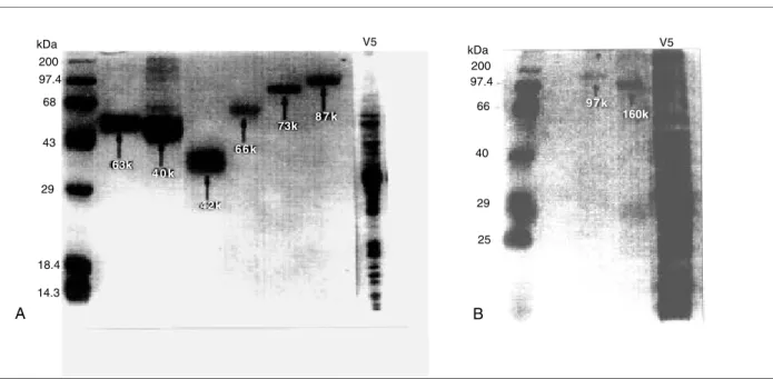
Eight proteins with estimated molecular weights of 42, 46, 63, 66, 73, 87, 97 and 160kD were purified from Leishvacin (Figure 1A,B).
All proteins were tested in vitro to stimulate peripheral blood monocyte cells (PBMC) from vaccinated mice in order to determine their ability to induce synthesis of IFN-γ. The results showed that the levels of IFN-γ, produced by protein stimulation in culture supernatants, were not statistically significant (P>0.05) ranging from
19.8UI/ml to 15.8UI/ml (Table 1). The levels of IFN-γ induced by each protein were comparable to those induced by Leishvacin. These proteins were also able to induce cell
proliferation and no statistically significant differences were observed between them with regard to the amplitude of proliferation induced (Table 1).
Vaccination of C57BL/10 mice with each of these purified proteins resulted in different levels of protection, as shown in Table 2. Lesion development was followed
Figure 1 - Proteins with estimated molecular weights of 63, 46, 42, 66, 73 and 87kD (A) and 97, 160kD (B) in 10% polyacrylamide gel electrophoresis (SDS-PAGE) purified from Leishvacin (V5). MW - molecular weight markers.
over a period of 180 days after challenge with 105
promastigotes of L. amazonensis. Non-vaccinated mice that received only C. parvum developed progressive infections on the footpads. The proteins of 63, 87 and 160kD induced protection of 4/7 (57.14%) mice, similar to the
protection obtained with Leishvacin. Protective rates
of 3/7 (42.86%) mice were obtained by those animals vaccinated with proteins of 46, 66, 73 and 97kD.The protein of 42kD induced protection of only 2/7 (28.57%) mice. In the other two control groups, all animals were infected.
kDa 200 97.4 68
43
29
18.4
14.3
A
63k 4 0 k
42k 66k
73k 8 7 k V5
kDa 200 97.4
66
40
29
25
97k 160k
V5
Good correlation was found between protection and LSI in the groups immunized with the purified proteins and in the group immunized with Leishvacin®, in
comparison with the other control groups (only CP and PBS). Such data could be observed for the proteins of 46, 63, 66, 87, 97 and 160kD, whereas the 42kD proteins produced only a slight protective effect (Table 1).
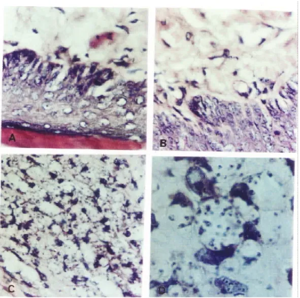
The presence of parasites in the footpad lesions of all mice was evaluated by histopathological studies involving serial sections of the footpad tissue. No parasites were found in vaccine-protected mice, while in the non-vaccinated ones parasites were observed inside macrophages or among the cells. Amastigote forms were found through histopathological examination in the control groups (Figure 2).
Table 1 – Gamma interferon (IFN-γ) production by human peripheral blood monocytes from vaccinated subjects and lymphocyte stimulation index (LSI) from vaccinated C%&BL/ 10 mice.
Group (7 mice) Protein (kD) IFN-γ (UI/ml) LSI (Ratio)
1 42 + CP 17.5 3.7
2 46 + CP 16.4 8.0
3 63 + CP 17.6 7.5
4 66 + CP 17.1 9.6
5 73 + CP 16.6 5.8
6 87 + CP 17.3 6.5
7 97 + CP 19.8 8.3
8 160 + CP 15.8 12.2
9 Leish + CP 14.4 11.2
10 CP 4.6 1.5
11 PBS 5.5 1.8
CP:Corynebacterium parvum; Leish: Leishvacin®; PBS: phosphate buffer saline
Table 2 – Protection of C57BL/10 mice induced by proteins purified from Leishvacin® after
challenge infection with 105 promastigotes of Leishmania (Leishmania) amazonensis
Group (7 mice) Protein (kD) Infection (Protection - %) Pearson (Chi square)
1 42 + CP 5/7 (28.57) P > 0.05
2 46 + CP 4/7 (42.86) P < 0.05
3 63 + CP 3/7 (57.14) P < 0.02
4 66 + CP 4/7 (42.86) P < 0.05
5 73 + CP 4/7 (42.86) P < 0.05
6 87 + CP 3/7 (57.14) P < 0.02
7 97 + CP 4/7 (42.86) P < 0.05
8 160 + CP 3/7 (57.14) P < 0.02
9 Leish + CP 3/7 (57.14) P < 0.02
10 CP 7/7 (00.00)
-11 PBS 7/7 (00.00)
-CP:Corynebacterium parvum; Leish: Leishvacin®; PBS: phosphate buffer saline
DISCUSSION In this study, we demonstrated that eight proteins,
isolated from Leishvacin, induced synthesis of IFN-γ by
lymphocyte from vaccinated mice and induced lymphocyte proliferation in vaccinated animals. Previous studies have shown that human individuals vaccinated with Leishvacin developed Leishmania-specific T cell
responses and protective immunity against this parasite. Characterization of cytokine production and lymphocyte phenotype involved in this immune response indicated that PBMC from vaccinated subjects induced IFN-γ but not IL-422. Moreover, in subjects vaccinated one year
before, CD8+ T cells proliferated preferentially in response toL. (V.) braziliensis antigen22.
Different levels of protection were observed in C57BL/ 10 mice vaccinated with each protein. C57BL/10 mice are resistant to L. major, but susceptible to L. amazonensis infection, developing a chronic infection31. A polarized Th1
response is detected after L. major infection, but this response is absent following L. amazonensis infection, indicating that parasite-associated factors are able to modify the T helper response profile induced in C57BL/ 10 mice30.
of infection. Th1 lymphocytes and synthesis of IFN-γ and IL-2 are associated with mild or self-healing disease, whereas the expansion of IL-4- and IL-10-producing Th2 lymphocytes is associated with disseminated infection31.
A similar distinction has been observed in patients with different disease manifestations. T cells present in lesions of patients with localized and self-healing cutaneous leishmaniasis predominantly secrete IFN-γand other Th1 cytokines, but not IL-45 27 31. CD8+ T cells also synthesized
IFN-γ, even though the protective role of these cells in leishmaniasis is not completely clear. The analysis of in vitro IFN-γ production in response to purified antigens therefore provides a reliable parameter for the ranking of immunodominant antigens.
Among the eight proteins evaluated, gp46 and gp63 have already received considerable attention and are
known to induce protective immunity in susceptible mice, as well as synthesis of IFN-γ. Furthermore, in a previous report, C57BL/10 mice were immunized with purified proteins from L. (L.) amazonensis and showed an induction of a protective immunity and an elevated synthesis of IFN-γ by spleen cells, suggesting a protection against the parasite23.
However, in our study, no positive correlation between levels of IFN-γ and protection was found. In addition, the amounts of IFN-γ production observed in susceptible mice were equivalent to the levels induced by Leishvacin. This
result also indicates a positive correlation between proliferation response and protection. These proteins are Leishmania immunodominant antigens and also important components of Leishvacin.
We must consider, however, that each protein fraction evaluated in this study may consist of a mixture of different proteins. Leishvacin is composed of five
different stocks of Leishmania, made from different species. Although high levels of homology may exist
among them, the presence of immunossupressive protein or epitopes cannot be ruled out24. Therefore,
further studies are required to investigate the immunogenic potential of these proteins isolated from theLeishmania strains that comprise Leishvacin.
REFERENCES
1. Afonso LCC, Scott P. Immune responses associated with susceptibility of C57BL/10 mice to Leishmania amazonensis. Infection and Immunity 61: 2952-2959, 1993.
2. Antunes CF, Mayrink W, Magalhães PA, Costa CA, Melo MN, Michalick MM, Williams P, Oliveira Lima A, Vieira JF, Schettini AM. Controlled field trials of a vaccine against New World Cutaneous Leishmaniasis. International Journal of Epidemiology 15: 572-580, 1986.
3. Ashford RW, Desjeux P, Raadt P. Estimation of population at risk of infection and number of cases of leishmaniasis. Parasitology Today 8: 104-105, 1992.
4. Bretscher PA, Wei G, Menon JN, Bielefeldt H. Establishment of stable, cell mediated immunity that makes “susceptible” mice resistant to Leishmania major. Science 257: 539-542, 1992.
5. Cáceres-Dittmar G, Tapia FJ, Sanches MA, Yamamura M, Uyemura K, Modlin RL, Bloom BR, Convit J. Determination of the cytokine profile in American Cutaneous Leishmaniasis using the polymerase chain reaction. Journal of Clinical Investigation 91: 500-505, 1993.
6. Chatelain R, Varkila K, Coffman R L. IL-4 induces a Th2 response inLeishmania major infected mice. Journal of Immunology 148:1182-1187, 1992.
7. Convit J, Ulrich M, Fernández CT. The clinical and immunological spectrum of American cutaneous leishmaniasis. Transaction of the Royal. Society of Tropical Medicine and Hygiene 87: 444-448, 1993.
8. De Luca PM, Mayrink W, Alves CA, Coutinho SG, Oliveira MP, Bertho AL, Toledo VPCP, Costa CA, Genaro O, Mendonça SCF. Evaluation of the stability and immunogenicity of autoclaved and non-autoclaved preparations of the vaccine against tegumentary leishmaniasis. Vaccine 17: 1179-1185, 1999.
9. Desjeux P. Aspects de Saluté publique et lutte. In: Dedet, JP (ed) Les Leishmanioses. Paris AUPELF-EREFF P, 219-238, 1999.
10. Dixon W, Massey JFJR. Introductin to Estatistical Analysis. Tokio: McGraw-Hill Booh, The normal distribution. Cap. 5. p.56, 1969.
11. Gazzinelli RT, Hakim FT, Hieny S, Shearer GM, Sher A Synergistic role of CD4+ and CD8+ T lymphocytes in IFN-γ production and protective immunity induced by an attenuated Toxoplasma gondii vaccine. Journal of Immunology 146:286-291, 1991.
12. Heinzel FP, Sadick MD, Mutha SS, Holaday BJ, Coffman RL, Locksley, RM. Reciprocal expression of interferon gamma or interleukin 4 during the resolution or progression of murine leishmaniasis. Journal of Experimental Medicine 169: 3149-3155, 1989.
13. Laemmli UK. Cleavage of structural proteins during assembly of the head of bacteriophage T4. Nature 227:680-685, 1970.
14. Liew FY. Cell-mediated immunity in experimental cutaneous leishmaniasis. Parasitology Today 2:264-270, 1986.
15. Locksley RM, Louis JA. Immunology of leishmaniasis. Current Opinion in Immunology 4: 413-218, 1992.
16. Lowry OH, Rosembrough NJ, Randall RJ. Protein measurement with folin reagent. Journal of Biological Chemistry 193:265-275, 1951.
17. Marzochi KB, Marzochi MA, Silva AF, Grativol N, Duarte R, Confort EM, Moddaber F. Phase I study of an inactivated vaccine against American tegumentary leishmaniasis in normal volunteers in Brazil. Memórias do Instituto Oswaldo Cruz 93: 205-212, 1998.
18. Mayrink W. Tratamento da Leishmaniose Tegumentar Americana utilizando vacina. Anais Brasileiro de Dermatologia 66:55-58, 1992.
19. Mayrink W, Da Costa CA, Magalhães PA, Melo MN, Dias M, Oliveira Lima A, Michalick MM, Williams P. A field trial of a vaccine against American dermal leishmaniasis. Transaction of the Royal Society of Tropical Medicine and Hygiene 73:385, 1979.
20. Mayrink W, Magalhães PA, Michalik MSM, Da Costa CA, Oliveira Lima A, Melo MN, Toledo VPCP, Nascimento E, Dias M, Genaro O, Hermeto MV, Williams. Immunotherapy as a treatment of American cutaneous leishmaniasis: preliminary studies in Brazil. Parasitologia 34: 159-165, 1992.
21. Mayrink W, Williams P, Da Costa CA, Magalhães PA, Melo MN, Dias M, Oliveira Lima A, Michalik MM, Carvalho EF, Barros GC, Sessa PA, Alencar JA An experimental vaccine against American dermal leishmaniasis: experience in the Espirito Santo, Brazil. Annals of Tropical Medical and Parasitology 79: 259-269, 1985.
22. Mendonça SE, De Luca PM, Mayrink W, Restom TG, Conceição-Silva F, Da Cruz AM, Bertho AL, Da Costa CA, Genaro O, Toledo VP, Coutinho SG. Characterization of human T lymphocyte-mediated immune response induced by vaccine against American tegumentary leishmaniasis. American Journal of Tropical Medicine and Hygiene 55:195-201, 1995.
23. Mora AM, Mayrink W, Da Costa RT, Da Costa CA, Genaro O, Nascimento, E. Protection of C57BL/10 mice by vaccination with association of purified proteins from Leishmania (L.) amazonensis. Revista do Instituto de Medicina Tropical de São Paulo 41: 243-248, 1999.
24. Nascimento E, Mayrink W, Da Costa CA, Michalick MM, Melo MN, Barros GC, Dias M, Antunes CMF, Lima MS, Toboada DC, Liu TY. Vaccination of humans against cutaneous leishmaniasis: cellular and humoral immune responses. Infection and Immunity 58:2198-2203, 1990.
25. Pessoa SB. Segunda nota sobre a vacinação preventiva na leishmaniose tegumentar americana com leptomonas mortas. Revista Paulista de Medicina 19: 1-9, 1941.
26. Pessoa SB, Pestana BR. Ensaio sobre a vacinação preventiva na leishmaniose tegumentar amaricana. Revista de Biologia e Higiene 10: 157-161, 1940.
27. Pirmez C, Yamamura M, Uyemura K, Paes-Oliveira M, Conceição-Silva F, Modlin RL. Cytokine patterns in the pathogenesis of human leishmaniasis. Journal of Clinical Investigation 91: 1390-1395, 1993.
29. Sadick MD, Heinzel BJ, Holaday BJ, Pu RT, Dawkins, RS, Locksley, RM. Cure of murine leishmaniasis with anti-interleukin-4 monoclonal antibody. Evidence for a T-cell dependent, interferon mechanism. Journal of Experimental Medicine 171:151-127, 1990.
30. Scott P. The role of Th1 and Th2 cells in experimental cutaneous leishmaniasis. Experimental Parasitology 68: 369-372, 1989.
31. Scott P. IFN-γ modulates the early development of Th1 and Th2 responses in a murine model of cutaneous leishmaniasis. Journal of Immunology 147: 3149-3155, 1991.
32. Scott P, Notovitz P, Coffman Rl, Pearce E, Sher A. Immunoregulation of cutaneous leishmaniasis. T cell lines that transfer protective immunity or exacerbation belong to different
T helper subsets and respond to distinct parasite antigens. Journal of Experimental Medicine 168: 1675-1684, 1988.
33. Sypek JP, Chung CL, Mayor SH, Subramanyan JM, Goldman SI, Sceburth DS, Wolf SF, Schaub RG. Resolution of cutaneous leishmaniasis: interleukin 12 initiated protective T helper type 1 immune response. Journal of Experimental Medicine 177:1797-1802, 1993.