Physiologically Low Dose
Leishmania major
Infection
Model after
a
GalCer Analog PBS57 Stimulation
Klaus G. Griewank1,2, Beate Lorenz1, Michael R. Fischer1, Louis Boon3, Susanna Lopez Kostka1, Esther von Stebut1*
1Department of Dermatology, University Medical Center, Johannes Gutenberg-University, Mainz, Germany,2Department of Dermatology, University Duisburg-Essen, University Hospital Essen, Essen, Germany,3Bioceros, Utrecht, The Netherlands
Abstract
Leishmaniasis is a parasitic infection affecting,12 million people worldwide, mostly in developing countries. Treatment options are limited and no effective vaccines exist to date. Natural Killer T (NKT) cells are a conserved innate-like lymphocyte population with immunomodulating effects in various settings. A number of reports state a role of NKT cells in different models ofLeishmaniainfection. Here, we investigated the effect of NKT cells in a physiologically relevant, intradermal low dose infection model. After inoculation of 103infectious-stageL. major, comparable numbers of skin-immigrating NKT cells
in both susceptible BALB/c mice and resistant C57BL/6 mice were noted. Compared to their wild type counterparts, NKT cell-deficient mice on a C57BL/6 background were better able to contain infection withL. majorand showed decreased IL-4 production in cytokine analysis performed 5 and 8 weeks after infection. Low doses of the NKT cell stimulatingaGalCer analog PBS57 applied at the time of infection led to disease exacerbation in C57BL/6 wild-type, but not NKT-deficient mice. The effect was dependent both on the timing and amount of PBS57 administered. The effect of NKT cell stimulation by PBS57 proved to be IL-4 dependent, as it was neutralized in IL-4-deficient C57BL/6 or anti-IL-4 antibody-treated wild-type mice. In contrast to C57BL/6 mice, administration of PBS57 in susceptible BALB/c mice resulted in an improved course of disease. Our results reveal a strain- and cytokine-dependent regulatory role of NKT cells in the development of immunity to low doseL. majorinfections. These effects, probably masked in previous studies using higher parasite inocula, should be considered in future therapy and immunization approaches.
Citation:Griewank KG, Lorenz B, Fischer MR, Boon L, Lopez Kostka S, et al. (2014) Immune Modulating Effects of NKT Cells in a Physiologically Low Dose Leishmania majorInfection Model afteraGalCer Analog PBS57 Stimulation. PLoS Negl Trop Dis 8(6): e2917. doi:10.1371/journal.pntd.0002917
Editor:Genevieve Milon, Institut Pasteur, France
ReceivedDecember 17, 2013;AcceptedApril 17, 2014;PublishedJune 26, 2014
Copyright:ß2014 Griewank et al. This is an open-access article distributed under the terms of the Creative Commons Attribution License, which permits unrestricted use, distribution, and reproduction in any medium, provided the original author and source are credited.
Funding:The authors received no specific funding for this study.
Competing Interests:The authors have declared that no competing interests exist.
* Email: [email protected]
Introduction
Leishmaniasis is a parasitic disease which is caused by a variety ofLeishmania spp.and affects about 12 million people worldwide. There are approximately 2 million people newly infected every year. Transmitted by a sand fly, it primarily affects people in (sub-) tropical climates. Depending on the genetic background and the immune status of the patient, the disease can have various clinical presentations. It can be primarily cutaneous, mucocutaneous, multilocular, chronic, or recurrent and in severe cases develop visceral forms. About 90% of infected individuals suffer from cutaneous leishmaniasis (CL), which often heals with disfiguring scars [1]. Primarily affecting poorer populations, there is little incentive for drug and vaccine development. Leishmaniasis is considered a neglected disease by the WHO.
Experimentally, leishmaniasis has long been used as an immunologic model. Infected mouse strains can either develop a Th1/Tc1-driven immune response successfully containing infec-tion or a Th2/Th17/Treg response, ultimately succumbing to uncontrolled parasite loads. A better understanding of the disease and the immunologic mechanisms involved will hopefully result in improved treatment options and an effective vaccine.
Natural killer T (NKT) cells are a subset of T cells first identified in the early 90s2. Obtaining their name because they express a subset of receptors primarily expressed on natural killer (NK) cells, they also have a number of other unique properties which distinguish them from conventional T cells. They harbor a restricted set of T cell receptors and recognize endogenous and exogenous lipid antigens presented by a MHC class 1b molecule named CD1d [2,3]. They also have an effector phenotype and function, readily able to secrete considerable amounts of both IL-4 and IFNcupon stimulation [4]. They have been shown to regulate and influence the immune response to a variety of infectious agents being activated either by direct recognition of bacterial lipid antigens or in a CD40-, IL-12-dependent manner by dendritic cells (DC) presenting endogenous lipids [5]. The strongest known ligand activating NKT cells is aGalactoysl-Ceramide (aGalCer)
[6]. Stimulation of NKT cells byaGalCer can lead to considerable
lipophosphoglycan and an impaired immune response in NKT deficient CD1d2/2 BALB/c mice was noted [18]. L. donovani
infection causing IFNcsecretion by NKT cells through liver SIRP
(signal regulatory proteina) upregulation was also reported [10]. In cutaneous leishmaniasis, infections with.106L. majorparasites (s.c. or i.v.) showed delayed parasite clearance in NKT cell-deficient CD1d2/2 and J
a182/2 mice [13]. In summary, the
results of these studies are variable and do not provide a coherent understanding of the role of NKT cells in leishmaniasis. Allin vivo
studies reported, with the exception of vaccine trials by Dondji et al. [19], used supraphysiological high dose parasite inocula, applying.106promastigotes at the time of infection.
In our study we analyzed the effect of NKT cells in a physiologically relevant model of cutaneous leishmaniasis applying 103infectious stageL. majorpromastigotes intradermally into the ear. Our results differ considerably from prior high dose inocula studies and show effects that point toward an important influence of NKT cells on the development of protective immunity againstLeishmania. This clear role of NKT cells for parasite clearance could be important for the development of new treatment options or an effective vaccine.
Materials and Methods
Mice
C57BL/6, Ja182/2, CD1d2/2, and IL-42/2mice (all C57BL/ 6 background), and BALB/c mice were housed under specific pathogen-free conditions in the animal care facility in Mainz.
Ethics statement
All animal experiments were conducted in accordance with federal guidelines and approved by the ethical committee of the state of Rheinland Pfalz (according to 18 Abs. 1 des Tierschutzgesetzes,
Landesuntersuchungsamt, approval#LUA 23 170–07/G07-1-022).
Parasites and infection
Metacyclic promastigotes ofL. majorclone VI (MHOM/IL/80/ Friedlin) were prepared as described previously [20]. Groups of
three to five mice were infected with low-dose (103) inocula in a volume of 20ml by intradermal injection into ear skin applying 0.3 mm diameter needles. Lesion volumes were measured weekly in three dimensions and are reported as ellipsoids: [(a/26b/26c/ 2)64/3p]. Parasite burdens were enumerated using a limiting
dilution assay [20]. All experiments used an artificial variant of
aGalCer termed PBS57 [21]. It was applied i.p. after dilution in 200ml PBS at the time of treatment or as referred to in the
manuscript. Control groups received 200ml PBS i.p. IL-4 neutralizing antibodies (clone 11B11) were applied i.p. at 1 mg/ 200ml, one day before and one day after infection.
Cytokine secretion profiles
To measure antigen-specific cytokine production, retroauricular LNs were isolated and single cell suspensions prepared. One million LN cells in 200ml complete RPMI 1640 medium
(BioWhittaker) were cultured in the presence of 25mg/ml SLA. Supernatants were harvested 48 h after stimulation and analyzed with ELISAs specific for IFNc (R&D Systems), IL-4 and IL-10 (BD). IFNcand IL-4 levels in serum samples were measured the
same way.
Single cell preparation
The livers were harvested, minced, and the tissue pieces strained through 70-mm mesh and pelleted by centrifugation. The cell
pellet was resuspended and lymphocytes isolated by Percoll gradient, spun at 200 g. Spleen and lymph node cells were isolated by mechanically grinding the tissue through 70-mm cell
strainers.
Cell isolation from infected mouse ears
Ears infected withL. majorwere excised, soaked in 70% ethanol, and washed with PBS. The ears were split into halves and placed in 0.5 mg/ml liberase (Sigma-Aldrich) diluted in RPMI 1640 with 5% penicillin/streptomycin for 1.5 h at 37uC. Liberase was inactivated by adding complete RPMI 1640 containing 5% FCS. The ears were put into 50mM Medicon homogenizers (BD) with 1 ml of complete
RPMI 1640 and homogenized in a Medimachine (BD) for 7 min. This was then passed through a 70mm-pore size filter and centrifuged at 200 g for 8 min. Cells were resuspended in PBS and labeled with antibodies for flow cytometry.
Antibodies and flow cytometry
The following antibodies were purchased from BD: PerCP-Cy5.5 rat anti–mouse CD8a (53–6.7), PE rat anti–mouse CD4 (L3T4). NKT cells were stained with APC-labeled, aGalCer
analog PBS57-loaded CD1d tetramers as previously described [22]. Flow cytometry analysis was performed using a FACSCa-libur or LSRII cytometer (BD) and FlowJo software (Tree Star). Cells were gated via FSC/SSC for viable cells, concentrating on lymphocytes.
Statistical analysis
Statistical analysis was performed using StatView software and Students t-test.
Results
Monitoring NKT cell numbers in different tissues during the course of infection after intradermalL. major
inoculation of C57BL/6 and BALB/c mice
Post intradermal inoculation of 1,000L. majorin the ear pinna of C57BL/6 and BALB/c mice, we assessed the number of NKT
Author Summary
Cutaneous leishmaniasis is a disease affecting about 12 million people worldwide. It is transmitted by a sand fly and primarily affects people in developing countries. To date there are no effective vaccines. Many of the treatments available have serious side effects and resis-tance mechanisms are becoming an increasingly prevalent problem. Natural killer T (NKT) cells are a unique T cell population recognizing glycolipids. Their role in immune processes, especially in infectious diseases, is incompletely understood. In the current study, we investigated the role of NKT cells in Leishmania infections in detail. We found that NKT cells can significantly alter the development of immunity, however in different directions depending on the host’s genetic background. Their natural effect on infection can be increased when applying the stimulating antigen alpha-Galactosyl-Ceramide (aGalCer) or its analogs (in our study PBS57). Our results show that the effect of these cells in resistant mice (which are generally reminis-cent of the situation in humans) is largely mediated by cytokine secretion, in particular IL-4, a Th2 cytokine. We conclude that NKT cells influence the course ofLeishmania
cells at different time points in the lymph nodes, spleen and liver (Figure 1, additional statistical information in Table S1). As expected, at later time points of infection, significant increases in
the amount of CD4 and CD8 T cells in the lymph nodes and ears of C57BL/6 and BALB/c groups were noted. NKT cell numbers were measured applying CD1d-PBS57 tetramers. Interestingly, in
Figure 2. Course of infection in NKT cell-deficient mice. A.Ear lesion volumes of wild type C57BL/6 and NKT cell deficient CD1d2/2and J a182/2
mice infected with physiological low dose inocula (103metacyclicL. majorpromastigotes).B
+C,Parasite loads in the ear and spleen were measured
at 5 and 8 weeks post infection.D–F,Levels of IFNc(D), IL-4 (E) and IL-10 (F) were assessed after restimulation with solubleLeishmaniaantigen (SLA).
Significant differences to C57BL/6 wild type mice are labeled with *, **, and *** referring to p-values of#0.05,#0.005, and#0.002, respectively (n$3 independent experiments with$3 animals per group (A, B, C) and$9 animals per group (D, E, F).
doi:10.1371/journal.pntd.0002917.g002
Figure 1. T and NKT cell numbers during the course of infection.The numbers of CD4, CD8, and NKT cells were analyzed during the course of low doseL. majorinfection in C57BL/6 and BALB/c mice. Cell frequencies were assessed by flow cytometry and numbers calculated for the ear, lymph node (LN), spleen, and liver in weeks 0, 3, 5, and 8. Significant differences between C57BL/6 and BALB/c mice at different time points are indicated by lines above bar graphs with *, **, and *** referring to p-values of#0.05,#0.005, and#0.002, respectively. Statistically significant changes in cell number compared to week 0 are shown as#, further statistical data is presented in Table S1 (n = 2 with 3–8 mice per group).
parallel to conventional T cells, NKT numbers were also increased at later time points in infected skin sites, the draining lymph nodes and the liver. Strain-dependent differences were not apparent.
Decreased severity and duration of disease in L. major -infected NKT cell-deficient Ja182/2and CD1d2/2C57BL/ 6 mice
The course of infection in Ja182/2and CD1d2/2compared to C57BL/6 wild-type mice was analyzed post intradermal injection of 1,000L. majorin the ear pinna. Both Ja182/2and CD1d2/2
mice lack the majority of NKT cells, with slight differences: Ja182/2 lack those containing the invariant Va14-Ja18 T cell receptor (TCR) alpha chain, CD1d2/2lack all T cells selected by and recognizing CD1d. Both mouse strains showed a less severe course of infection than wild-type C57BL/6 controls as assessed by significantly decreased lesion volumes at different time points (Figure 2A). Parasite burdens assessed in the ear and spleen showed a similar trend with lower parasite levels in NKT cell-deficient mice. Differences between Ja182/2 and C57BL/6 groups reached statistical significance (Figure 2B and 2C). Antigen-specific cytokine production was assessed after restimula-tion of draining LN cells (Figure 2D, 2E, and 2F respectively). A consistent decrease of IL-4 secretion in both Ja182/2 and
CD1d2/2 LN cells compared with C57BL/6 cells was noted in both weeks 5 and 8.
IntraperitonealaGalCer analog PBS57 administration leads to disease exacerbation inL. major-infected C57BL/ 6 mice
To analyze the effect of NKT cell stimulation on the course of infection, we injected 100 ng of aGalCer analog PBS57 at the
time of infection. Interestingly, this led to a dramatic worsening of the course of disease, with lesion sizes reaching twice the size of the control group. Healing was also considerably delayed, prolonging the course of disease (Figure 3A). The effects correlated with parasite burdens over time, which were higher in the PBS57-treated group both in the ear (week 5) and spleen (week 8) (Figure 3B+C). Consistent with a primarily CD1d-mediated effect, PBS57 administration had only minor effects on the course of infection in CD1d2/2mice (Figure 3D), where parasite burdens assessed in week 5 and 8 demonstrated only smaller changes (Figure 3E+F). Although not significant, antigen-specific cytokine levels showed a trend to a decrease of IL-4 production by CD1d2/2 LN cells than wild type counterparts and to an increase of IL-4 production in the C57BL/6 mice which had received aGalCer analog PBS57 (Figure 3G).
Effect ofaGalCer analog PBS57 is dependent on dose and time of administration
Interested in assessing potential dose-dependent effects on the course of disease after intradermal L. major infection, aGalCer analog PBS57 was applied in doses of 10 ng, 100 ng, and 2mg.
Disease exacerbation upon PBS57 administration did show dose dependency with lesion size increases and prolongation of resolution being highest in the 2mg-treated group and weakest in the 10 ng-treated group (Figure 4A). Administration of a subsequent dose of 100mg at 6 weeks elicited a considerable effect with a rapid increase in lesion sizes and additional delay in healing. Finally, the control group treated with PBS57 only 6 weeks after infection showed a similar effect, considerably worsening the course of disease compared to the untreated group (Figure 4B).
To further delineate the time variable of PBS57 effects, an additional experiment with groups where 100 ng of PBS57 was
applied 1 week before and 3 weeks after infection was initiated. Interestingly, while giving PBS57 one week before injection did slightly influence the course of infection, the effect was stronger in mice receiving PBS57 3 weeks after infection (Figure 4C). This confirmed the result of earlier experiments indicating that effects of
aGalCer analog PBS57 were strongest if applied at the time of or post
infection.
aGalCer analog PBS57-induced effects are strain dependent and can improve disease in BALB/c mice
In contrast to resistant C57BL/6 mice, susceptible BALB/c mice routinely develop a Th2/Th17 response to L. major not allowing them to contain the parasite and ultimately leading to death due to extremely high visceral parasite loads. To analyze if
aGalCer analog PBS57 administration in BALB/c mice has
similar effects as in C57BL/6 mice, a range of concentrations were tested. Unexpectedly, the dose of 100 ng of PBS57 led to an improvement of disease. It did not allow the mice to contain infection, but significantly delayed the growth of lesions, resulting in prolonged survival (Figure 5A). This effect was not only seen in terms of lesion size, but also mirrored in parasite burdens, which were measured again at week 5 and 8 in ear and spleen (Figure 5B). IFNc, IL-4, and IL-10 cytokine levels showed no significant
differences (Figure 5C).
Application of a second dose of 100 ng of PBS57 led to a slight worsening of disease. In contrast, a single dose of 2mg given at the
time of infection showed only a small window of improvement from week 3–5 in terms of lesion sizes. At week 6, the lesion sizes were once again comparable to untreated controls (Figure 5A).
aGalCer analog PBS57-induced cytokine secretion is strain-dependent
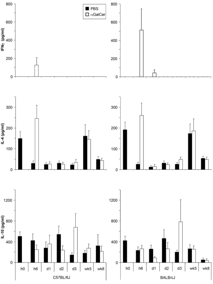
To determine if the differences seen in C57BL/6 and BALB/c infected mice were due to variations in the early cytokine response, we measured these in the serum shortly afterL. majorinfection in PBS57 and untreated controls. PBS57 treatment led to an acute increase of serum levels of IFNcand IL-4 at 6 hrs, which was not
observed in untreated, infected controls. PBS57-treated mice also showed higher IL-10 levels 3 days post infection. The only clear observed strain-specific difference was a higher IFNclevel 6 hrs post infection in BALB/c than in C57BL/6 serum (Figure 6).
Effects ofaGalCer analog PBS57 in C57BL/6 mice are IL-4 dependent
To further analyze the role of early cytokine secretion in the observed aGalCer analog PBS57-mediated effects on infection,
experiments were performed eliminating IL-4. In one set of experiments, IL-42/2 mice were infected with L. major and subsequently treated with PBS57. However, whereas controls showed the known exacerbation of disease after application of PBS57, this was not seen in IL-42/2 mice (Figure 7A [23]). In another set of experiments, IL-4 neutralizing antibodies were applied to infected C57BL/6 mice. Here, similar results were observed, with PBS57 application no longer changing the course of infection (Figure 7B). Anti-IL-4-treated mice also exhibited significantly lower parasite loads. PBS57 administration no longer affected the course of infection in these mice (Figure 7C), supporting an important role of IL-4 in
aGalCer analog PBS57-mediated disease exacerbation.
Discussion
model. Mice on a C57BL/6 background lacking NKT cells showed an improved course of disease. Correspondingly, NKT cell stimulation withaGalCer analog PBS57 worsened the course of
disease. Effects of stimulation were time, dose, and strain dependent. The effect of PBS57-stimulated NKT cells appeared to be IL-4 mediated, as neutralizing this cytokine abrogated the observed effectsin vivo. Our results show that NKT cells should be considered both when treating activeLeishmaniainfection as well as in the development of vaccines.
The reported effects ofL. major-activated NKT cells observed in various models ofLeishmaniainfection have been variable and often conflicting [10–16]. Most of this is probably due to both different infection models andLeishmaniastrains applied. In our model, 103 infectious stage parasites are inoculated intradermally in the ear. This is considerably lower than in most other studies usingL. major, which additionally often infect the foot pads instead of ear skin. Studies by Mattner et al. [13] and Ishikawa et al. [12] presented very different effects of NKT cells on the course of L. major
infection than those we observed. In their hands, high doses of parasites (106–108) given intradermally or even intravenously showed a protective effect of NKT cells, with C57BL/6 mice having lower parasite loads than their NKT cell-deficient counterparts. We and others [24] argue that such high parasite doses are far removed from the actual real life scenario, where infection is initiated by small amounts of parasites leaving the sand fly during its feeding on the host. We therefore believe our findings might be more representative of most infection settings in humans. Both NKT cell deficient mice strains on a C57BL/6 background controlledL. major infection significantly better than wild-type counterparts. CD1d2/2mice lack all NKT cells. J
a182/2
mice do not have the larger invariant Va14-Ja18 TCR subset, however still maintain a smaller NKT cell subset which recognizes CD1d with a different TCRs. Although there was a trend toward better disease control for Ja182/2than CD1d2/2mice (Figure 2) this was not statistically significant, nor were substantial differences in cytokine secretion between the two strains noted. Future studies would be needed to elucidate the role of different NKT cell subsets in
Leishmaniainfection.
Stimulating NKT cells by applying aGalCer analog PBS57 significantly altered the course of disease. The effect was time- and dose-dependent. As initiating a Th1 immune response is critical for the control of Leishmaniainfection, we hypothesized that the mechanism through which NKT cells have a negative effect on the course of infection in C57BL/6 mice, could be through secretion of Th2 cytokines, in particular IL-4. When IL-4 was neutralized, either in IL-4 deficient mice or by applying IL-4 binding antibodies, the effect of PBS57 on the course of infection was alleviated. We believe this proves IL-4 secretion by NKT cells is the relevant factor negatively influencing control of L. major in C576BL/6 mice.
It is noteworthy that aGalCer analog PBS57 administration
showed strain-dependent differences. While leading to disease exacerbation in C57BL/6, it improved the course of disease in BALB/c mice (at least in smaller doses of 100 ng). The higher amount of IFNcsecretion in the serum of PBS57-treated BALB/c we noted compared to C57BL/6 mice might play a relevant role.
Figure 3.aGalCer analog PBS57 application alters the course of infection. A,D.Ear lesion volumes of wild type C57BL/6 (A) and NKT
cell-deficient CD1d2/2(D) mice infected with low doseL. majorpromastigotes with or without additionally receiving 100 ng
aGalCer at the time of
infection.B, C, E, F.Parasite loads in the ear (B,E) and spleen (C, F) measured at 5 and 8 weeks post infection in C57BL/6 (B, C) or CD1d2/2(E, F) mice
treated with or withoutaGalCer analog PBS57. G. Levels of IFNc, IL-4, and IL-10 measured after restimulation with SLA. Significant differences to
C57BL/6 control mice are labeled with *, **, and *** referring to p-values#0.05,#0.005, and#0.002, respectively (n$3 independent experiments with$3 animals per group).
doi:10.1371/journal.pntd.0002917.g003
Figure 4.aGalCer analog PBS57 effects are dependent on dose
and time of administration. A.Ear lesion volumes of wild type C57BL/6 mice infected with low dose promastigotes and receiving 10, 100, or 2000 ng.B.Ear lesion volumes during the course of infection with 100 ng ofaGalCer analog PBS57 applied at the time of infection, 6
weeks after infection, or at both time points.C.As in B, only here PBS57 was applied at 1 week before and 3 weeks after infection. Significant differences between groups at each time point are labeled with *, **, and *** referring to p-values#0.05,#0.005, and#0.002 respectively (n = 2 independent experiments with$4 mice/group).
Figure 5.aGalCer analog PBS57 effects are strain dependent, improving disease in BALB/c. A.Ear lesion volumes of wild type BALB/c
mice infected with low dose promastigotes and receiving 100 ng or 2000 ng at the time point of infection or 100 ng at the time point of infection and 6 weeks later. Significance is indicated as compared to control mice.B.Parasite burdens measured in the ear and spleen of control or 100 ng
aGalCer analog PBS57 treated mice at week 5 and 8 post infection.C.Levels of IFNc, IL-4 and IL-10 measured from lymphocytes restimulated with
SLA, 5 and 8 weeks post infection. Results shown are from.1 experiment with a minimum of 4 animals per group. Significant differences between groups are labeled with *, **, and *** referring to p-values#0.05,#0.005, and#0.002 respectively.
Figure 6. Cytokine secretion induced byaGalCer analog PBS57 is strain dependent.Serum levels of IFNc, IL-4, and IL-10 were measured in
the serum of C57BL/6 and BALB/c mice at different time points after infection. Mice received either PBS or 100 ngaGalCer analog PBS57 i.p. at the
time point of infection with 103L. majorpromastigotes (n = 3 independent experiments with
$8 mice per group). None of the differences noted were found to be statistically significant.
Figure 7.aGalCer analog PBS57 effects on C57BL/6 are IL-4 dependent. A.Ear lesion volumes of C57BL/6 mice infected with low dose
promastigotes with or without 100 ngaGalCer analog PBS57 at the time point of infection. Wild type groups are shown on the left, IL-4-deficient
mice on the right (n = 2 independent experiments,$4 mice per group).B.Ear lesion volumes of a similar experiment where mice received IL-4 neutralizing antibody (shown on the right, n = 1,$4 mice per group).C.Parasite burdens of mice from the experiment shown in B, 5 weeks post infection. Significant differences between groups are labeled with * referring to p-values#0.05.
Additionally, we assume the effect could be strongly context-dependent, with the same cytokines (IFNcand IL-4, both secreted by NKT cells), resulting in a slight Th1 shift in the developing Th2 response of BALB/c, compared to a Th2 shift in the Th1 setting of C57BL/6.
The strain specific observations are important to consider when trying to extrapulate what effect administration ofaGalCer or its
analogs might have in a human infection setting. Generally, it is believed that the course of infection seen in C57BL/6 mice better mirrors the situation seen in most humans with cutaneous leishmaniasis. As such, NKT cell activation through aGalCer analog PBS57 during the course of infection is most likely non-beneficial to the host and should be avoided. On the other hand,
aGalCer has shown a very beneficial effect as an adjuvant inL. major vaccination studies [19]. This could mean that while applyingaGalCer in a vaccination setting appears very promising,
one should be careful before applying such a vaccine in the setting of an existing infection, as it could potentially lead to disease worsening.
A potential additional strategy would be to test other NKT cell stimulating glycolipids. PBS57, the compound used in our study, was shown to stimulate slightly higher amounts of IL-4 and IFN-c
secretion than the originalaGalCer compound, KRN7000 [21].
Several versions of aGalCer and other glycolipids have been synthetically generated and vary in terms of cytokine response generated, favoring more of a Th-1 or Th-2 response [7,25]. Potentially applying other aGalCer variants or glycolipid com-pounds which elicit a more pronounced Th1 profile cytokine
stimulation could prove valuable, both in terms of treatment and vaccination forLeishmaniainfections.
In summary, our findings demonstrate that NKT cells influence the course ofL. majorinfection in a physiological low dose model. The effects were strain-dependent and could be augmented through aGalCer analog PBS57 stimulation, leading to disease worsening inLeishmaniaresistant C57BL/6 mice. Our results make it apparent that immune response modulating effects of NKT cells should be considered both when treating active Leishmania
infection as well as developing vaccines.
Supporting Information
Table S1 Statistical analysis of data in Figure 1; frequency of CD4, CD8 and NKT cell populations in the course of infection.
(PDF)
Acknowledgments
We thank Albert Bendelac, Luc Teyton, and Paul B. Savage for providing CD1d tetramers and PBS57, Uwe Klemm for providing Ja182/2 and CD1d2/2mice, and Kerstin Steinbrink for IL-42/2mice.
Author Contributions
Conceived and designed the experiments: KGG BL MRF LB SLK EvS. Performed the experiments: KGG BL MRF SLK. Analyzed the data: KGG BL MRF SLK EvS. Contributed reagents/materials/analysis tools: LB. Wrote the paper: KGG MRF LB EvS.
References
1. von Stebut E (2007) Cutaneous Leishmania infection: progress in pathogenesis research and experimental therapy. Exp Dermatol 16: 340–346.
2. Bendelac A, Lantz O, Quimby ME, Yewdell JW, Bennink JR, et al. (1995) CD1 recognition by mouse NK1+T lymphocytes. Science 268: 863–865. 3. Kawano T, Cui J, Koezuka Y, Toura I, Kaneko Y, et al. (1997) CD1d-restricted
and TCR-mediated activation of valpha14 NKT cells by glycosylceramides. Science 278: 1626–1629.
4. Bendelac A, Savage PB, Teyton L (2007) The biology of NKT cells. Annu Rev Immunol 25: 297–336.
5. Mattner J, Debord KL, Ismail N, Goff RD, Cantu C, 3rd, et al. (2005) Exogenous and endogenous glycolipid antigens activate NKT cells during microbial infections. Nature 434: 525–529.
6. Morita M, Motoki K, Akimoto K, Natori T, Sakai T, et al. (1995) Structure-activity relationship of alpha-galactosylceramides against B16-bearing mice. J Med Chem 38: 2176–2187.
7. Van Kaer L (2005) alpha-Galactosylceramide therapy for autoimmune diseases: prospects and obstacles. Nat Rev Immunol 5: 31–42.
8. Vivier E, Ugolini S, Blaise D, Chabannon C, Brossay L (2012) Targeting natural killer cells and natural killer T cells in cancer. Nat Rev Immunol 12: 239–252. 9. Van Kaer L, Parekh VV, Wu L (2011) Invariant NK T cells: potential for immunotherapeutic targeting with glycolipid antigens. Immunotherapy 3: 59– 75.
10. Beattie L, Svensson M, Bune A, Brown N, Maroof A, et al. (2010) Leishmania donovani-induced expression of signal regulatory protein alpha on Kupffer cells enhances hepatic invariant NKT-cell activation. Eur J Immunol 40: 117–123. 11. Campos-Martin Y, Colmenares M, Gozalbo-Lopez B, Lopez-Nunez M, Savage
PB, et al. (2006) Immature human dendritic cells infected with Leishmania infantum are resistant to NK-mediated cytolysis but are efficiently recognized by NKT cells. J Immunol 176: 6172–6179.
12. Ishikawa H, Hisaeda H, Taniguchi M, Nakayama T, Sakai T, et al. (2000) CD4(+) v(alpha)14 NKT cells play a crucial role in an early stage of protective immunity against infection with Leishmania major. Int Immunol 12: 1267– 1274.
13. Mattner J, Donhauser N, Werner-Felmayer G, Bogdan C (2006) NKT cells mediate organ-specific resistance against Leishmania major infection. Microbes Infect 8: 354–362.
14. Robert-Gangneux F, Drogoul AS, Rostan O, Piquet-Pellorce C, Cayon J, et al. (2012) Invariant NKT cells drive hepatic cytokinic microenvironment favoring
efficient granuloma formation and early control of Leishmania donovani infection. PLoS One 7: e33413.
15. Stanley AC, Zhou Y, Amante FH, Randall LM, Haque A, et al. (2008) Activation of invariant NKT cells exacerbates experimental visceral leishman-iasis. PLoS Pathog 4: e1000028.
16. Wiethe C, Debus A, Mohrs M, Steinkasserer A, Lutz M, et al. (2008) Dendritic cell differentiation state and their interaction with NKT cells determine Th1/ Th2 differentiation in the murine model of Leishmania major infection. J Immunol 180: 4371–4381.
17. Joyee AG, Uzonna J, Yang X (2010) Invariant NKT cells preferentially modulate the function of CD8 alpha+dendritic cell subset in inducing type 1 immunity against infection. J Immunol 184: 2095–2106.
18. Amprey JL, Im JS, Turco SJ, Murray HW, Illarionov PA, et al. (2004) A subset of liver NK T cells is activated during Leishmania donovani infection by CD1d-bound lipophosphoglycan. J Exp Med 200: 895–904.
19. Dondji B, Deak E, Goldsmith-Pestana K, Perez-Jimenez E, Esteban M, et al. (2008) Intradermal NKT cell activation during DNA priming in heterologous prime-boost vaccination enhances T cell responses and protection against Leishmania. Eur J Immunol 38: 706–719.
20. Von Stebut E, Ehrchen JM, Belkaid Y, Kostka SL, Molle K, et al. (2003) Interleukin 1alpha promotes Th1 differentiation and inhibits disease progression in Leishmania major-susceptible BALB/c mice. J Exp Med 198: 191–199. 21. Liu Y, Goff RD, Zhou D, Mattner J, Sullivan BA, et al. (2006) A modified
alpha-galactosyl ceramide for staining and stimulating natural killer T cells. J Immunol Methods 312: 34–39.
22. Benlagha K, Weiss A, Beavis A, Teyton L, Bendelac A (2000) In vivo identification of glycolipid antigen-specific T cells using fluorescent CD1d tetramers. J Exp Med 191: 1895–1903.
23. Matthews DJ, Emson CL, McKenzie GJ, Jolin HE, Blackwell JM, et al. (2000) IL-13 is a susceptibility factor for Leishmania major infection. J Immunol 164: 1458–1462.
24. Belkaid Y, Mendez S, Lira R, Kadambi N, Milon G, et al. (2000) A natural model of Leishmania major infection reveals a prolonged ‘‘silent’’ phase of parasite amplification in the skin before the onset of lesion formation and immunity. J Immunol 165: 969–977.