Ц К А MEDICAL TREATMENT
А ИЈА
Ѓ
А ИЈА А А
А А
И
ИЈА А А
и ија В А1
ора ЈА ВА2
а Ф В 1
икица А А А ВА1
1 Г ш ц „8 “, О
ј , О ј , ј ,
. М ј
2 „ . К М ј“,
, И ј , ј , .
М ј
CORRELATION BETWEEN TYMPANIC
MEMBRANE PERFORATION AND HEARING
LOSS
Lidija RISTOVSKA1 Zora JACHOVA2 Rade FILIPOVSKI1 Nikica ATANASOVA1
1 Citв General Hospital “8 September”, Department of
Otorhinolaryngology, Division of Audiology, Skopje, Republic of Macedonia
2 Universitв “Ss Cвril and MetСodius”, Facultв of
Philosophy, Institute of Special Education and Rehabilitation, Skopje, Republic of Macedonia
: 30.11.2015 : 19.01.2016 UDK: 616.285:616.28-008.14
Recived: 30.11.2015 Accepted: 19.01.2016
Original Article
Abstract
Во : ј
-, ј .
-ј
-ј њ
ј њ
-џ .
Ц :
ј ј
ј
.
М р ј о :
-,
-ј 218
, 114 (52,3%) 104
(47,7%), 9 75 (
47,9 ),
2012 2015
.
-
ј p<0,05.
Introduction: Perforation of the tympanic
membrane primarily results from middle ear infections, trauma or iatrogenic causes. The perforation causes conductive hearing loss by reducing the surface area available for sound transmission to the ossicular chain.
Objective: The objective was to analyze the
characteristics of tympanic membrane perforations in relation to hearing loss and to determine the type and degree of hearing loss.
Materials and methods: We analyzed
audiometric, otoscopic findings and medical reports of 218 patients, 114 males (52.3%) and 104 females (47.7%), aged 9 to 75 years (mean age of 47.9 years), examined during the period of November 2012 to October 2015. For statistical data analysis we used Chi-square test with level of significance p<0.05.
ј :
ј И В А
„8 “,
ј , ј
, 1000 ј , . ј
: 0038902 3087612 Email: [email protected]
Corresponding address:
Lidija RISTOVSKA
CТtв GОnОrКХ HospТtКХ “8 SОptОЦЛОr”, DОpКrtЦОnt oП
Otorhinolaryngology, Division of Audiology, Pariska NN. 1000 Skopje, Republic of Macedonia.
hone: 0038902 3087612
Ф Ш А ИЈА И А И А 2016; 17(1–2):36–49 37
: ј ј
(89%)
ј
-њ pКrs tОnsК (37,2%).
23,9 НB.
ј ј 250
Hг. ј ј (73,1%)
ј (p=0,032) 21 40 НB.
З чо :
ј
ѓ
-ј . ј
-ј ј .
њ ј .
ѓ -
-ј .
Results: Most of the patients had unilateral
perforations (89%) with right ear predominance and involvement of two quadrants of pars tensa (37.2%). Mean air-bone gap was 23.9 dB. The largest air-bone gap was at frequency of 250 Hz. Most of the patients (73.1%) had mixed hearing loss (p=0.032), and average hearing thresholds from 21 to 40 dB.
Conclusion: Mean air-bone gap is largest at the
lower frequencies, and decreases as frequency increases. Size of the perforation has effect on hearing loss. Mean air-bone gap increases with increasing size of the perforation. There is no big difference between the mean air-bone gap in posterior versus anterior perforations.
Клуч р : ј ,
-ј , Keywordsmembrane : hearing loss, perforation, tympanic
В
Introduction
: pКrs
tensК, ј ј
pКrs ПХКММТНК (1).
-ј
,
ј (2).
-ј
.
(3). ј
(4).
, ,
-,
.
ј
њ ј
њ
џ (5).
ј
50 НB (6).
ј
ј ј
.
ј
(7).
ј ј ј
ј
ј
-ј (8).
ј
-ј ј ј .
Ш
( . -
-)
ј
(6).
ј ј (9).
ќ
,
ј (10).
-ј њ
ј (11).
њ
ј ј
ј .
Hearing loss in tympanic membrane perforation is related to the size of perforation and the degree of middle ear and mastoid pneu-matization (8). Whether location of the perfo-ration had effect on hearing loss is debatable. Widely held clinical view was that perforations over the region of the round window (i.e., postero-inferior quadrant) result in significantly greater hearing loss than anterior perforations (6).
Perforations as a result of acute otitis media and trauma heal spontaneously in the majority of cases (9). Perforations that do not heal spontaneously may require surgical repair, particularly when associated with recurrent infections or hearing loss (10). The recon-struction may involve the harvesting autologous graft materials (11).
The aim of the study was to analyzethe chara-cteristics of tympanic membrane perforations in relation to hearing loss and to determine the type and degree ofhearing loss.
а
ја
Materials and
Methods
ј
218 , 114 (52,3%)
104 (47,7%), 9 75
-,
-ј , „8
-“, ј , ј ,
2012 2015
. њ :
ј
pКrs tОnsК ј . ј
,
њ , ј
.
-њ (
, ј
-) .
,
-ј
-.
-ј BОХХ PХus
(InvОntТs, ј ) -
TОХОpСonТМs TDH-39 „ “.
: 125, 250, 500, 1000, 2000, 4000
Ф Ш А ИЈА И А И А 2016; 17(1–2):36–49 39
8000 Hг.
≤20 НB
250 8000
Hг.
-
-ј p<0,05. ј
: 4709/2015.
НОПТnОН Кs tСrОsСoХНs ≤20 НB СОКrТnР ХОvОХ (HL) at audiometric frequencies from 250 to 8000 Hz. For statistical data analysis we used Chi-square test with level of significance p<0.05. Protocol number of Ethical approval: 4709/ 2015.
а
Results
ј
-ј 218, 114 (52,3%)
104 (47,7%), 9 75
( 47,9 ).
242 ј
њ .
18 (
12,8 ).
-ј
-( 1).
The total number of patients surveyed in our study was 218, 114 males (52.3%) and 104 females (47.7%), aged 9 to 75 years (mean age of 47.9 years). Twenty-four patients had bilateral perforations and total of 242 ears met the inclusion criteria. Seventeen patients were younger than 18 years (mean age of 12.8 years). Distribution of perforations according to affected ear was displayed (Table 1).
T 1. ј Table 1. Distribution of perforations according to
affected ear
/ Affected ear / Males / Females / Total
No % No % No %
/ Right ear 54 24.8 51 23.4 105 48.2 / Left ear 46 21.1 43 19.7 89 40.8 / Bilateral 14 6.4 10 4.6 24 11
p=0.82 * / Total 114 52.3 104 47.7 218 100 * - / Chi-square test
ј ј
ј
(48,2%),
ј
(χ²=0,396,
df=2, p=0,82). 194 (89%)
, 100
(45,9%) 94 (43,1%).
ј 24
(11%), 14 (6,4%) 10 (4,6%).
129
(53,3%), 68 (28,1%) ј
61 (25,2%) ј .
113
(46,7%), 60 (24,8%) ј
53 (21,9%) ј .
. ј 209 (86,4%)
, 198
(81,8%) 11 (4,5%)
. (8,3%)
. ј 10 ,
-Most of the patients had unilateral perforations with right ear predominance (48.2%), but there is no significant difference in distribution of the perforations according to affected ear and the gender (χ²=0.396, df=2, p=0.82). A total of 194 patients (89%) had unilateral perforations, 100 males (45.9%), and 94 females (43.1%). Bilateral perforations were present in 24 patients (11%), 14 males (6.4%), and 10 females (4.6%). Right ear was affected in 129 cases (53.3%), 68 perforations (28.1%) were in males, and 61 (25.2%) were in females. Left ear was affected in 113 cases (46.7%), 60 perforations (24.8%) were in males, and 53 (21.9%) in females.
ј
( ),
њ ,
,
, ј
њ њ ј
.
(4,5%)
ј .
(0,8%) ј ,
њ њ
2003 ( 1).
from them, the perforation was caused by open-handed slap across the ear (violence), in five cases injury with cotton swabs, in two cases patients were accidentally slapped, one patient had blast injury, one perforation occurred during swimming and diving, and one perforation after fall on ice on street. Eleven patients (4.5%) had perforations occurred many years ago with unknown etiology. Only two perforations (0.8%) were iatrogenic, after tympanostomy tubes placement in 2003 (Figure 1).
1. ј Figure 1.Etiology of tympanic membrane
perforations
Ј ј
pКrs tОnsК :
( ), ( ),
( ), ( )
( 2). PКrs tОnsК
њ ј
ЦКnuЛrТuЦ ЦКХОus
ј uЦЛo, ј
.
: ,
pКrs tОnsК
ј , ,
,
,
-, ,
pКrs tОnsК .
Ф Ш А ИЈА И А И А 2016; 17(1–2):36–49 41
2. И
pКrs tОnsК ј Table 2. tympanic membrane perforationInvolvement of pars tensa quadrants in
/ Quadrant
/ Males / Females / Total
No % N % No %
/ AS 9 3.7 8 3.3 17 7
/ AI 12 5 11 4.5 23 9.5
/ PS 10 4.1 8 3.3 18 7.4
/ PI 12 5 8 3.3 20 8.3
+ / AS+AI 14 5.8 16 6.6 30 12.4
+ / PS+PI 14 5.8 13 5.4 27 11.2
+ / AI+PI 18 7.4 15 6.2 33 13.6
+ + / AS+AI+PI
15 6.2 14 5.8 29 12
+ + + /
AS+AI+PS+PI
24 9.9 21 8.7 45 18.6
/ Total 128 52.9 114 47.1 242 100
ј ј
(29,5%
-)
. 180
90
-, 16,7%,
35%, 15%
33,3%. 78
(32,2%) , 90
(37,2%) , 29 (12%)
45 (18,6%)
-.
ј 177 ,
ј 172 .
ј
ј ј .
-- 250,
500, 1000, 2000 4000 Hг.
-
23,9 НB.
- ј
17,1 НB, ј
23,5 НB, ј 28,5
НB ј 33,4
НB. ј
- .
-ј ј :
33,6 НB 250 Hг, 29,7 НB 500 Hг, 21,9 НB 1000 Hг, 15,2 НB 2000 Hг 18,9 НB
4000 Hг. ј
-ј 250 Hг.
-
: I 0-10 НB, II
11-20 НB, III 21-30 НB IV >30
НB ( 3).
Most of the small perforations were in AI quadrant (29.5% from all small perforations), and medium perforations were predominantly in inferior quadrants. From the total of 180 involved quadrants in 90 medium perforations, AS quadrant was involved in 16.7%, AI quadrant in 35%, PS quadrant in 15%, and PI quadrant in 33.3%. A total of 78 perforations (32.2%) were small, 90 perforations (37.2%) were medium, 29 perforations (12%) were large, and 45 perforations (18.6%) were subtotal. Anterior quadrants were affected in 177 perforations, and posterior quadrants in 172 perforations.
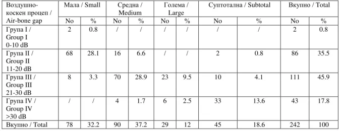
The effect of the size of perforation on hearing loss was analyzed. Mean air-bone gap at frequencies 250, 500, 1000, 2000, and 4000Hz was calculated. Mean air-bone gap in all frequencies was 23.9 dB. Mean air-bone gap in small perforations was 17.1 dB, in medium perforations 23.5 dB, in large perforations 28.5 dB, and in subtotal perforations was 33.4 dB. Subtotal perforations had the largest air-bone gap. Separately calculated mean air-bone gap in each frequency was as follows: 33.6 dB in 250 Hz, 29.7 dB in 500 Hz, 21.9 dB in 1000 Hz, 15.2 dB in 2000 Hz, and 18.9 dB in 4000 Hz. The largest air-bone gap was at frequency of 250 Hz.
3.
-ј
Table 3. Mean air-bone gap according to size of tympanic membrane perforation
/
Тr-bone gap
/ Small / Medium
/ Large
/ Subtotal / Total
No % No % No % No % No %
I / Group I 0-10 dB
2 0.8 / / / / / / 2 0.8
II / Group II 11-20 dB
68 28.1 16 6.6 / / 2 0.8 86 35.5
III / Group III 21-30 dB
8 3.3 70 28.9 23 9.5 10 4.1 111 45.9
IV / Group IV >30 dB
/ / 4 1.7 6 2.5 33 13.6 43 17.8
/ Total 78 32.2 90 37.2 29 12 45 18.6 242 100
ј ј
-
11 20 НB. ј ј
-21 30 НB
-- >30 НB
ј .
ј ј
ј ј
ѓ
- ј
-.
-ј 78 (32,2%) 90
(37,2%) 20,5 НB.
-- ј
21,2 НB, ј
19,8 НB.
-ј ј
-
-ј
-.
ј
њ
ј .
ј ј
ј
( 4). ј
. ј
ј
. ј ј
ј (73,1%).
ѓ
Most of the small perforations had mean air-bone gap from 11 to 20 dB. Most of the medium and large perforations had mean air-bone gap from 21 to 30 dB, and mean air-air-bone gap >30 dB was predominant in subtotal perforations.
Ф Ш А ИЈА И А И А 2016; 17(1–2):36–49 43
ј
-ј (χ²=4,598, df=1, p=0,032).
number of ears with conductive and mixed hearing loss (χ²=4.598, df=1, p=0.032).
4. ј ј
ј Table 4. membrane perforation Type of hearing loss in ears with tympanic
ј
/ Type of hearing loss
/ Males / Females / Total
No % No % No %
/ Conductive
27 11.2 38 15.7 65 26.9
/ Mixed
101 41.7 76 31.4 177 73.1
p=0.032*
/ Total 128 52.9 114 47.1 242 100
* - / Chi-square test
ј
ј ј
( 5).
500, 1000, 2000 4000 Hг.
ј ј ј
21 40 НB HL (46,3%).
Degree of hearing loss in ears with tympanic membrane perforation was determined (Table 5). Average hearing threshold at speech frequencies 500, 1000, 2000, and 4000 Hz was calculated. In most cases hearing loss was 21-40 dB HL (46.3%).
5. ј ј
ј Table 5. tympanic membrane perforation Degree of hearing loss in ears with
ј
/ Degree of hearing loss
/ Males / Females / Total
No % No % No %
0-20 dB HL 8 3.3 6 2.5 14 5.8
21-40 dB HL 56 23.1 56 23.1 112 46.3
41-60 dB HL 37 15.3 41 16.9 78 32.2
61-95 dB HL 27 11.2 11 4.5 38 15.7
p=0.92* / Total 128 52.9 114 47.1 242 100 * - / Chi-square test
ѓ ј
ј
ј (χ²=6,439,
df=3, p=0,92).
ј
9 18 (
12,8 ).
17 , 10 .
11 9 14 .
19
њ (7,9%
).
-ј , 15 ,
ј
, ј
There is no significant difference between number of ears with different degree of hearing loss and gender distribution (χ²=6.439, df=3, p=0.92).
( ) ј
-њ њ
њ . ,
,
-,
-ј . ј
ј 19 .
ј , ј
14 ј 21 - 40
НB HL, ј
0 - 20 НB HL
ј , 41
- 60 dB HL.
traumatic (slap injury), and one child had both tympanic membrane perforated after tympanostomy tube placement. According to the size, four perforations were small, nine perforations were medium, five were large, and one perforation was subtotal. The type of hearing loss in all 19 cases was conductive. In terms of degree of hearing loss, in 14 cases hearing loss was 21-40 dB HL, in four ears average hearing thresholds were 0-20 dB HL, and one ear had greater hearing loss, 41-60 dB HL.
ја
Discussion
-ј ј ј
. ,
ј
.
(2, 12). SСКrЦК .
ј (13). ј
ј
-, . Fukuchi
. ј
(14).
њ ј ј
ј
.
-.
ј
ј
(15).
. OХoаookОrО
. 6%
-ј (16). ј 80%
-њ .
ј
-ј (17-20).
.
-.
ј ј ј
(21).
ѕ
-јќ ј (22).
.
ј .
Determinants of hearing loss in patients with tympanic membrane perforation were analyzed. According to the gender, there was slight male predominance in our sample. It is similar to other authors' findings (2, 12). Sharma et al. reported slight female predominance (13). Most of the patients had unilateral perforations, more frequently in right ear. Fukuchi et al. also reported right ear predominance (14).
Ф Ш А ИЈА И А И А 2016; 17(1–2):36–49 45
.
(23). RТtОnour .
ј ј
16%
(24). ј
-, њ
њ . ј
ј
(25).
ј ј
, .
њ ѓ њ
-њ .
ј
-ѓ
- ј
-. MОСtК . ј
ј ј . ј
ј , ,
(6). IЛОkаО . ј
ј ј
-ј ј
,
-ј ј
(26). MКСКrУКn .
-ј ј
ј ј .
-ј (27). њ
њ .
, ј ј
,
њ .
-ќ
ќ
.
-ј 23,9 НB. AnsКrТ .
-- 20 40 НB
ј (28). ј
-ј ѓ ј
ј .
-
ј .
ј
-.
ј
-ј 250 Hг.
- ј
ѓ
-over the left ear. One patient in our sample had blast injury. Primary injures to the ear due to blast trauma are caused by high pressure wave followed by a negative phase (23). Ritenour et al. concluded that perforations occur in approximately 16% of the patients wounded in combat explosions (24). Only two perforations were iatrogenic, after tympanostomy tubes insertion. Residual perforation of the tympanic membrane after this intervention is not uncommon (25). In our sample there were no other iatrogenic cases of tympanic membrane perforation, for example, caused during foreign body removal or irrigation of the external auditory canal for removal of impacted cerumen.
In terms of the location of the perforations, there was no big difference in mean air-bone gap in posterior versus anterior perforations. Mehta et al. concluded that hearing loss did not vary substantially with location of the perforation. Effects of location, if any, were small (6). Ibekwe et al. reported that the location of perforation has no effect on the magnitude of hearing loss in acute tympanic membrane perforations, but, it has significant impact in chronic tympanic membrane perforations (26). According to Maharjan et al. the location of perforation had significant effect on hearing loss. Posterior placed perforations had greater degree of loss (27). There are different opinions regarding this issue.
According to the size, most of the perforations in our sample were medium, involving two quadrants. Inferior quadrants were more affected than superior and anterior more than posterior quadrants.
њ ј .
-- 2000 Hг. LОrut
. ј
-, „ V “
2000Hг. 2000Hг,
2000 Hг ј
.
ј
( ј )
2000 Hг, ј (29).
ј
2000 Hг,
ј ј (30).
ј
ј (6, 8, 31-35). RТЛОТro .
ј ѓ
ј ј ј
sТЦpХОб (36).
-
-,
PКrk . (8),
ј
KКsХТаКХ . (37).
ј ј
-ј .
ѓ ј
ј
(p=0,032). ј ј
ј
21 - 40 НB HL. ј
-ј . ј ј
ј ,
. ј
ј
-. ј
-њ
ј
-ј . ј
ј ј
.
ј
(7).
-ј ј
ј
ј
ј .
reported a consistent frequency pattern, similar to Кn “ТnvОrtОН V sСКpО” oП tСО КuНТoРrКЦ аТtС a turning point around 2000Hz. Below 2000Hz, the air-bone gap is larger for the lower frequencies, and above 2000 Hz, the air-bone gap gets bigger again in the higher frequencies. They concluded that the human middle ear had the least loss of sound transmission (or best hearing) around 2000 Hz, independently of the pathology (29). The inherent frequency of the tympanic membrane had been calculated to be at 2000 Hz, the tympanic membrane vibrates the most at this frequency (30). Many authors reported that hearing loss increased with increasing size of the perforation (6, 8, 31-35). Ribeiro et al. did not find the correlation between the size of tympanic membrane perforation and hearing loss in simple chronic otitis media (36).
According to the mean air bone gap, we divided the perforations into four groups similar to Park et al. (8), and we determined the size of the perforations in terms of involved quadrants similar to Kasliwal et al. (37).
Ф Ш А ИЈА И А И А 2016; 17(1–2):36–49 47
ќ
(37, 38). MoСsТn .
ј
њ ј
(39). DО AгОvОНo .
ј ј
13%
. ј
-,
-њ њ (40).
, ј ј
18
.
ј
.
ј ѓ
ј
. 18
ј .
the lower frequencies (37, 38). According to Mohsin et al. risk of sensorineural hearing loss increases with increase in duration of ear discharge (39). De Azevedo et al. reported occurrence of sensorineural hearing loss in 13% of the patients with chronic suppurative otitis media. It was correlated with an age increase, but not with longer duration of ear disease (40). Similar to adults, most of the patients younger than 18 years had chronic otitis media, and traumatic perforations were also less common in this subgroup. This sample is too small to draw any conclusions. We can only notice that there was difference between children and adults in terms of the type of hearing loss. All patients younger than 18 years had conductive hearing loss.
а
Conclusion
ј
ѓ ј .
ј ј ј
.
-њ ј .
-ј ј ј
-ј ,
ѓ - ј
-.
Mean air-bone gap is largest at the lower frequencies, and decreases as frequency increases. Size of the perforation has effect on hearing loss. Mean air-bone gap increases with increasing size of the perforation. In terms of the location of the perforation and the effect on hearing loss, there was no big difference between the mean air-bone gap in posterior versus anterior perforations.
а
ања а
а
ањ
Limitations of the Study
њј ј
,
-ј
-њ њ ј
.
We don't have precise measurement of the size of perforation in tympanic membrane photograph, data on degree of middle ear and mastoid pneumatization, and duration of disease in cases of chronic otitis media.
К
а
Conflict of interests
ј
.
Authors declare no conflict of interests.
/ References
1. Villar-Fernandez MA, Lopez-Escamez JA. Outlook for tissue engineering of the tympanic membrane.
Audiol Res, 2015; 5(1): 117.
hearing function. Acta Otorhinolaryngol Ital, 2011; 31(6): 366-371.
3. ErkorkЦКг о, ВıХЦКг MS, GüvОn M, KКвЦКг R.
Determination of factors that impact patient satisfaction following tympanoplasty. J Int Adv Otol
2014; 10(3): 264-269.
4. Stenfeldt K, Johansson C, Hellström S. The collagen structure of the tympanic membrane: collagen types I, II, and III in the healthy tympanic membrane, during healing of a perforation, and during infection.
Arch Otolaryngol Head Neck Surg, 2006; 132(3): 293-298.
5. Isaacson JE, Vora NM. Diferential diagnosis and treatment of hearing loss. Am Fam Physician 2003; 68(6): 1125-1132.
6. Mehta RP, Rosowski JJ, Voss SE, O'Neil E, Merchant SN. Determinants of hearing loss in perforations of the tympanic membrane. Otol Neurotol 2006; 27(2): 136-143.
7. Kumar N, Chilke D, Puttewar MP. Clinical profile of tubotympanic CSOM and its management with special reference to site and size of tympanic membrane perforation. Eustachian tube function and three flap tympanoplasty. Indian J Otolaryngol Head
Neck Surg 2012; 64(1): 5-12.
8. Park H, Hong SN, Kim HS et al. Determinants of conductive hearing loss in tympanic membrane perforation. Clin Exp Otorhinolaryngol 2015; 8(2): 92-96.
9. Debnath M, Khanna S. A comparative study of closure of tympanic membrane perforation between chemical cauterization and fat plug myringoplasty.
International Journal of Otolaryngology and Head & Neck Surgery, 2013; 2(6): 248-252.
10. Saliba I, Froehlich P. Hyaluronic acid fat graft myringoplasty, An office based technique adapted to children. Arch Otolaryngol Head Neck Surg, 2011; 137(12): 1203-1209.
11. Hong P, Bance M, Gratzer PF. Repair of tympanic membrane perforation using novel adjuvant therapies: a contemporary review of experimental and tissue engineering studies. Int J Pediatr Otorhinolaryngol, 2013; 77(1): 3-12.
12. Rafique M, Farrukh MS, Shaikh AA. Assessment of hearing loss in tympanic membrane perforation at tertiary care hospitals. Journal of Liaquat University of Medical and Health Sciences, 2014; 13(1): 32-36.
13. Sharma K, Manjari M, Salaria N. Middle ear cleft in chronic otitis media: a clinicohistopatological study.
Indian J Otolaryngol Head Neck Surg, 2013; 65(Suppl 3): S493-S497.
14. Fukuchi I, Cerchiari DP, Garcia E, Rezende CEB, Rapoport PB. Tympanoplasty: surgical results and comparison of the factors that may interfere in their success. Braz J Otorhinolaryngol 2006; 72(2): 267-271.
15. Schroeder A, Darrow DH. Management of the draining ear in children. Pediatr Ann 2004; 33(12): 843-853.
16. Olowookere SA, Ibekwe TS, Adeosun AA. Pattern of tympanic membrane perforation in Ibadan: a retrospective study. Ann Ib Postgrad Med 2008; 6(2):
31-33.
17. Singh B, Baka N, Kumar N, Purohit JP. Study of various grafts in closure of tympanic membrane perforation. Scholars Journal of Applied Medical Sciences 2015; 3(3G): 1509-1515.
18. Bhadouriya S, Srivastava M, Gaur S, Lavania A, Saxena R. A study of chemical cauterization of tympanic membrane perforations by using trichloroacetic acid. International Journal of Institutional Pharmacy and Life Sciences 2012; 2(2): 195-204.
19. Al-Juboori AN. Evaluation of spontaneous healing of traumatic tympanic membrane perforation. General Med 2014; 2(1): 129.
20. Dawood MR. Spontaneous healing of traumatic tympanic membrane perforation. Mustansiriya Medical Journal 2015; 14(1): 24-29.
21. Afolabi OA, Aremu SK, Alabi BS, Segun-Busary S. Traumatic tympanic membrane perforation: An aetiological profile. BMC Res Notes 2009; 2: 232. 22. Sarojamma DSR, Raj S, Satish HS. A clinical study
of traumatic perforation of tympanic membrane.
IOSR Journal of Dental and Medical Sciences 2014; 13(4): 24-28.
23. Abbas S, Arshad M, Ghani S. Tympanic membrane perforations secondary to blast trauma - an experience of 74 affected ears. Isra Medical Journal
2014; 6(4): 267-269.
24. Ritenour AE, Wickley A, Ritenour JS et al. Tym-panic membrane perforation and hearing loss from blast overpressure in Operation Enduring Freedom and Operation Iraqi Freedom wounded. J Trauma
2008; 64(2 Suppl): S174-S178.
25. Johnston LC, Feldman HM, Paradise JL et al. Tym-panic membrane abnormalities and hearing levels at the ages of 5 and 6 years in relation to persistent otitis media and tympanostomy tube insertion in the first 3 years of life: a prospective study incorporating a randomized clinical trial. Pediatrics 2004; 114(1): e58-e67.
26. Ibekwe TS, Nwaorgu OG, Ijaduola TG. Correlating the site of tympanic membrane perforation with hearing loss. BMC Ear Nose Throat Disord 2009; 9: 1.
27. Maharjan M, Kafle P, Bista M, Shrestha S, Toran KC. Observation of hearing loss in patients with chronic suppurative otitis media tubotympanic type.
Kathmandu Univ Med J (KUMJ) 2009; 7(4): 397-401.
28. Ansari MA, Khayani IAM, Farrukh MS, Kashmiri ZA, Farooq MU. Outcome of bilateral myringoplasty in dry central perforation - an appraisal. J Dow Univ Health Sci 2014; 8(1): 16-20.
29. Lerut B, Pfammatter A, Moons J, Linder T. Functional correlations of tympanic membrane perforation size. Otol Neurotol 2012; 33(3): 379-386. 30. Nahata V, Patil CY, Patil RK, Gattani G. Tympanic membrane perforations: Its correlation with hearing loss and frequency affected – An analytical study.
Indian Journal of Otology 2014; 20(1): 10-15. 31. Elhaj AsHA, Abdalla MB, Abdalla HA. The effect of
Ф Ш А ИЈА И А И А 2016; 17(1–2):36–49 49
Journal of Health Sciences 2008; 4(1): 1-12. 32. Bhusal CL, Guragain RPS, Shrivastav RP. Size of
tympanic membrane perforation and hearing loss.
JNMA J Nepal Med Assoc 2006; 45(161): 167-172. 33. Pannu KK, Chadha S, Kumar D, Preeti. Evaluation
of hearing loss in tympanic membrane perforation.
Indian JOtolaryngol Head Neck Surg 2011; 63(3): 208-213.
34. Santhi T, Rajan KV. A study of closure of tympanic membrane perforations by chemical cauterization.
Indian JOtolaryngol Head Neck Surg 2012; 64(4): 389-392.
35. Islam MS, Islam R, Bhuiyan MAR, Rashid S, Datta PG. Pattern and degree of hearing loss in chronic suppurative otitis media. Bangladesh Journal of Otorhinolaryngology 2010; 16(2): 96-105.
36. Ribeiro FdeAQ, Gaudino VRR, Pinheiro CD, Marçal GJ, Mitre EI. Objective comparison between perforation and hearing loss. Braz J Otorhinolaryngol 2014; 80(5): 386-389.
37. Kasliwal N, Joshi S, Pareek SM. Determinants of sensorineural hearing loss in chronic middle-ear disease. Indian J Otolaryngol Head Neck Surg 2004; 56(4): 269-273.
38. Barman D, Dutta M, Mukherjee M, Sarkar A, Shit S, Sarkar A. Evaluation of cochlear function in safe type of chronic otitis media. Bengal Journal of Otolaryngology and Head Neck Surgery 2013; 21(1): 8-11.
39. Mohsin MA, Kumar MR, Reddy BGN, Ravikumar D. Sensorineural hearing loss in chronic suppurative otitis media of tubotympanic variety. National Journal of Otorhinolaryngology and Head & Neck Surgery 2013; 1(10): 3-4.