Host-Specific Adhesion to Erythrocytes
Muriel Vayssier-Taussat1,2*, Danielle Le Rhun1, Hong Kuan Deng1, Francis Biville3, Sandra Cescau4, Antoine Danchin5, Genevie`ve Marignac1, Evelyne Lenaour1, Henri Jean Boulouis3, Maria Mavris1, Lionel Arnaud6, Huanming Yang7, Jing Wang8, Maxime Quebatte2, Philipp Engel2, Henri Saenz2, Christoph Dehio2*
1Unite´ Sous Contrat Bartonella, INRA, Maisons-Alfort, France,2Focal Area Infection Biology, Biozentrum, University of Basel, Basel, Switzerland,3UMR BIPAR, Equipe des Bacte´ries Zoonotiques He´motropes, ENVA/AFSSA, Maisons-Alfort, France,4Unite´ des Ge´nomes Bacte´riens, Institut Pasteur, CNRS URA 2171, Paris, France,5AMAbiotics, Evry, France,6Institut de Transfusion Sanguine, Paris, France,7Beijing Genomics Institute, Shenzhen, China,8Institute of Psychology, Chinese Academy of Sciences, Beijing, China
Abstract
Bacterial pathogens typically infect only a limited range of hosts; however, the genetic mechanisms governing host-specificity are poorly understood. The a-proteobacterial genusBartonella comprises 21 species that cause host-specific intraerythrocytic bacteremia as hallmark of infection in their respective mammalian reservoirs, including the human-specific pathogensBartonella quintanaandBartonella bacilliformisthat cause trench fever and Oroya fever, respectively. Here, we have identified bacterial factors that mediate host-specific erythrocyte colonization in the mammalian reservoirs. Using mouse-specific Bartonella birtlesii, human-specific Bartonella quintana, cat-specific Bartonella henselae and rat-specific
Bartonella tribocorum, we establishedin vitroadhesion and invasion assays with isolated erythrocytes that fully reproduce the host-specificity of erythrocyte infection as observedin vivo. By signature-tagged mutagenesis ofB. birtlesiiand mutant selection in a mouse infection model we identified mutants impaired in establishing intraerythrocytic bacteremia. Among 45 abacteremic mutants, five failed to adhere to and invade mouse erythrocytesin vitro. The corresponding genes encode components of the type IV secretion system (T4SS) Trw, demonstrating that this virulence factor laterally acquired by the
Bartonellalineage is directly involved in adherence to erythrocytes. Strikingly, ectopic expression of Trw of rat-specificB. tribocorumin cat-specificB. henselaeor human-specificB. quintanaexpanded their host range for erythrocyte infection to rat, demonstrating that Trw mediates host-specific erythrocyte infection. A molecular evolutionary analysis of thetrwlocus further indicated that the variable, surface-located TrwL and TrwJ might represent the T4SS components that determine host-specificity of erythrocyte parasitism. In conclusion, we show that the laterally acquired Trw T4SS diversified in the
Bartonellalineage to facilitate host-restricted adhesion to erythrocytes in a wide range of mammals.
Citation:Vayssier-Taussat M, Le Rhun D, Deng HK, Biville F, Cescau S, et al. (2010) The Trw Type IV Secretion System ofBartonellaMediates Host-Specific Adhesion to Erythrocytes. PLoS Pathog 6(6): e1000946. doi:10.1371/journal.ppat.1000946
Editor:Craig R. Roy, Yale University School of Medicine, United States of America
ReceivedNovember 6, 2009;AcceptedMay 10, 2010;PublishedJune 10, 2010
Copyright:ß2010 Vayssier-Taussat et al. This is an open-access article distributed under the terms of the Creative Commons Attribution License, which permits unrestricted use, distribution, and reproduction in any medium, provided the original author and source are credited.
Funding:This work was supported by grant Hem-Bart06-08 from Agence Nationale de la Recherche (to M.V.-T.), grant 31003A-109925 from the Swiss National Science Foundation (to C.D.), grant 55005501 from the Howard Hughes Medical Institute (to C.D.), and grant 51RT-0_126008 (InfectX) in the frame of the SystemsX.ch Swiss Initiative for Systems Biology (to C.D.). The funders had no role in study design, data collection and analysis, decision to publish, or preparation of the manuscript.
Competing Interests:The authors have declared that no competing interests exist.
* E-mail: [email protected] (CD); [email protected] (MVT)
Introduction
The successful infection of a mammalian host by a bacterial pathogen typically involves a series of intimate host-pathogen interactions. On the molecular level this is reflected by specific receptor-ligand interactions between bacterial virulence factors and their targeted host factors [1]. Adaptation of a bacterial virulence factor to a host factor that displays variability within the host population can restrict the host range that is susceptible to infection. The resulting host-specificity is an inherent feature of most bacterial pathogens of humans, including Helicobacter pylori,
Listeria monocytogenes, Neisseria gonorrhoae,Salmonella typhi,Streptococcus pyogenes and Staphylococcus aureus. However, remarkably little is known about the molecular determinants of host specificity in bacterial infections, with the only exception ofL. monocytogenesfor
the characteristic intraerythrocytic stages of these pathogens. The other 19 species cause intraerythrocytic infections in various non-primate mammalian reservoirs. At least seven of them are recognized as zoonotic pathogens which incidentally infect humans. Commonly, B. henselae is associated with cat scratch disease [7].
The life cycle of Bartonella in the reservoir host has been analyzed in detail in rats experimentally infected withB. tribocorum
[8]. Following intravenous inoculation, bacteria initially infect a primary niche outside of circulating blood, which is considered to comprise the vascular endothelium and possibly other cell types. Approximately on day five of infection, large numbers of bacteria are released into the bloodstream where they bind to and invade mature erythrocytes. Bacteria then replicate in a membrane-bound compartment until reaching a critical number. For the remaining life span of the erythrocytes the intracellular bacteria remain in a non-dividing state [8]. Monitoring of bacteremia in other animal models, such as the B. birtlesii-mouse [9] and B. henselae-cat models [10], or in captive naturally infected animals has yielded results that match those observed in theB. tribocorum -rat model, suggesting a common mode of infection of the different species in their respective animal reservoirs [11]. The only exception is B. bacilliformis, which causes lysis of the infected human erythrocytes, eventually resulting in a severe hemolytic anemia.
The B. tribocorum-rat model was further explored to identify bacterial pathogenicity factors that are required for colonization of the mammalian reservoir host. A signature-tagged mutagenesis (STM) screen identified 98 essential bacterial loci [12], including genes encoding components of two distinct type IV secretion systems (T4SS), VirB/VirD4 and Trw, the invasion-associated locus B (IalB) protein, the trimeric autotransporter adhesin BadA, as well as further members of the autotransporter family [6]. Whether any of the identified genes is critical for host-specificity is unknown, although it is conceivable to assume that host-specificity loci are essential for infection and may thus be represented among the hits of the performed STM screen.
Experimental infections of different mammalian hosts by a given Bartonella strain have reproduced the species-specificity of erythrocyte invasion as observed in natural infections [11,13,14,15]. However, despite their availability,in vitro erythro-cyte infection assays [16,17] have not been investigated for the study of host specificity. Here, we demonstrate for the first time that host specificity is reflected by the exclusive capacity of
Bartonella species to adhere to erythrocytes isolated from their natural host(s). Second, by performing STM inBartonella birtlesii
followed by screening in micein vivoand in isolated erythrocytesin vitrowe identified the T4SS Trw as the molecular determinant of host-specific erythrocyte infection.
Results
Anin vitroerythrocyte colonization assay to study host-restricted infection
Based on described in vitro models of human and feline erythrocyte infection byB. bacilliformisandB. henselae, respectively [16,17], we established forB. birtlesiianin vitroinfection model for erythrocytes isolated from the murine reservoir host. Balb/C mice were used as the source of erythrocytes as they are known to develop a long lasting intraerythrocytic infection upon experi-mental infection withB. birtlesii[9]. The intraerythrocytic presence of bacteria was evaluated over a period of three days using the gentamicin protection assay (Fig. 1A). Bacterial entry into erythrocytes was dependent on the number of bacteria per erythrocyte (multiplicity of infection, MOI; tested MOI range: 0.01 to 10) and time of infection (days post infection, DPI; tested time range: 1 to 3 DPI). The highest intraerythrocytic bacterial content over time was obtained for MOI = 0.1 and 1, with approximately 26105 colony forming units (CFU) per 1010 erythrocytes (<0.002% infected erythrocytes) at 3 DPI. Given
that mouse blood contains approximately 1010 erythrocytes/ml, this value corresponds well to the bacteremia reported for experimentally infected Balb/C mice (<16105CFU/ml; 0.001% infected erythrocytes) [9]. For MOI = 10, erythrocytes were infected at 1 DPI, but lysed entirely by 3 DPI. At MOI = 0.01, only low numbers of intraerythrocytic bacteria were detected over time. Based on these data, MOI = 1 was used for all subsequent erythrocyte infection assays. To evaluate whether the increase of intraerythrocytic bacteria over time was mainly due to continued bacterial invasion, or to intraerythrocytic bacterial multiplication, or to a combination of both, erythrocytes were infected withB. birtlesiifor one day in the absence of gentamicin, followed by incubation in the continuous presence of gentamicin to kill extracellular bacteria. Fig. 1B shows that the number of intracellular bacteria increased over time in the presence of gentamicin, albeit to a lesser extent than in the untreated control. Bacteria thus appear to enter erythrocytes beyond 1 DPI and, moreover, to replicate in an intra-erythrocytic location.
Invasion of erythrocytes by Bartonella is preceded by bacterial adhesion to the erythrocyte surface [18]. To quantify erythrocytes infected by adherent extracellular and/or intracellular bacteria, we used GFP-expressing bacteria in combination with flow cytometry (Fig. 1C, D.). Similar as described for intraerythrocytic bacteria in the gentamicin protection assay, erythrocyte coloniza-tion revealed by flow cytometry was dependent on time (Fig. 1.C) and MOI (Fig. 1.D). However, the rate of erythrocyte colonization evaluated by flow cytometry (55% for MOI = 1 at 3 DPI) was approximately 209000-fold higher than erythrocytes invasion determined by the gentamicin protection assay (compare Fig. 1.A), indicating that the vast majority of bacteria detected by flow cytometry were associated extracellularly with erythro-Author Summary
Pathogens are—as the result of adaptive evolution in their principal host(s)—typically limited in the range of hosts that they can infect successfully. However, infrequently such host-restricted pathogens may undergo a spontane-ous host switch, which can lead to the evolution of pathogens with altered host specificity. Most human pathogens evolved this way, and animal-specific patho-gens have thus to be considered as an important reservoir for the emergence of novel human pathogens. Despite host-specificity representing a common feature of patho-gens, the underlying molecular mechanisms are largely unknown. In this study we have used bacterial pathogens of the genus Bartonella to identify bacterial factors involved in the determination of host specificity. The bartonellae represent an excellent model to study host-specificity as each species is adapted to cause an intracellular infection of erythrocytes exclusively in its respective reservoir host(s). Using a genetic approach in combination with erythrocyte infection modelsin vitroand
cytes. Confocal microscopy confirmed the predominant extracel-lular localization of erythrocyte-associated bacteria (Fig. 1.E).
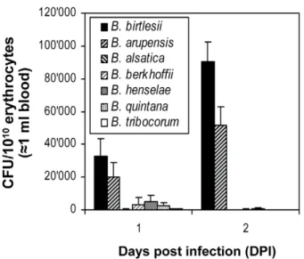
Next we investigated whetherBartonella species differ in their capacity to interact in vitro with erythrocytes of different mammalian origin, and whether this capacity may reflect the host-restriction displayed during natural infection. First, mouse erythrocytes were infected with eitherB. birtlesii,B. vinsonii arupensis
(both mouse-specific),B. alsatica(rabbit-specific),B. vinsonii berkhoffii
(dog-specific),B. henselae(cat-specific),B. quintana(human-specific), orB. tribocorum(rat-specific). Erythrocyte invasion was quantified by the gentamicin protection assay (Fig. 2). B. vinsonii arupensis
displayed invasion rates similar to B. birtlesii, while none of the other strains tested resulted in significant erythrocyte invasion. Using a corresponding set of strains expressing GFP, consistent results were obtained for the flow cytometric determination of bacterial adhesion to mouse erythrocytes (Fig. 3 and Fig. S1). These findings indicate that specificity for the mouse reservoirin vivo correlates with efficient adhesion to and invasion of mouse erythrocytesin vitro.
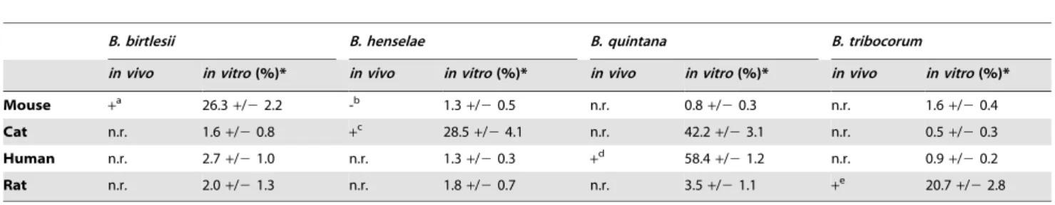
Next, we tested whether - similarly as observed for mouse erythrocytes andB.birtlesii– the capacity ofB. henselae, B. quintana
andB. tribocorumto adhere to erythrocytesin vitrois also restricted to erythrocytes from their natural reservoir host, i.e. cat, human and rat, respectively. GFP-expressing bacteria were used for erythrocyte infection, and adhesion was quantified by flow cytometric analysis. Fig. 3 and Table 1 illustrate that all tested
Bartonella species were able to efficiently adhere to erythrocytes isolated from their respective reservoir hosts, while they essentially did not adhere to erythrocytes from non-reservoir hosts. The only exception is B. quintana, which further to erythrocytes from the human reservoir also colonized cat erythrocytes. Together, these data indicate that the established in vitro model of erythrocyte colonization reflects well the host restriction as observed during natural infection.
B. birtlesiigenes required for intra-erythrocytic bacteremia in mice
As a basis for identifying genetic factors involved in host-restricted erythrocyte colonization, we identified a comprehensive set of B. birtlesiigenes required for establishing intraerythrocytic bacteremia in mice. To this end, an STM library ofB. birtlesiiwas constructed as previously described for B. tribocorum [6,12,19].
Figure 1.B. birtlesiiinvades murine erythrocytesin vitro.(A,B) Time- and bacterial number-dependency ofB. birtlesiiinvasion of murine erythrocytes determined by the gentamicin protection assay. (A) Freshly isolated murine erythrocytes were infected withB. birtlesiiat the indicated multiplicity of infection (MOI) and the numbers of intra-erythrocytic bacteria (colony forming units, CFU) was determined by the gentamicin protection assay at 1, 2 and 3 days post infection (DPI); n = 6, mean+/-SD; *, **: significant difference of data compared to 1 DPI. (B) Freshly isolated murine erythrocytes were infected withB. birtlesiiat MOI = 1. At 1 DPI, gentamicin was added to half of the samples, and growth was continued in the continous presence of gentamicin through 3 DPI to kill extracellular bacteria. The other half of the infected erythrcyotes was not treated with gentamicin. For both untreated and gentamicin treated samples, numbers of intra-erythrocytic bacteria were determined by the gentamicin protection assay at 1, 2 and 3 DPI (n = 6; mean+/2SD, *, **: significant difference in gentamicin treated samples compared to 1 DPI). (C) Time- and (D) bacterial number-dependency ofB. birtlesiiassociated to murine erythrocytes determined by flow cytometry. Freshly isolated murine erythrocytes were infected withB. birtlesii-gfp(MOI = 1, detection at 1 and 3 DPI in C and MOI = 0.1 or 1, detection at 3 DPI in D). The percentage of erythrocytes associated with bacteria were quantified by flow cytometric analysis at 2 and 3 DPI. Representative data for the fluorescence (FL-1) of 109000 erythrocytes are shown as histogram plots. (E) Confocal microscopic analysis of murine erythrocytes infected for 2 days with GFP-expressingB. birtlesii(MOI = 1). Arrows point to bacteria found in close association with erythrocytes.
From each conjugation assay, we selected 96 single kanamycin-resistant colonies and assembled an STM mutant library of 3456 mutants. We then identified mutants that have lost the capacity to cause intraerythrocytic bacteremia by screening the library in the mouse infection model [9]. Of 1456 mutants tested in the input
pools, 98 were not detected in the output pools from mice at days 7 and 14 post infection and were thus classified as abacteremic mutant candidates. All 98 abacteremic mutant candidates were retested by reassembling them into 49 pools of 9 mutants, each pool containing two abacteremic mutant candidates and an invariable set of seven mutants displaying wild-type behavior (bacteremic mutants). The rescreen confirmed an abacteremic phenotype for 48 of the initial 98 abacteremic mutant candidates, corresponding to 3.3% of the total number of mutants screened. Growth of all of the 48 confirmed abacteremic mutants on solid media was similar to the parental wild-type strain (data not shown).
We determined the transposon insertion sites for all 48 abacteremic mutants by direct sequencing out of the transposon into the flanking chromosomal region and mapping of the derived sequences onto the draft genome sequence ofB. birtlesii(S. Cescau, H.M. Yang, J. Wang, M. Vayssier-Taussat, A. Danchin, and F. Biville, unpublished data). Three mutants harboring two separate transposon insertions were not considered further in the analysis.
Table S1lists the loci inactivated by single transposon insertion in the remaining 45 abacteremic mutants. Five mutants carried the transposon insertion in an intergenic region: one (83D04) was near a gene encoding a tRNA; three of them (04A01, 86C05, 69B07) were upstream of genes encoding proteins of unknown function and one (69C09) was in proximity to a putative transcriptional regulator gene. In these mutants, the transposon may have thus disrupted a promoter or another regulatory sequence. 40 transposon insertions were mapped to the coding region of 38 different protein-encoding genes. In 8 mutants the insertions were found in genes encoding a conserved protein of unknown function, among them three putative surface proteins. Sixteen mutants carried insertions in genes previously implicated in bacterial
Figure 2. Efficiency ofin vitroinvasion of murine erythrocytes by differentBartonellaspecies.Freshly isolated murine erythrocytes were infected with the indicatedBartonellaspecies with a MOI = 1. The numbers of intra-erythrocytic bacteria (colony forming units, CFU) was determined by the gentamicin protection assay at 1 and 2 days post infection (DPI); mean+/-SD of triplicate samples.
doi:10.1371/journal.ppat.1000946.g002
Figure 3. Efficiency of interaction between erythrocyte andBartonellasp. according to host origin andBartonellaspecies.Freshly isolated erythrocytes from mouse, cat, human or rat were infected withgfp-expressing bacteria of the indicatedBartonellaspecies (MOI = 1). The percentages of infected erythrocytes were determined by flow cytometry at two DPI. Representative histogram plots for GFP-fluorescence (FL-1) of 109000 erythrocytes are shown.
pathogenicity, either in Bartonella (virB/D4, trw, ialA/B, badA,
omp43, iba) or other pathogenic bacteria (i.e. loci encoding heat shock proteins) [5]. Moreover, mutant genes encoding proteins involved in transport and metabolism, as well as phage-related function were also identified.
B. birtlesiigenes required for erythrocytic infection
in vitro
The 45 confirmed abacteremic mutants with single transposon insertion were individually tested for their capacity to invade murine erythrocytes using the gentamicin protection assay (Table S1). Nine mutants were found to be impaired in murine erythrocyte invasion (Fig. 4). Complementary erythrocyte adhe-sion assays based on flow cytometric analysis of antibody-stained bacteria demonstrated that seven of these nine invasion-deficient mutants are also deficient in erythrocytes adhesion. All seven mutants harbor a mutation in the operon encoding the T4SS Trw (two intrwD,trwE,trwF, trwJ2, trwL1, trwL2), which was previously
shown to be important for establishing an intraerythrocytic bacteremia in B. tribocorum [20]. Compared to wild-type, both
trwDmutants (04B03 and 41C12) showed a five-fold decrease in invasion/adhesion efficiency. All othertrwmutants failed to invade erythrocytes and were severely impaired in their capacity to adhere to erythrocytes (Fig. 4). These data demonstrate that the Trw system is required for erythrocyte invasion by mediating specific adhesion to erythrocytes. In contrast, mutants harboring an insertion in the invasion-associated locusialA/Bshowed normal erythrocyte adhesion but impaired invasion (10-fold reduced, p,0.01), confirming the previously suggested role of this locus in erythrocyte invasion [21,22]. Equally, an insertion mutant (25A02) inactivating livG (encoding an amino acid ABC-transporter) showed normal adhesion but a specific defect in invasion (4-fold, p,0.05) compared to wild-type (Fig. 4). None of the other abacteremic mutants appeared to be involved in erythrocytes invasion indicating that they probably are required for an earlier step of infection, i.e. for colonization of the primary niche.
Table 1.Efficiency of erythrocyte colonization according to host origin andBartonellaspecies.
B. birtlesii B. henselae B. quintana B. tribocorum
in vivo in vitro(%)* in vivo in vitro(%)* in vivo in vitro(%)* in vivo in vitro(%)*
Mouse +a 26.3+/22.2 -b 1.3
+/20.5 n.r. 0.8+/20.3 n.r. 1.6+/20.4
Cat n.r. 1.6+/20.8 +c 28.5+/24.1 n.r. 42.2+/23.1 n.r. 0.5+/20.3
Human n.r. 2.7+/21.0 n.r. 1.3+/20.3 +d 58.4+/21.2 n.r. 0.9+/20.2
Rat n.r. 2.0+/21.3 n.r. 1.8+/20.7 n.r. 3.5+/21.1 +e 20.7+/22.8
a[9],b[15],c[41,42],d[8,43,44],e[8].
*Freshly isolated erythrocytes from mouse, cat, human or rat were infected withgfp-expressing bacteria of the indicatedBartonellaspecies (MOI = 1). The percentages of colonized erythrocytes were determined by flow cytometry at day two post infection. Data for 109000 erythrocytes per time-point were analyzed (n = 6 for tests with homologous species and n = 3 for tests with heterologous species, mean+/2SD). For previously described infections of the respective mammalian hosts with the indicatedBartonellaspecies the presence (+) or absence (-) of intraerythrocytic bacteremia is indicated (n.r. = not reported).
doi:10.1371/journal.ppat.1000946.t001
Figure 4. Role of Trw in erythrocyte infection.Efficiency ofin vitroinvasion of murine erythrocyte by abacteremic mutants ofB. birtlesii. Thein vitroerythrocyte adhesion or invasion phenotype of abacteremic mutants identified in the STM screen was evaluated at 2 DPI by flow cytometry after immunocytochemical staining of bacteria (see Figure S2) or at 1 DPI by the gentamicin protection assay, respectively. The efficiency of erythrocyte adhesion or invasion of each tested mutant is expressed as percentage of erythrocyte adhesion or invasion of the isogenic wild-type strain (mean +/-SD of triplicate samples). All mutants listed inTable S1that do not appear in this figure display wild-type phenotype in regard ofin vitroerythrocyte invasion.
Role of Trw T4SS in host-specific infection of erythrocytes
Next we tested whether the Trw system shown here to be essential for erythrocyte infectionin vitroandin vivomay be directly involved in determining host-specificity. To this end we introduced pAB2, a plasmid encoding thetrwlocus of rat-specificB. tribocorum
[20], into cat-specificB. henselae, human-specificB. quintanaandB. tribocorum (control). We then compared the capacity of these recombinant strains to infect rat erythrocytes with their parental strains. Among the parental strains, only B. tribocorum mediated invasion of rat erythrocytes (Fig. 5), which is consistent with the erythrocyte adhesion data presented in Fig. 2. The pAB2-mediated ectopic over-expression of trw inB. tribocorum resulted only in a slight increase of invasion, indicating that the endogenous level of trw expression is sufficient to mediate efficient bacterial entry. Strikingly, ectopic expression of theB. tribocorum trwlocus in
B. henselae and B. quintana rendered these pathogens capable of infecting rat erythrocytes. These data clearly demonstrate a direct role of the Trw system in determining host-specificity of erythrocyte infection.
To further assess which components of the Trw T4SS may mediate host specificity we analyzed the molecular evolution of different trw genes of B. birtlesii and related species. Candidate genes for mediating host specificity are surface exposed compo-nents, i.e. the T4SS pilus components TrwL and TrwJ. As shown by Nystedt et al. [23] for other Bartonella species, trwL and trwJ
genes have been amplified and diversified several times during evolution. Thetrwlocus ofB. birtlesiialso displays amplification of
trwL(five copies) and co-amplification oftrwJtogether withtrwH and trwI(two copies) (Fig. S3, panel A). Phylogenetic analyses and calculation of the non-synonymous (dN) and synonymous (dS) substitution frequencies of differenttrwgenes further showed that
trwJand trwL homologs have diversified to much higher degree than other components of the Trw T4SS, within and among different species [Fig. S3, panel B-G, and [23]].
Discussion
Host-specificity is a prominent feature of pathogenic bacteria that reflects the host range susceptible to infection. Subtle changes in the molecular mechanisms that govern host-specificity may result in spontaneous host shifts, which represent a major risk for the emergence of novel human pathogens from animal reservoirs. Striking examples for this evolutionary scenario are the barto-nellae, which cause host-restricted intra-erythrocytic infections in their mammalian reservoirs. In conjunction with repeated host shifts, the large number ofBartonella species evolved by adaptive radiation [6], including the human-specific pathogenB. quintana
that evolved from cat-specificB. henselae[24]. Here we explored the bacterial genetic basis for host-restricted infection of erythrocytes. The establishment of anin vitromodel of erythrocyte adherence and invasion allowed us to demonstrate for the first time a direct correlation of host-restricted erythrocyte infectionin vivoand in vitro, demonstrating that host-specificity is determined by the capacity of bacteria to adhere to erythrocytes. In order to identify the bacterial factors critical for host-restricted erythrocyte infection we have used a two-step experimental protocol. First, we performed an STM screen forB. birtlesiiin mice which allowed us to identify 45 abacteremic mutants defective in establishing intra-erythrocytic infection. Among the corresponding set of 38 protein-encoding genes, 13 loci were also indentified in a similar STM screen performed in theB. tribocorum-rat model [6]. This indicates extensive similarities in the repertoire of pathogenesis factors in these closely related organisms as well as robustness of the performed genetic screens. Second, rescreening of the entire set of 45 abacteremicB. birtlesiimutants in thein vitromouse erythrocyte infection model resulted in the identification of nine mutants impaired in erythrocte invasion. The other mutants (36 of 45 = 80%) displaying a wild-type phenotype in this assay are therefore not directly involved in erythrocyte infection, but rather may contribute to the establishment of infection in the primary niche. Prominent examples are thevirB/virD4genes encoding the VirB/VirD4 T4SS, which is known to be required for primary niche infection in the B. tribocorum/rat model (Schulein, 2002). Moreover, a recent study inferred the VirB/VirD4 T4SS as major bacterial factor facilitating bacterial adaptation to novel hosts [6]. The nine mutants impaired inin vitroerythrocyte invasion differ in their capacity to adhere to erythrocytes. Transposon insertions in the invasion locus (ialA/B) previously implicated in erythrocyte invasion [21,22] and livG encoding an amino acid ABC-transporter displayed wild-type like adherence to erythrocytes. IalA/B and LivG should thus represent invasion factors. The remaining seven invasion-deficient transposon mutants were all severely impaired in erythrocyte adhesion. Strikingly, all these mutants carry insertions in components encoding the T4SS Trw, which thus represents an erythrocyte adherence system that is critical for erythrocyte invasion. Trw is known to be required for establishing intra-erythrocytic infection in the B. tribocorum-rat model [5,20,25], however, evidence for a direct role of the Trw system in erythrocyte adhesion as provided here was lacking so far. Based on the presumable surface location of components of Trw [20] this T4SS may directly interact with the erythrocyte surface and thus may restrict the host range of erythrocyte infection. To test the hypothesis that Trw determines host range we have expressed Trw of rat-specificB. tribocorumin cat-specificB. henselae
and human-specificB. quintana. Strikingly, this genetic manipula-tion resulted in an extension of the host range for in vitro
erythrocyte infection towards rats, demonstrating that Trw indeed represents a major determinant of host-specificity of erythrocyte infection. Thus, this finding establishes a new experimental model
Figure 5. Role of Trw T4SS in mediating host-specific erythrocyte invasion.Freshly isolated rat erythrocytes were infected withB. tribocorum, B. tribocorum(pAB2),B. henselae,B. henselae(pAB2), B. quintana, or B. quintana (pAB2) at a MOI = 1. Intra-erythrocytic bacteria were enumerated at 1, 2 and 3 days post infection (DPI) by the gentamicin protection assay (n = 3; mean+/2SD).
to study the molecular mechanisms governing host restriction -further to the molecular paradigm of host-specificity exemplified by the interaction of two surface proteins ofL. monocytogenes, InlA and InlB, with their respective host receptors [2,3,4].
The Trw locus was laterally acquired during evolution of the bartonellae. It is present in the largest sub-branch of the genus tree, comprising 13 species that are adapted to diverse mammalian reservoir hosts, while it is absent from human-specific B. bacilliformis, cat-specific Bartonella clarridgeiae and the species of the ruminent-specific sub-branch, which all diverted early during evolution of the bartonellae [6]. Interestingly, the acquisition of Trw by the modern lineage correlates with the loss of flagella, which are know to represent a major pathogencity factor for the invasion of erythroctes by B. bacilliformis and probably other flagellated bartonellae [25]. The Trw system of
Bartonella represents an interesting example of a pathogenesis-related T4SS that evolved rather recently by functional diversification of a laterally acquired bacterial conjugation system. The trw locus displays characteristic features of a pathogenicity island and shares extensive similarity with thetrw
locus of IncW broad-host range plasmid R388 encoding a genuine conjugation system. Thetrwloci ofBartonellaand R388 are colinear, except for multiple tandem gene duplications of
trwL and trwJ-trwH in Bartonella. Complementation of R388 derivatives carrying mutations in different trwgenes with their
Bartonella homologues allowed to demonstrate functional inter-changability for some T4SS components [20,26], underscoring the structural and functional conservation of individual subunits of these functionally diversified T4SSs. However, a major difference between these homologous systems is the lack of the coupling protein TrwB inBartonella, which in R388 is required for export of T4SS substrates. The lack of TrwB inBartonellathus indicates that its Trw system may not be capable of translocating substrates. However, the multiple copies oftrwLandtrwJin the
Bartonella trwlocus encode variant forms of surface-exposed pilus components, which probably are all co-expressed [20], indicat-ing that the primary function of theBartonellaTrw system may be the formation of variant pilus forms [25]. Based on the essential role of the Trw system for adhesion to erythrocyte and its role in determining host range it is conceivable to assume that these variant pili may facilitate the specific interaction with polymor-phic erythrocyte receptors, either within the reservoir host population (e.g. different blood group antigens), or among different reservoir hosts. Phylogenetic analyses and calculation of the non-synonymous (dN) and synonymous (dS) substitution frequencies of differenttrwgenes indeed demonstrated thattrwJ
andtrwLhomologs have diversified to much higher degree than other components of the Trw T4SS, within and among different species [23]. Together with the notion that the number of tandem repeats oftrwLandtrwJIHare variable among different
Bartonellaspecies these findings indicate thattrwLandtrwJgenes have been amplified and diversified several times during evolution. Horizontal transfer of such genes from a different bartonellae – similarly as we have demonstrated here for the entire trw operon of rat-specific B. tribocorum resulting in an extension of the host range of cat-specificB. henselaeor human-specific B. quintana to rat – or alternatively pre-adaption of superfluous copies of trwL and trwJ may represent realistic molecular evolutionary scenarios for host shifts and thereby the evolution of pathogens with an altered host-specificity as it has happened repeatedly during the evolution of the bartonellae. Future studies should identify the nature of the erythrocyte receptors targeted by the Trw system and their specific interaction that facilitate host-specific erythrocyte infection.
Materials and Methods
Ethics statement
Animals were handled in strict accordance with good animal practice as defined by the relevant European (European standards of welfare for animals in research), national (Information and guidelines for animal experiments and alternative methods, Federal Veterinary Office of Switzerland) and/or local animal welfare bodies. Animal work performed at the Biozentrum of the University of Basel was approved by the Veterinary Office of the Canton Basel City on June 2003 (licence no. 1741), and animal work performed at the Ecole Nationale Ve´te´rinaire d’Alfort (ENVA/AFSSA) was approved by the institute’s ethics committee on September 2005.
Bacterial strains and growth conditions
B. alsatica(IBS 382T, CIP 105477T) [27],B. birtlesii(IBS 135T, CIP 106691T) [28], B. henselae (Houston-1, ATCC 49882T), B. quintana(FullerT, ATCC VR-358T),B. tribocorum(IBS 506T, CIP 105476T) [29],B. vinsonii subsp. berkhoffii(ATCC 51672T),B. vinsonii subsp arupensis (ATCC 700727) [30] were grown for 5 days on Columbia agar containing 5% defibrinated sheep blood (CBA) in a humidified atmosphere with 5% C02at 35uC.
Construction of bacterial strains
B. tribocorum-gfp containing a chromosomally-integrated gfp -expression cassette [8] was used as GFP-expressingB. tribocorum
strain. GFP-expressing bacteria of other Bartonella species were obtained by electroporation with plasmid pJMBGFP as previously described [31,32]. This plasmid was extracted and purified from
B. quintanausing a Midi Prep Kit (Qiagen). The electroporation procedures was described previously [31]. Transformed bacteria were selected by plating on CBA-Km. A signature-tagged mutant library ofB. birtlesiiIBS135Twas constructed as described forB. tribocorum[6,12]. Cosmid pAB2 encoding the entiretrwlocus ofB. tribocorum[20,33,34] was introduced intoB.henselaeandB. quintana
by three parental mating [34,35].
In vitroinfection of erythrocytes
Erythrocytes from peripheral blood of mice (Balb/C), cats, rats (Wistar) and humans were isolated and purified by Ficoll gradient centrifugation. After washing in PBS, they were maintained in F12 modified medium [supplemented with 10% fetal calf serum, 2 mM glutamine, 1 mM sodium pyruvate, 0.1 mM Hepes, 257 mM histidine, 0.1 mg/ml hematin/histidine, non-essential amino acid (Gibco, FRANCE)] at 26108/ml. Forin vitroinfection experiments,
Bartonellaspecies were grown on CBA or CBA-km (Bartonella-gfpand STM mutants) plates. After 5 days of culture (10 days for GFP-expressingBartonella), bacteria were harvested, washed, suspended in PBS, and added to erythrocytes at a multiplicity of infection (MOI, calculation based on 1 OD600 nm= 36109 bacteria/ml) varying from 0.01 to 10 and incubated at 35uC in 5% C02 for various periods of time (from 1 to 3 days).
Detection of erythrocyte-associated bacteria
Erythro-cytes were then washed three times in PBS to remove the antibiotic and intracellular bacteria were released from erythro-cytes by hypotonic lyses of erythroerythro-cytes in 10ml of sterile water by freezing at220uC for 15 min. After thawing, serial dilutions of bacteria in PBS were inoculated onto CBA plates and incubated at 35uC for 5 days before being counted. For data presentation, all measurements were expressed as the number of CFU/1010 erythrocytes (corresponding to<1 ml of blood).
For flow cytometric detection of erythrocyte-associated with GFP-expressing bacteria, measurements were performed at day 1, 2, 3 after in vitro infection of erythrocytes with ten days old bacterial cultures. 100ml of the infection mixtures was washed 3 times in PBS and fixed for 10 min with 0.8% paraformaldehyde and 0.025% glutaraldehyde. After fixing, erythrocytes were analyzed by flow cytometry (FACScan, Becton Dickinson Bioscience, France). For flow cytometric detection of erythrocytes associated with bacteria that do not express GFP (abacteremic mutants), measurements were performed at day 2 after in vitro
infection. 100ml of the infection mixtures was washed 3 times in PBS and fixed for 10 min with 0.8% paraformaldehyde and 0.025% glutaraldehyde. After fixing, erythroctes/mutants associ-ation was revealed with mouse anti-B. birtlesii serum and anti-mouse FITC antibodies (Santa Cruz Biotechnology, Santa Cruz, CA,USA) and analyzed by flow cytomtry. Data were analyzed using the CellQuestPro software, version 4.0.2. Data for 109000 gated erythrocytes were collected and analyzed.
For confocal microscopy, 100ml of the infection mixtures was washed three times in PBS and the erythrocytes cell surface was stained using goat anti-mouse GPA antibodies (Santa Cruz Biotechnology, California, USA) and labelled with anti- goat-PE-antibodies (ImmunoQuest Antibody, North Yorkshire, UK). Samples were viewed with a Nikon Eclipse C1 Plus confocal laser scanning microscope (Nikon, Amstelveen, Netherlands) with detection in channel 1 (GFP fluorescence) and channel 2 (PE fluorescence) at original magnification x100.
STM library
The transposon vectors pHS006-Tag-001 to pHS006-Tag-036 each contained an oriT for conjugative transfer, the Himar1
transposon, a kanamycin resistant marker, a hyperactive trans-posase and one of 36 distinct signature-tags [6]. These 36 signature-tagged mariner transposon vectors were separately transferred fromE. colib2155 toB. birtlesiiby two-parental mating as previously described [35]. From each mating, 96 single kanamycin-resistant B. birtlesii transconjugants were transferred to a 96-well plate with cryo-medium and stored at280uC.
Mouse infections
Eight weeks old female Balb/C mice from Charles River Laboratories were housed in an animal facility (2 animals/cage) and allowed to acclimate to the facility and the diet for at least 5 days prior infection. Food and water were provided ad libitum. 36 differently signature-tagged mutants were grown separately from the transposon library for each input pool. They were pooled in PBS immediately before infection, and used to infect two mice with a total inoculum of 56107colony forming units (10ml of OD595= 1) in the ear dermis of Balb/C mice. The remainder of the input pools was heated at 100uC for 10 min and used as template for PCR detection. Fifty ml of blood were taken from the tail vein of the infected mice when bacteremia is peaking (days 7 and 14 post-infection) [9]. Bacteria released from erythrocytes by a freeze/thaw cycle were plated on CBA-km. After 10 days, bacterial colonies (output pool) were counted, harvested in PBS, suspended to OD595= 1 and heated at 100uC for 10 min to be used as template
for PCR detection. The rescreen was done following the same protocol using pools of nine mutants (two abacteremic mutants and seven mutants displaying a wild-type phenotype).
PCR detection of abacteremic mutants
For signature-tag identification, the generic primer Srev01 corresponding to a sequence in the transposon and a set of tag-specific primer were used for amplification of a fragment of approximately 600 bp [6]. The conditions for the PCR were as follows: a first denaturation step at 95uC for 5 min, followed by 30 cycles of PCR with denaturation at 95uC for 1 min, annealing for 30 s at 52uC, and extension at 72uC for 1 min. The program was completed by an extension step at 72uC for 5 min. The amplified fragments were displayed on a 1% agarose gel. Mutants that were detected in the input pools and absent from the out put pools (days 7 and 14) in both mice were considered as abacteremic mutants.
Identification and analysis of transposon insertion sites
Genomic DNA from abacteremic mutants, regrown from the library, was prepared with the ROCHE Genomic DNA Isolation Kit. Genomic DNA was sent to QIAGEN for sequencing with primers Tnstart and Tnend [6]. The sequences obtained by the genomic sequencing were compared by BlastN to the nr data base of NCBI (http://blast.ncbi.nlm.nih.gov/Blast.cgi). The exact transposon insertion sites were found by comparing the genomic sequences to contigs of the ongoingB. birtlesiigenome sequencing project by BlastN.
Screening of abacteremic mutants for their capacity to infect murine erythrocytes
Mutants displaying an abacteremic phenotype were tested for their capacity to invade murine erythrocytes using the gentamicin protection assay. Each mutant was tested at MOI = 1 in at least two independent experiments performed in triplicate samples. For mutants displaying an impaired erythrocyte invasion phenotype, invasion assays were performed at least three times in triplicate samples and adhesion assays were tested at day 2 post infection by flow cytometric detection once in triplicate samples.
Statistical analysis
Numerical data are reported as the mean of at least 3 replicate samples+/- standard errors of the means. Statistical significance of the data was measured by use of Student’s t test. A p-value,0.05 was considered significant.
Phylogenetic and evolutionary analysis
The sequence of theB. birtlesii trwlocus was deposited under the EMBL-EBI accession no. FN555106. Sequence alignments were calculated with ClustalW as implemented in MEGA4. Phyloge-netic trees were inferred by maximum likelihood methods with Paup 4.0 [36] and 100 bootstrap replicates were calculated. To select an appropriate substitution model the Akaike information criterion of Modeltest 3.7 was used [37]. The models obtained were general time reversible (GTR) + I for trwFED and trwN, transversion model (TVM)+I fortrwI, and TVM+I+G fortrwJ
andtrwL. Nonsynonymous (dN) and synonymous (dS) substitution frequencies were calculated using the method of Yang and Nielson [38] as implemented in the PAML package [39,40].
Supporting Information
with Bartonella sp.-GFP (MOI = 1, detection at two DPI). The percentage of erythrocytes associated with bacteria were quanti-fied by flow cytometric analysis. Representative data for the fluorescence (FL-1) of 10’000 erythrocytes are shown as histogram plots.
Found at: doi:10.1371/journal.ppat.1000946.s001 (0.08 MB TIF)
Figure S2 Efficiency of invitroadhesion of murine erythrocytes to abacteremic mutants. Freshly isolated murine erythrocytes were infected withB. birtlesiiabacteremic mutants (MOI = 1, detection 2 DPI). Association between erythrocytes and bacteria was revealed with mouse anti-B. birtlesiipolyclonal serum and labelled with anti-mouse FITC antibody. The percentage of erythrocyte associated with bacteria was quantified by flow cytometric analysis. Representative data for the fluorescence (FL-1) of 10’000 erythrocytes are shown as histogram plots.
Found at: doi:10.1371/journal.ppat.1000946.s002 (0.20 MB TIF)
Figure S3 Genetic organization of theBartonella trw locus, and phylogenies and synonymous (dS) vs. nonsynonymous (dN) substitution frequencies of the encoded trw genes. (A) Gene order structure of the trw locus ofB. birtlesiiand comparison to other
Bartonellaspecies. The copy number of amplified genes or segments in otherBartonellaspecies is indicated within brackets. Maximum Likelihood phylogenies of (B) the concatenated nucleotide alignments oftrwF, trwE, andtrwD, the nucleotide alignments of (C)trwJcopies, (D)trwI, (E)trwLcopies, and (F)trwNofB. birtlesii (Bb), B. grahamii (Bg), B. henselae (Bh), B. quintana (Bq), and B. tribocorum (Bt). FortrwJ(C) andtrwL(E), the range of pairwisedN/
dS ratios of different phylogenetic subclusters (shaded areas) are indicated at the upper right of each cluster. FortrwL1, the range of pairwise dN/dS ratios is indicated as well, although they do not cluster. (G) The pairwisedN/dS ratios of orthologous trw genes and the two adjacent genes ubiH and sdhA of B. birtlesii and B. grahamii, B. henselae, B. quintana, or B. tribocorum are plotted
according to their gene order. For the tandem repeated genes
trwL, trwJ, trwI, andtrwHonlytrwL5, trwJ1, trwI1, andtrwH1are shown, since ortholog assignment is difficult for the others due to copy number variation and the occurrence of recombination among different species [23].
Found at: doi:10.1371/journal.ppat.1000946.s003 (0.46 MB JPG)
Table S1 Genotypic characterization of abacteremic mutants of
B. birtlesiiobtained by signature-tagged mutagenesis (STM). The columns BARBAKC, BH, BQ and BT list the extensions of systematic names of orthologous genes from the published genomes of B. bacilliformis (accession no. CP000524), B. henselae
(accession no. BX897699),B. quintana(accession no. 897700) and
B. tribocorum(accession no. AM260525), respectively. * Thein vitro
erythrocate invasion phenotype of each mutant was determined by the gentamicin protection assay after 1 day of infection (triplicate samples) and categorized as normal (.70% of wild-type), reduced (,70% of wild-type but.1% of wild-type) or none (,1% of wild-type). Mutants with reduced or none in vitro invasion were tested again (n = 3) and the resulting mean and SD of all three experiments are represented in Figure 4.
Found at: doi:10.1371/journal.ppat.1000946.s004 (0.15 MB DOC)
Acknowledgments
We thank Dr. Arto Pulliainen for critically reading of the manuscript and Drs Didier Raoult and Pierre Edouard Fournier for providing the pJMB-GFP plasmid.
Author Contributions
Conceived and designed the experiments: MVT CD. Performed the experiments: MVT DLR HKD GM MM LA MQ HS. Analyzed the data: MVT EL PE HS CD. Contributed reagents/materials/analysis tools: MVT FB SC AD HJB HY JW PE HS CD. Wrote the paper: MVT CD.
References
1. Finlay BB, Cossart P (1997) Exploitation of mammalian host cell functions by bacterial pathogens. Science 276: 718–725.
2. Disson O, Grayo S, Huillet E, Nikitas G, Langa-Vives F, et al. (2008) Conjugated action of two species-specific invasion proteins for fetoplacental listeriosis. Nature 455: 1114–1118.
3. Khelef N, Lecuit M, Bierne H, Cossart P (2006) Species specificity of theListeria monocytogenesInlB protein. Cell Microbiol 8: 457–470.
4. Lecuit M, Dramsi S, Gottardi C, Fedor-Chaiken M, Gumbiner B, et al. (1999) A single amino acid in E-cadherin responsible for host specificity towards the human pathogenListeria monocytogenes. Embo J 18: 3956–3963.
5. Dehio C (2004) Molecular and cellular basis ofBartonellapathogenesis. Annu Rev Microbiol 58: 365–390.
6. Saenz HL, Engel P, Stoeckli MC, Lanz C, Raddatz G, et al. (2007) Genomic analysis ofBartonellaidentifies type IV secretion systems as host adaptability factors. Nat Genet 39: 1469–1476.
7. Dehio C (2005)Bartonella-host-cell interactions and vascular tumour formation. Nat Rev Microbiol 3: 621–631.
8. Schulein R, Seubert A, Gille C, Lanz C, Hansmann Y, et al. (2001) Invasion and persistent intracellular colonization of erythrocytes. A unique parasitic strategy of the emerging pathogenBartonella. J Exp Med 193: 1077–1086.
9. Boulouis HJ, Barrat F, Bermond D, Bernex F, Thibault D, et al. (2001) Kinetics ofBartonella birtlesiiinfection in experimentally infected mice and pathogenic effect on reproductive functions. Infect Immun 69: 5313–5317.
10. Yamamoto K, Chomel BB, Kasten RW, Hew CM, Weber DK, et al. (2002) Experimental infection of specific pathogen free (SPF) cats with two different strains ofBartonella henselaetype I: a comparative study. Vet Res 33: 669–684. 11. Chomel BB, Boulouis HJ, Breitschwerdt EB, Kasten RW, Vayssier-Taussat M,
et al. (2009) Ecological fitness and strategies of adaptation ofBartonellaspecies to their hosts and vectors. Vet Res 40: 29.
12. Saenz HL, Dehio C (2005) Signature-tagged mutagenesis: technical advances in a negative selection method for virulence gene identification. Curr Opin Microbiol 8: 612–619.
13. Kosoy MY, Saito EK, Green D, Marston EL, Jones DC, et al. (2000) Experimental evidence of host specificity ofBartonellainfection in rodents. Comp Immunol Microbiol Infect Dis 23: 221–238.
14. Chomel BB, Henn JB, Kasten RW, Nieto NC, Foley J, et al. (2009) Dogs are more permissive than cats or guinea pigs to experimental infection with a human isolate ofBartonella rochalimae. Vet Res 40: 27.
15. Kabeya H, Tsunoda E, Maruyama S, Mikami T (2003) Immune responses of immunocompetent and immunocompromised mice experimentally infected with
Bartonella henselae. J Vet Med Sci 65: 479–484.
16. Mehock JR, Greene CE, Gherardini FC, Hahn TW, Krause DC (1998)
Bartonella henselae invasion of feline erythrocytes in vitro. Infect Immun 66: 3462–3466.
17. Scherer DC, DeBuron-Connors I, Minnick MF (1993) Characterization of
Bartonella bacilliformisflagella and effect of antiflagellin antibodies on invasion of human erythrocytes. Infect Immun 61: 4962–4971.
18. Schulein R, Guye P, Rhomberg TA, Schmid MC, Schroder G, et al. (2005) A bipartite signal mediates the transfer of type IV secretion substrates ofBartonella henselaeinto human cells. Proc Natl Acad Sci U S A 102: 856–861.
19. Mavris M, Saenz H, Monteil M, Boulouis HJ, Dehio C, et al. (2005) Characterization of genes involved in long-term bacteremia in mice byBartonella birtlesii. Ann N Y Acad Sci 1063: 312–314.
20. Seubert A, Hiestand R, de la Cruz F, Dehio C (2003) A bacterial conjugation machinery recruited for pathogenesis. Mol Microbiol 49: 1253–1266. 21. Coleman SA, Minnick MF (2001) Establishing a direct role for theBartonella
bacilliformis invasion- associated locus B (ialB) protein in human erythrocyte parasitism. Infect Immun 69: 4373–4381.
22. Mitchell SJ, Minnick MF (1995) Characterization of a two-gene locus from
Bartonella bacilliformisassociated with the ability to invade human erythrocytes. Infect Immun 63: 1552–1562.
23. Nystedt B, Frank AC, Thollesson M, Andersson SG (2008) Diversifying selection and concerted evolution of a type IV secretion system inBartonella. Mol Biol Evol 25: 287–300.
24. Alsmark CM, Frank AC, Karlberg EO, Legault BA, Ardell DH, et al. (2004) The louse-borne human pathogenBartonella quintanais a genomic derivative of the zoonotic agentBartonella henselae. Proc Natl Acad Sci U S A 101: 9716– 9721.
26. de Paz HD, Sangari FJ, Bolland S, Garcia-Lobo JM, Dehio C, et al. (2005) Functional interactions between type IV secretion systems involved in DNA transfer and virulence. Microbiology 151: 3505–3516.
27. Heller R, Kubina M, Mariet P, Riegel P, Delacour G, et al. (1999)Bartonella alsaticasp. nov., a newBartonellaspecies isolated from the blood of wild rabbits. Int J Syst Bacteriol 49: 283–288.
28. Bermond D, Heller R, Barrat F, Delacour G, Dehio C, et al. (2000)Bartonella birtlesiisp. nov., isolated from small mammals (Apodemusspp.). Int J Syst Evol Microbiol 50: 1973–1979.
29. Heller R, Riegel P, Hansmann Y, Delacour G, Bermond D, et al. (1998)
Bartonella tribocorumsp. nov., a newBartonellaspecies isolated from the blood of wild rats. Int J Syst Bacteriol 48: 1333–1339.
30. Welch DF, Carroll KC, Hofmeister EK, Persing DH, Robison DA, et al. (1999) Isolation of a new subspecies,Bartonella vinsoniisubsp. arupensis, from a cattle rancher: identity with isolates found in conjunction withBorrelia burgdorferiand
Babesia microtiamong naturally infected mice. J Clin Microbiol 37: 2598–2601. 31. Fournier PE, Minnick MF, Lepidi H, Salvo E, Raoult D (2001) Experimental model of human body louse infection using green fluorescent protein-expressing
Bartonella quintana. Infect Immun 69: 1876–1879.
32. Kovach ME, Elzer PH, Hill DS, Robertson GT, Farris MA, et al. (1995) Four new derivatives of the broad-host-range cloning vector pBBR1MCS, carrying different antibiotic-resistance cassettes. Gene 166: 175–176.
33. de Paz HD, Sangari FJ, Bolland S, Garcia-Lobo JM, Dehio C, et al. (2005) Functional interactions between type IV secretion systems involved in DNA transfer and virulence. Microbiology 151: 3505–3516.
34. Dehio M, Knorre A, Lanz C, Dehio C (1998) Construction of versatile high-level expression vectors forBartonella henselaeand the use of green fluorescent protein as a new expression marker. Gene 215: 223–229.
35. Dehio C, Meyer M (1997) Maintenance of broad-host-range incompatibility group P and group Q plasmids and transposition of Tn5 inBartonella henselae
following conjugal plasmid transfer from Escherichia coli. J Bacteriol 179: 538–540.
36. Wilgenbusch JC, Swofford D (2003) Inferring evolutionary trees with PAUP*. Curr Protoc Bioinformatics Chapter 6: Unit 6 4.
37. Posada D, Crandall KA (1998) MODELTEST: testing the model of DNA substitution. Bioinformatics 14: 817–818.
38. Yang Z, Nielsen R (2000) Estimating synonymous and nonsynonymous substitution rates under realistic evolutionary models. Mol Biol Evol 17: 32–43. 39. Yang Z (1997) PAML: a program package for phylogenetic analysis by
maximum likelihood. Comput Appl Biosci 13: 555–556.
40. Yang Z (2007) PAML 4: phylogenetic analysis by maximum likelihood. Mol Biol Evol 24: 1586–1591.
41. Rolain JM, La Scola B, Liang Z, Davoust B, Raoult D (2001) Immunofluo-rescent detection of intraerythrocyticBartonella henselaein naturally infected cats. J Clin Microbiol 39: 2978–2980.
42. Rolain JM, Locatelli C, Chabanne L, Davoust B, Raoult D (2004) Prevalence of
Bartonella clarridgeiae andBartonella henselaein domestic cats from France and detection of the organisms in erythrocytes by immunofluorescence. Clin Diagn Lab Immunol 11: 423–425.
43. Foucault C, Rolain JM, Raoult D, Brouqui P (2004) Detection ofBartonella quintanaby direct immunofluorescence examination of blood smears of a patient with acute trench fever. J Clin Microbiol 42: 4904–4906.