Staphylococcus aureus
Sortase A-Mediated
Incorporation of Peptides: Effect of Peptide
Modification on Incorporation
Silvie Hansenová Maňásková1,2*, Kamran Nazmi1, Wim van‘t Hof1, Alex van Belkum2¤, Nathaniel I. Martin3, Floris J. Bikker1, Willem J. B. van Wamel2, Enno C. I. Veerman1
1Department of Periodontology and Oral Biochemistry, Academic Centre for Dentistry Amsterdam (ACTA), University of Amsterdam and VU University Amsterdam, Amsterdam, The Netherlands,2Department of Medical Microbiology and Infectious Diseases, Erasmus MC, Rotterdam, The Netherlands,3Department of Medicinal Chemistry and Chemical Biology, Utrecht Institute for Pharmaceutical Sciences, Utrecht University, Utrecht, The Netherlands
¤ Current address: Microbiology Research and Development Unit, BioMérieux, La Balme les Grottes, France *[email protected]
Abstract
The endogenousStaphylococcus aureussortase A (SrtA) transpeptidase covalently anchors cell wall-anchored (CWA) proteins equipped with a specific recognition motif (LPXTG) into the peptidoglycan layer of the staphylococcal cell wall. Previousin situ experi-ments have shown that SrtA is also able to incorporate exogenous, fluorescently labelled, synthetic substrates equipped with the LPXTG motif (K(FITC)LPETG-amide) into the bacte-rial cell wall, albeit at high concentrations of 500μM to 1 mM. In the present study, we have
evaluated the effect of substrate modification on the incorporation efficiency. This revealed that (i) by elongation of LPETG-amide with a sequence of positively charged amino acids, derived from the C-terminal domain of physiological SrtA substrates, the incorporation effi-ciency was increased by 20-fold at 10μM, 100μM and 250μM; (ii) Substituting aspartic
acid (E) for methionine increased the incorporation of the resulting K(FITC)LPMTG-amide approximately three times at all concentrations tested; (iii) conjugation of the lipid II binding antibiotic vancomycin to K(FITC)LPMTG-amide resulted in the same incorporation levels as K(FITC)LPETG-amide, but much more efficient at an impressive 500-fold lower sub-strate concentration. These newly developed synthetic subsub-strates can potentially find broad applications in for example thein situimaging of bacteria; the incorporation of antibody recruiting moieties; the targeted delivery and covalent incorporation of antimicrobial com-pounds into the bacterial cell wall.
Introduction
Staphylococcus aureusinfections are complex events that occur at the interface of the host and the pathogen [1]. From a bacterial perspective, infections require expression of virulence fac-tors including cell wall-anchored (CWA) proteins [2]. The C-terminal region of staphylococcal
a11111
OPEN ACCESS
Citation:Hansenová Maňásková S, Nazmi K, van‘t Hof W, van Belkum A, Martin NI, Bikker FJ, et al. (2016)Staphylococcus aureusSortase A-Mediated Incorporation of Peptides: Effect of Peptide Modification on Incorporation. PLoS ONE 11(1): e0147401. doi:10.1371/journal.pone.0147401
Editor:Herminia de Lencastre, Rockefeller University, UNITED STATES
Received:May 19, 2015
Accepted:January 4, 2016
Published:January 22, 2016
Copyright:© 2016 Hansenová Maňásková et al. This is an open access article distributed under the terms of theCreative Commons Attribution License, which permits unrestricted use, distribution, and reproduction in any medium, provided the original author and source are credited.
Data Availability Statement:All relevant data are within the paper.
Funding:This work was supported by ZonMW; grant title: Exploiting Staphylococcus aureus sortase for anti-infective purposes with the project number: 50-51700-98-055. The funding organisation was involved in financial support for conducting the research only.
CWA proteins contains a cluster of positively charged amino acids, which positions the CWA protein precursors to the cytoplasmic side of the bacterial cell membrane, and a hydrophobic transmembrane domain that immobilizes CWA protein precursors within the cell membrane, facilitating further processing by membrane-associated sortase A (SrtA) [3,4]. The SrtA enzyme recognizes the LPXTG motif of these proteins and catalyses their cleavage between threonine and glycine residues [5]. Next, SrtA catalyses a transpeptidation reaction by which the proteins are covalently linked to the peptidoglycan precursor lipid II [6–8]. Then, the pepti-doglycan precursor moiety that is coupled to lipid II is incorporated following transglycosyla-tion. SrtA is therefore closely involved in the control ofS.aureusvirulence [9–12].
In previous studies, it has been demonstrated that endogenous SrtA is also able to incorpo-rate exogenously added K(FITC)LPETG-amide, a fluorescently labelled artificial SrtA sub-strate, into the cell wall ofS.aureusstrains and of other Gram-positive bacteria. This required, however, high substrate concentrations, in the mM range [13,14]. The present study was aimed at the development of novel SrtA substrates exhibiting improved incorporation effi-ciency. In our view, such substrates might find applications in for examplein situimaging, the incorporation of antibody recruiting moieties, and the targeted delivery and covalent incorpo-ration of antimicrobial compounds into the bacterial cell wall.
In the present study a number of novel SrtA substrates were developed and their covalent incorporation into the staphylococcal cell wall was characterisedin situ. Three different types of substrate modifications were explored: (i) variants of the K(FITC)LPETG-amide, which had been elongated with sequences derived from physiological SrtA substrates—sequences contain-ing a hydrophobic membrane spanncontain-ing domain linked to a stretch of positively charged amino acids; (ii) a substrate (K(FITC)LPMTG-amide) in which glutamate (E) was substituted for methionine (M), which appeared to be better processed by SrtAin vitro[15] and (iii) K(FITC) LPMTG-amide conjugated to vancomycin, designed to potentially improve the targeting to lipid II, the primary acceptor for physiological SrtA substrates inS.aureus.
Materials and Methods
Solid-phase peptide synthesis
SrtA substrates were manufactured by solid-phase peptide synthesis using 9-fluorenyl-methox-ycarbonyl (Fmoc)-chemistry with a Syro II synthesizer (Biotage EU Customer Service, Upp-sala, Sweden) essentially as described previously [13,14,16]. Since the presence of a carboxylic acid group at the C-terminus of the SrtA recognition motif (LPXTG) may inhibit SrtA activity, an amide group was incorporated at this position [17]. Peptide synthesis grade solvents were obtained from Actu-All Chemicals (Oss, The Netherlands), the preloaded NovaSyn TGA resins from NovaBiochem (Merck Schuchardt, Hohenbrunn, Germany) and the N-α-Fmoc-amino
acids from Orpegen Pharma (Heidelberg, Germany) and Iris Biotech (Marktredwitz, Ger-many). The composition of all synthetic substrates (substrates1–13) is shown inTable 1.
FITC- labelling
Peptides were labelled with FITC (Fluorescein isothiocyanate) as the final step of the solid-phase synthesis by coupling FITC to theε-aminogroup of the N-terminal lysine residue which
was introduced using Boc-Lys(εFmoc)-OH [14].
‘
Click
’
addition of vancomycin
A previously-described alkyne-modified vancomycin building block was prepared by coupling
C-terminus [18].N-(3-(2-(2-(3-aminopropoxy) ethoxy)ethoxy)propyl)pent-4-ynamide was prepared by first mono-Boc protecting one amino group of 4,7,10-Trioxa-1,13-tridecanedia-mine with 1 equiv. Boc according to literature protocols [19] after which 4-pentynoic acid was coupled [20]. Removal of the Boc group was accomplished using standard conditions (1:1 tri-fluoroacetic acid/ dichloromethane (TFA/DCM), 1 hr) to provide the TFA salt of the desired product as a light yellow oil that was used directly after evaporation of TFA and DCM. Vanco-mycin (AppliChem, Darmstadt, Germany) was conjugated to the amino-alkyne spacer as fol-lows:N-(3-(2-(2-(3-aminopropoxy) ethoxy)ethoxy)propyl)pent-4-ynamide (250 mg, 0.63 mmol, 3 equiv.) was dissolved in DMF (10 ml) followed by addition of triethylamine (351μl,
2.52 mmol, 4 equiv.), vancomycin-HCl (936 mg, 0.63 mmol, 1.0 equiv.), EDAC (133 mg, 0.69 mmol, 1.1 equiv.) and HOBt (102 mg, 0.76 mmol, 1.2 equiv). The reaction mixture was stirred at ambient temperature for 2 days after which the DMF was removed in a vacuum concentrator (CHRIST, Osterode am Harz, Germany) and the residue was purified using preparative RP-HPLC as described below for peptide purification. The vancomycin-alkyne conjugate was next coupled to the azide-containing peptide by means of Cu(I) catalyzed‘click’chemistry fol-lowing a procedure described previously [21]. The azido peptide was prepared by introducing L-azidolysine (Fmoc-Lys(N3)-OH) (obtained from Chiralix, Nijmegen, The Netherlands). To
perform the‘click’reaction, the vancomycin alkyne was added in equimolar concentrations to the resin-bound azido peptide in a 20 ml syringe, containing DMF (5 ml), 2 equiv. of 2,6-luti-dine, 2 equiv. 2,2-bipyri2,6-luti-dine, 1 equiv. CuBr and 2 equiv. of sodium ascorbate (all peptide grade, Actu-All Chemicals) and flushed for 1 min with N2. After incubation for 24 hrs at ambient
temperature the mixture was flushed sequentially with DMF, H2O, MeOH, EDTA (100 mM),
H2O, and DMF followed by washing and drying by flushing three times with isopropanol and
DCM each. Next the peptide was detached from the resin and purified as described below for peptide purification.
Peptide purification
Peptides were purified by preparative RP-HPLC (Dionex Ultimate 3000, Thermo Scientific, Breda, The Netherlands) on a Grace Spring column (250 mm x 25 mm, Grace, Deerfield, IL, USA) containing Vydac C18 TP beads,10μm (Vydac, Hesperia, CA, USA). Elution was
per-formed with a linear gradient from 15 to 45% AcN containing 0.1% TFA in 20 min at a flow
Table 1. Synthetic substrate amino acid sequences used in this study [13].
Substrate number N-term Substrate sequence C-term
1 K(FITC) LPETG amide
2 K(FITC) EGTLP amide
3 K(FITC) AKKSELPETGGEESTNKGMLFGGLFSILGLALLRRNKKNHK amide
4 K(FITC) AKKSELPETGGEESTNKRRNKKNHKAGMLFGGLFSILGLALL amide
5 K(FITC) AKKSELPETGGEESTNKRRNKLKNHK amide
6 K(FITC) LPETGGEESTNKRKKW amide
7 K(FITC) LPMTG amide
8 K(FITC) K(Vancomycin)LPMTG amide
9 K(FITC) K(Vancomycin)MGTLP amide
10 K(FITC) LPMTGG-K(vancomycin) amide
11 K(FITC) Vancomycin-K-AKKSELPETGGEESTNKRRNKLKNHK amide
12 K(Vancomycin)LPMTG amide
13 K(Vancomycin)MGTLP amide
rate of 20 ml/min. The absorbance of the column effluent was monitored at 214 nm, and peak fractions were pooled and lyophilized. Re-analysis by RP-HPLC on an analytic Vydac C18-col-umn (218MS54) developed with a similar gradient at a flow rate of 1 ml/min revealed a purity of at least 95%. The authenticity was confirmed by mass spectrometry with a Microflex LRF MALDI-TOF, equipped with a gridless reflectron (Bruker Daltonik GmbH, Bremen, Germany).
Bacterial strains
TheS.aureus8325–4 wild type (WT) strain and its isogenicsrtAdeletion mutant (srtAKO) were used in this study. ThesrtAmutant was generated bysrtA:ermCallele transduction, as previously described [9,13]. ThesrtAKO mutant was selected and maintained on brain-heart infusion agar (BHA, BD (Becton Dickinson and Company)-Difco, Etten-Leur, The Nether-lands) plates supplemented with 3μg/ml erythromycin (Abbott Laboratories, North Chicago,
Illinois, U.S.).
In situ
incorporation of SrtA substrates
WT andsrtAKO strains were grown overnight on respectively BHA and BHA supplemented with 3μg/ml erythromycin. Subsequently, bacteria were resuspended in Luria-Bertani medium
(LB, BD-Difco, Etten-Leur, The Netherlands), to an OD600nmof 0.1. Twenty-five microliters of
bacterial suspension was mixed with 25μl LB containing up to 1 mM substrate (Table 1).
Bac-teria were then cultured in 96 wells-plates until the late stationary phase (approximately 24 hrs) in the dark at 37°C with continuous shaking at 230 rpm. As additional controls, we used 0.5 or 1μM vancomycin and a combination of vancomycin with substrate7at 0.5 or 1μM.
Flow cytometry
The bacteria were washed four times with PBS followed by centrifugation for 5 min at 3,700 x
g. To remove the non-covalently bound and intracellular substrate, bacteria were washed with 1% of sodium dodecyl sulphate (SDS, Merck-Schuchardt OHG, Hohenbrunn, Germany) at 60°C for 5 min. Subsequently, bacteria were washed four times with PBS followed by centrifu-gation for 5 min at 3,700 xg. Labelled bacteria were resuspended in 100μl of 4% formaldehyde
(Sigma-Aldrich, Zwijndrecht, The Netherlands) in PBS and incubated for 45 min at ambient temperature with continuous agitation. After 3 washes with PBS, the bacteria were resuspended in 0.5 ml PBS. The bacteria-associated fluorescence was measured on a FACS Canto II™flow cytometer and analysed using FacsDiva™software (BD Biosciences, Breda, The Netherlands) [13].
Effect of growth phase on substrate 5 incorporation
Substrate5(Table 1) incorporation was characterized in greater detail due to its enhanced incorporation levels. Bacteria were cultured in LB medium in the continuous presence of either (i) 1 mM substrate1or (ii) 250μM substrate5until either a) exponential growth phase (OD600nm0.5), b) post-logarithmic growth phase (OD600nm1), c) late exponential growth
phase (OD600nm1.2) or d) late stationary growth phase (24 hrs incubation) in the dark at 37°C
with continuous shaking (230 rpm). After the various growth phases had been reached, bacte-ria were harvested and the incorporation of substrates was established by determining the pres-ence of FITC using FACS analysis.
washed 3 times with PBS, and then incubated with either; i) SrtA buffer (50 mM Tris, 150 mM NaCl, 10 mM CaCl2, pH 7.5), ii) substrate1, or iii) substrate5(Table 1) in SrtA buffer. Bacteria
were incubated in the presence of 0 to 1 mM of these substrates using a final volume of 50μl
and 96 wells-plates. Incubation was performed for 17 hrs in the dark at 37°C with continuous shaking (230 rpm).
Determination of the minimal inhibitory concentration (MIC) of
vancomycin and vancomycin-conjugates
WT andsrtAKO bacteria were cultured in LB medium in a 96-wells plate in the presence of serial dilutions (0.1 to 100μM) of either vancomycin (Sigma-Aldrich, Zwijndrecht, The
Neth-erlands) or substrate8;9;12or13at 37°C with continuous shaking (230 rpm). After 24 hrs, the wells were visually inspected and the first clear well was designated as the MIC.
All experiments were repeated independently three times and the standard error of the mean (SEM) was calculated.
Statistical analysis
Statistical analysis was performed using the Prism 5.0 package (GraphPad software, San Diego, CA, USA). We used one-way ANOVA testing with Bonferroni correction, considering P<0.05 as being statistically significant.
Results
Optimization of synthetic SrtA substrates
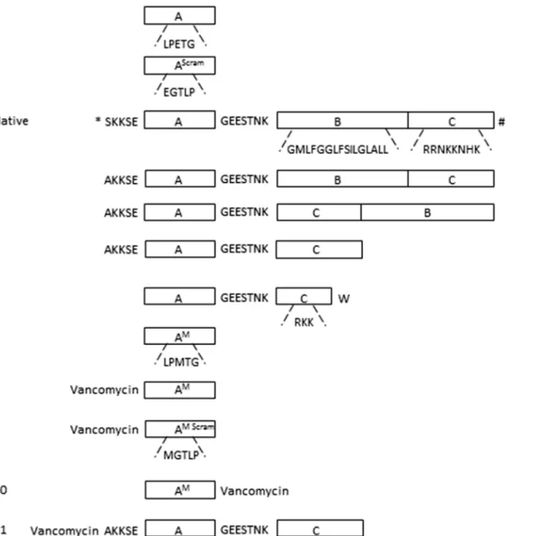
In vivothe incorporation of physiological SrtA substrates is determined by a recognition signal in their C-terminus. The C-terminus contains three motifs: (A) the LPXTG SrtA recognition sig-nal, (B) a hydrophobic transmembrane domain and (C) a cluster of cationic amino acid residues (SeeFig 1, native substrate). The LPXTG motif protrudes from the outside of the bacterium, and is linked via a hydrophilic negatively charged spacer GEESTNK to the transmembrane domain, which in turn is connected to the cationic cluster at the cytosolic side of the membrane [4,22, 23]. In a previous study, we found that an artificial motif (K(FITC)LPETG) (substrate1,Fig 1, Table 1) is covalently incorporated in a SrtA-dependent manner into the cell wall ofS.aureus
[13]. In the present study, we first tested a series of artificial SrtA substrates equipped with the additional domains B and C, to examine the effect on incorporation. To monitor their incorpo-ration in the cell wall by FACS, the substrates were linked via an additional lysine (K) residue to FITC.S.aureusWT and its isogenicsrtAKO (control) were cultured in the presence of sub-strates for 24 hrs. After washing, to remove non-covalently bound subsub-strates, the cell-associated fluorescence was determined by FACS analysis. A scrambled version of substrate1(K(FITC) EGTLP, substrate2,Fig 1,Table 1) was used as a negative control.S.aureus(WT) cultured in the presence of 1 mM substrate1showed a mean fluorescence level of 840 FU (Fluorescence Units). In thesrtAKO strain little if any incorporation of substrate1was observed (70 FU). Cul-turing in the presence of the scrambled substrate2did not result in any substantial incorpo-ration, neither in the WT nor in thesrtAKO strain (75 FU and 50 FU for WT andsrtAKO strains, respectively) (Fig 2). This confirms previous studies [13,14] that the incorporation of substrate1was mediated for>90% by SrtA. We next tested a set of substrates that included the native sequence of the C-terminal region of the fibronectin-binding protein (FnBP) ofS.aureus
functions as a hydrophilic spacer. Substrate3is flanked at the N-terminal part with a sequence of non-repetitive region of cell wall-spanning domain AKKSE to increase the targeting towards the cell-wall. Incorporation of substrate3inS.aureusWT was comparable to that observed with substrate1, with a mean fluorescence intensity per cell of 800 FU at 1 mM. In contrast to sub-strate1, substrate3labelled also thesrtAKO strain (400 FU at 1 mM) (Fig 2).
The position of the motifs A, B and C in substrate3is tailored for transferring physiological SrtA substrates from the inside to the outside of the bacteriain situ. This configuration may
Fig 1. Schematic representation of the C-terminal region of FnBP precursor and synthetic substrates used in this study. 1: the minimal substrate used, containing the SrtA recognition motif LPETG only (designated A).2: the scrambled version of substrate1, EGTLP.Native: the sorting motif of the FnBP precursor ofS.aureus, containing the SrtA recognition motif (A), a hydrophobic domain (B) and a positively charged region (C). The full sequences of the separate regions are depicted under the blocks. Domain (A) and (B) are linked via a hydrophilic spacer GEESTNK [24]. The N-terminus and the C-terminus are indicated with*and # respectively.3: schematic representation of substrate3, based on the complete sorting signal of the FnBP precursor.4: schematic representation of substrate4, in which domain (B) and (C) are interchanged.5: schematic representation of substrate5, in which domain B is removed.6: schematic representation of substrate6containing domain (A), truncated domain (C) and a C-terminal tryptophan.7: schematic representation of substrate7 containing domain (A), with glutamate (E) substituted for methionine (M).8: schematic representation of substrate8, composed of substrate7with its N-terminus linked to vancomycin.9: schematic representation of substrate9, the scrambled version of substrate8, with an MGTLP motif instead of an LPMTG motif.10: schematic representation of substrate10, composed of substrate7with its C-terminus linked to vancomycin.11: schematic representation of substrate11, composed of substrate5with its N-terminus linked to vancomycin. All substrates were at the N-terminus labelled with a FITC group.
therefore be sub-optimal for the incorporation of exogenous substrates, which target the endogenous SrtA from theoutside. We therefore examined whether interchanging the posi-tions of the membrane-spanning domain (B) and the cationic cluster (C) (substrate4,Fig 1, Table 1) would result in a higher incorporation. This peptide, however, labelledS.aureusWT andS.aureus srtAKO to the same extent (1600 versus 1400 FU) (Fig 2). Substrates3and4 formed precipitates during incubation, due to their hydrophobic character, which could not be completely removed in the washing steps and stayed attached to the bacteria. We envisage that this has caused the SrtA-independent background labelling of theS.aureus srtAKO strain. Because of the poor solubility, substrates3and4were not further examined. We next tested substrate5(Fig 1,Table 1), which lacks the hydrophobic membrane-spanning domain (B). This substrate was incorporated at a higher efficiency compared to substrate1(Fig 2). Between 10 and 250μM the incorporation of substrate5increased almost linearly with the
concentra-tion. In this concentration range, substrate5showed an approximately 20-fold higher incorpo-ration than substrate1. At concentrations>250μM, the incorporation of substrate5levelled off, suggesting that the process became saturated. Substrate5revealed a higher background in
srtAKO strain than substrate1(860 vs 70 fluorescence units at 1 mM,Fig 2). This is probably caused by SrtA independent electrostatic interactions between the cationic cluster and the bac-terial cell surface. Still, the incorporation of substrate5in theS.aureusWT strain was 10 to 20-fold higher than in theS.aureus srtAKO strain, indicating that>90% of the incorporation of substrate5was mediated by SrtA, similar to that of substrate1. In an attempt to further min-imize the substrate, a variant (LPETGGEESTNKRKKW (substrate6,Fig 1,Table 1)) was developed composed of the SrtA recognition motif LPETG, linked via the GEESTNK linker to
Fig 2. Titrations of the SrtA synthetic substrates.Cultures of 8325–4 WT andsrtAKO strains were incubated in LB medium during 24 hrs in the presence of either substrate1;2;3;4;5or6[13] at 10μM, 100μM, 250μM, 500μM or 1 mM and analysed by FACS. The synthetic substrate concentrations used are
depicted on the X-axis and the mean fluorescence, as a measure for the level of substrate incorporation into the bacterial cell wall, was plotted on the Y-axis. Substrate1signal at 100μM and 250μM was significantly lower than the signals obtained with substrate5at the same concentration (P<0.0001). Substrate
1signal obtained at 1 mM was significantly lower than the signal obtained with substrate5at 100μM (P<0.0001). Only the most relevant significant differences are presented.
a minimal cationic domain C (RKK) and terminated at the C-terminus by a tryptophan (W) residue. As this residue has a preference for the membrane-water interface [25,26], we rea-soned that it might target the substrate to the bacterial membrane. The efficiency of this pep-tide, however, did not differ from that of substrate1(700 FU; 1 mM), with the background labelling of 160 FU at 1 mM (Fig 2).
Effect of growth phase on substrate incorporation
To analyse the effect of growth phase on the cumulative incorporation of the SrtA substrates,S.
aureus(WT andsrtAKO strains) were cultured in the presence of substrate1and5, at a con-centration that yielded the maximal incorporation level (1 mM and 250μM, respectively).
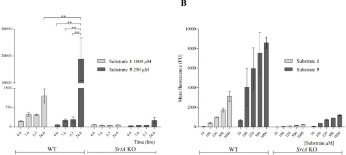
Bac-teria were harvested after 4 hr (exponential phase), 7 hr (post-logarithmic phase), 8.5 hr (late exponential phase) and 24 hr (late stationary phase) and analysed by FACS (Fig 3A). After 8.5 hrs both substrates showed comparable incorporation (approximately 500 and 300 FU, respec-tively). Between 8.5 and 24 hrs the incorporation of substrate1increased approximately three-fold, whereas that of substrate5increased more than 60-fold (from 300 to 19,000 FU;Fig 3A). We then explored the incorporation of substrate5in late stationaryS.aureus. To do so,S.
aureuswas cultured until late stationary phase in the absence of substrates. After washing, bac-teria were transferred into SrtA reaction buffer (conditions where the bacbac-terial metabolism is suppressed) supplemented with either substrates1or5at concentrations varying between 10μM to 1 mM. After 17 hrs, the reaction was stopped and incorporation was determined by
FACS; bacteria treated with substrate5displayed a 4 to 10 times higher fluorescence than those incubated with substrate1(Fig 3B). This supported that substrate5was incorporated with a higher efficiency than substrate1.
Fig 3. Kinetics determination of substrate 5.8325–4 WT andsrtAKO strains were incubated in the presence of (A) either 1 mM of substrate1or 250μM of
substrate5until either exponential (4 hrs), post-logarithmic (7 hrs), late exponential (8.5 hrs) or late stationary phase (24 hrs) in LB medium. The mean fluorescence of substrate1and substrate5is depicted on y-axis. (B) Washed and diluted (OD600nm0.5) late stationary grown WT andsrtAKO bacteria were incubated with either 10μM, 100μM, 250μM, 500μM or 1 mM substrate1or substrate5in SrtA buffer during 17 hrs. The mean fluorescence was
determined by FACS. The signal obtained with substrate1at 1 mM after 24 hrs of incubation was significantly lower than the signal obtained with substrate5 at 250μM after 24 hrs (P<0.001). Substrate5signals at time points 4 hrs, 7 hrs and 8 hrs were all significantly lower in comparison to the signal obtained with the same substrate at 24 hrs time point at 250μM (P<0.001).
Effect of glutamate to methionine substitution
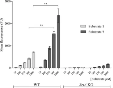
The third position in the SrtA recognition motif (LPXTG) is tolerant to various amino acids substitutions. Still, some residues are preferred over others,e.g. LPMTG is more rapidly pro-cessed than LPETG by isolated recombinant SrtAin vitro[15]. Therefore, we examined if substituting E for M (substrate7,Fig 1,Table 1) would also enhance thein situprocessing of exogenous SrtA substrates. Substrate7was incorporated significantly better than substrate1 into the WT bacterial surface at 500μM and 1 mM (Fig 4). Substrate7showed little if any
incorporation insrtAKO strain (150 FU), indicating that its incorporation inS.aureusWT required a functional SrtA.
Effect of vancomycin conjugation on incorporation
The glycopeptide antibiotic vancomycin targets the D-ala-D-ala motif of lipid II, whichin vivo
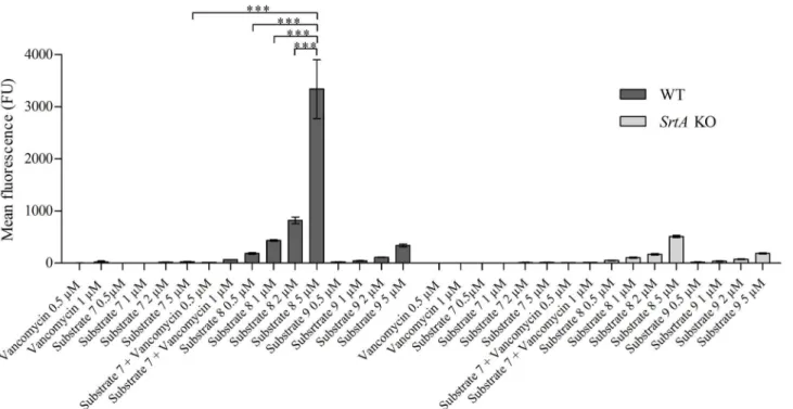
functions as an acceptor for SrtA-processed CWA proteins. Vancomycin binds specifically to lipid II and decreases its accessibility, thereby interfering with SrtA-dependent anchoring of CWA proteins [7,27,28]. Conjugation of vancomycin to substrate7was therefore considered as an attractive strategy to enhance the targeting of these substrates to the vicinity of SrtA. To do so, vancomycin was coupled to substrate7at either the penultimate N-terminal position to yield K(FITC)K(vancomycin)LPMTG (substrate8,Fig 1,Table 1) or the C-terminal site of the peptide, to yield K(FITC)LPMTGGK(vancomycin) (substrate10,Fig 1,Table 1). As a control served substrate9(K(FITC)K(vancomycin)MGTLP), a scrambled variant of substrate8(Fig 1, Table 1). Thein situincorporation of the vancomycin-LPMTG conjugates was assessed by cul-turingS.aureusin LB medium supplemented with the substrates at concentrations varying from 0.5μM—5μM (Fig 5). This revealed that the vancomycin-modified substrates8and10
(the latter not shown) were incorporated to the same extent as the parent peptide7, but at a 200-fold lower concentration (Fig 5). Culturing in the presence of equimolar concentrations of vancomycin and substrate7did not result in labelling ofS.aureus, ruling out that vancomycin by itself stimulated the incorporation of substrate8and10(Fig 5). At the highest
Fig 4. Incorporation of substrate 7.WT andsrtAKOS.aureusbacteria were incubated in the presence of either substrate1or substrate7at 10μM, 100μM, 250μM, 500μM or 1 mM concentrations in LB medium
during 24 hrs and analysed by FACS. The incorporation of substrate7was significantly higher at 500μM and
at 1 mM compared to substrate1at the same concentrations after 24 hrs in WT bacteria (P<0.001).
concentration tested (5μM), background labelling of substrate8with thesrtAKO strain was
observed, possibly due to formation of non-covalent complexes of vancomycin with its D-ala-D-ala ligand. Similarly, the control substrate9also showed background staining with both the WT and thesrtAKO strain at higher substrate concentration. The specific substrate incorpo-ration of substrate8in WT bacteria was 10- fold higher than in theS.aureus srtAKO strain incubated with substrate8andsrtAKO and WT strains incubated with control substrate9. These data indicate that>90% of the incorporation of substrate8was mediated by SrtA. Con-jugation of vancomycin to substrate5(substrate11,Fig 1,Table 1) did not result in the same increased incorporation observed for conjugates8and10(data not shown).
Apparently, coupling of LPMTG to vancomycin enhanced the processing by SrtA. We won-dered if, reciprocally, LPMTG would enhance the targeting of vancomycin to lipid II, and henceforth its antibacterial activity. Determination of the MIC-values of substrate8and9, however, showed that conjugation of vancomycin with LPMTG negatively affects its antimi-crobial activity, causing an increase in the MIC of vancomycin from 1μM to either 50 or
100μM for substrate8and9, respectively, in both the WT and thesrtAKO strain (Fig 6). To
test the effect of vancomycin modification on MIC increase within the substrate8and9, we generated substrate12and13lacking the FITC molecule. The MIC of these non-FITCylated variants were 2μM (substrate12) and 50μM (substrate13) (Fig 6). These data suggest, that
the addition of FITC molecule decreases the antimicrobial activity of the vancomycin unit within the synthetic substrate.
Discussion
In the present study, we have examined the effect of various modifications on the incorporation efficiency of artificial SrtA substrates. Physiological SrtA substrates contain, besides the
Fig 5. Incorporation of vancomycin—conjugated substrate.8325–4 WT andsrtAKO bacteria were incubated in the presence of either 0.5μM or 1μM of
1) vancomycin; 2) substrate7+ vancomycin; or 0.5μM; 1μM; 2μM or 5μM of 3) substrate7; 4) substrate8or 3) substrate9in LB medium during 24 hrs and
analysed by FACS (mean fluorescence). (***: P<0.0001).
LPXTG motif, a hydrophobic transmembrane domain and a cluster of positively charged amino acids, which together retain the protein in the vicinity of the bacterial membrane [4].In vivo, this prevents leakage of key CWA surface proteins into the bacterial surroundings, and facilitates efficient processing by SrtA. By elongating the LPXTG motif with these domains, we aimed to improve thein situprocessing of synthetic SrtA substrates. However, substrates3and 4equipped with both the hydrophobic membrane-spanning and the cationic domain, were
poorly soluble and formed visible aggregates. This probably caused high non-specific adsorp-tion to bacterial surfaces (seeFig 2). Omitting the hydrophobic domain (substrate5), however, resulted in 5-fold better incorporation than the parent substrate. Also substrate5exhibited more a-specific binding to thesrtAKO strain than the parent peptide1, probably due to a strong electrostatic binding with the bacterial surface. The incorporation of substrate5reached saturation at concentrations250μM. This suggests that under these conditions another
fac-tor, likely the number of accessible pentaglycine acceptors, had become limiting [29,30]. Strik-ingly, even in the presence of these saturating concentrations of exogenous substrate, the cell-wall expression of the endogenous substrate SpA was not decreased (Hansenova Manaskova
et al, unpublished observations), indicating that this physiological substrate is preferentially processed by SrtA. In line, incorporation of SpA predominantly occurs in the exponential growth phase [31], whereas incorporation of substrates5(Fig 3) and substrate1[13] mainly takes place in the stationary phase, when the synthesis of CWAs is subdued [31].
Substitution of E by M within the LPXTG motif (substrate7) resulted in a 3-fold higher incorporation. This is in a close agreement with the results of a previous study, which found that LPMTGin vitrois cleaved by recombinant SrtA approximately 2.5 times faster than
Fig 6. MIC determination of vancomycin-conjugated substrates.WT andsrtAKO bacteria were cultured in LB medium in the presence of serial dilutions (0.1 to 100μM) of either vancomycin or substrate8;9;12or13at 37°C with continuous shaking (230 rpm) during 24 hrs. The MIC-value (first clear well) was
visually determined after 24 hrs and is indicated with an arrow.
LPETG [15]. Interestingly, the SrtA recognition motifs of different native CWA proteins con-tain various different amino acids at the ambiguous third position: lysine, glutamic acid, aspar-tic acid, alanine or glutamine, but none of them contains methionine [32].
Conjugation of LPMTG to vancomycin enhanced the incorporation efficiency drastically: the same level of incorporation was obtained at 200- to 500-fold lower concentrations than with the unconjugated substrate (Fig 5). SrtA-independent labelling of theS.aureusWT and theS.aureus srtAKO strain was observed with substrate8and the scrambled substrate9, pos-sibly caused by vancomycin direct binding to D-ala-D-ala. Interestingly, conjugation of the peptide moiety to vancomycin increased the MIC value 50- to 100-fold, in line with similar decreases in antibiotic activity observed for other fluorescently-labelled vancomycin probes, which despite a>100-fold increase in MIC value, labelled the bacterial cell wall at sub-MIC concentrations [33]. The fluorescent label in vancomycin substrates seems to be responsible for the MIC increase, as substrate12(substrate8variant without FITC) had comparable MIC value to vancomycin molecule only (2μM and 1μM, respectively).
In summary, we have demonstrated that modification of SrtA synthetic substrates is a feasi-ble approach to enhance theirin situincorporation by endogenousS.aureusSrtA. Interest-ingly, we found that vancomycin-LPMTG conjugates are incorporated in the cell wall ofS.
aureuswith high efficiency. Such molecules may form a platform for targeted modification of the bacterial surface for e.g. antimicrobial or imaging purposes.
Acknowledgments
We kindly thank Dr. John Hays (Department of Medical Microbiology & Infectious Diseases, Erasmus MC) for correcting the manuscript. We are grateful to the Department of Gastroen-terology and Hepatology, Erasmus MC, in particular Anthonie Groothuismink and Dr. André Boonstra, for use of the FACS machine and their helpful assistance.
Author Contributions
Conceived and designed the experiments: SHM WH NIM FJB WJBW ECIV. Performed the experiments: SHM KN. Analyzed the data: SHM. Contributed reagents/materials/analysis tools: KN NIM. Wrote the paper: SHM WH AB NIM FJB WJBW ECIV.
References
1. Lowy FD. Staphylococcus aureus infections. N Engl J Med. 1998; 339(8):520–32. Epub 1998/08/26. doi:10.1056/NEJM199808203390806
2. Foster TJ, Geoghegan JA, Ganesh VK, Hook M. Adhesion, invasion and evasion: the many functions of the surface proteins of Staphylococcus aureus. Nature reviews Microbiology. 2014; 12(1):49–62. Epub 2013/12/18. doi:10.1038/nrmicro3161PMID:24336184.
3. Mazmanian SK, Liu G, Ton-That H, Schneewind O. Staphylococcus aureus sortase, an enzyme that anchors surface proteins to the cell wall. Science. 1999; 285(5428):760–3. Epub 1999/07/31. PMID:
10427003.
4. Schneewind O, Model P, Fischetti VA. Sorting of protein A to the staphylococcal cell wall. Cell. 1992; 70(2):267–81. Epub 1992/07/24. 0092-8674(92)90101-H [pii]. PMID:1638631.
5. Navarre WW, Schneewind O. Proteolytic cleavage and cell wall anchoring at the LPXTG motif of sur-face proteins in gram-positive bacteria. Mol Microbiol. 1994; 14(1):115–21. Epub 1994/10/01. PMID:
7830549.
6. Schneewind O, Fowler A, Faull KF. Structure of the cell wall anchor of surface proteins in Staphylococ-cus aureus. Science. 1995; 268(5207):103–6. Epub 1995/04/07. PMID:7701329.
8. Ruzin A, Severin A, Ritacco F, Tabei K, Singh G, Bradford PA, et al. Further evidence that a cell wall precursor [C(55)-MurNAc-(peptide)-GlcNAc] serves as an acceptor in a sorting reaction. J Bacteriol. 2002; 184(8):2141–7. Epub 2002/03/27. PMID:11914345; PubMed Central PMCID: PMC134952. 9. Mazmanian SK, Liu G, Jensen ER, Lenoy E, Schneewind O. Staphylococcus aureus sortase mutants
defective in the display of surface proteins and in the pathogenesis of animal infections. Proc Natl Acad Sci U S A. 2000; 97(10):5510–5. Epub 2000/05/11. doi:10.1073/pnas.080520697[pii]. PMID:
10805806; PubMed Central PMCID: PMC25859.
10. Jonsson IM, Mazmanian SK, Schneewind O, Verdrengh M, Bremell T, Tarkowski A. On the role of Staphylococcus aureus sortase and sortase-catalyzed surface protein anchoring in murine septic arthritis. J Infect Dis. 2002; 185(10):1417–24. Epub 2002/05/07. JID010772 [pii] doi:10.1086/340503
PMID:11992276.
11. Jonsson IM, Mazmanian SK, Schneewind O, Bremell T, Tarkowski A. The role of Staphylococcus aureus sortase A and sortase B in murine arthritis. Microbes Infect. 2003; 5(9):775–80. Epub 2003/07/ 10. S1286457903001436 [pii]. PMID:12850203.
12. Weiss WJ, Lenoy E, Murphy T, Tardio L, Burgio P, Projan SJ, et al. Effect of srtA and srtB gene expres-sion on the virulence of Staphylococcus aureus in animal models of infection. J Antimicrob Chemother. 2004; 53(3):480–6. Epub 2004/02/06. doi:10.1093/jac/dkh078[pii]. PMID:14762051.
13. Hansenova Manaskova S, Nazmi K, van Belkum A, Bikker FJ, van Wamel WJ, Veerman EC. Synthetic LPETG-containing peptide incorporation in the Staphylococcus aureus cell-wall in a sortase A- and growth phase-dependent manner. PLoS One. 2014; 9(2):e89260. Epub 2014/03/04. doi:10.1371/ journal.pone.0089260[pii]. PMID:24586638; PubMed Central PMCID: PMC3929722.
14. Nelson JW, Chamessian AG, McEnaney PJ, Murelli RP, Kazmierczak BI, Spiegel DA. A biosynthetic strategy for re-engineering the Staphylococcus aureus cell wall with non-native small molecules. ACS Chem Biol. 2010; 5(12):1147–55. Epub 2010/10/07. doi:10.1021/cb100195dPMID:20923200; PubMed Central PMCID: PMC3003768.
15. Kruger RG, Otvos B, Frankel BA, Bentley M, Dostal P, McCafferty DG. Analysis of the substrate speci-ficity of the Staphylococcus aureus sortase transpeptidase SrtA. Biochemistry. 2004; 43(6):1541–51. Epub 2004/02/11. doi:10.1021/bi035920jPMID:14769030.
16. Bolscher JG, Oudhoff MJ, Nazmi K, Antos JM, Guimaraes CP, Spooner E, et al. Sortase A as a tool for high-yield histatin cyclization. FASEB J. 2011; 25(8):2650–8. Epub 2011/04/29. fj.11-182212 [pii] doi:
10.1096/fj.11-182212PMID:21525488.
17. Popp MW, Antos JM, Ploegh HL. Site-specific protein labeling via sortase-mediated transpeptidation. Curr Protoc Protein Sci. 2009; Chapter 15:Unit 15 3. Epub 2009/04/15. doi:10.1002/0471140864. ps1503s56PMID:19365788.
18. Arnusch CJ, Bonvin AM, Verel AM, Jansen WT, Liskamp RM, de Kruijff B, et al. The vancomycin-nisin (1–12) hybrid restores activity against vancomycin resistant Enterococci. Biochemistry. 2008; 47 (48):12661–3. Epub 2008/11/08. doi:10.1021/bi801597bPMID:18989934.
19. Zhang L, Wu Y, Brunsveld L. A synthetic supramolecular construct modulating protein assembly in cells. Angew Chem Int Ed Engl. 2007; 46(11):1798–802. Epub 2007/04/24. doi:10.1002/anie. 200604222PMID:17450607.
20. Pera NP, Kouki A, Haataja S, Branderhorst HM, Liskamp RM, Visser GM, et al. Detection of pathogenic Streptococcus suis bacteria using magnetic glycoparticles. Org Biomol Chem. 2010; 8(10):2425–9. Epub 2010/05/08. doi:10.1039/c000819bPMID:20448902.
21. Punna S, Kaltgrad E, Finn MG. "Clickable" agarose for affinity chromatography. Bioconjug Chem. 2005; 16(6):1536–41. Epub 2005/11/17. doi:10.1021/bc0501496PMID:16287252.
22. Fischetti VA, Pancholi V, Schneewind O. Conservation of a hexapeptide sequence in the anchor region of surface proteins from gram-positive cocci. Mol Microbiol. 1990; 4(9):1603–5. Epub 1990/09/01. PMID:2287281.
23. Schneewind O, Mihaylova-Petkov D, Model P. Cell wall sorting signals in surface proteins of gram-posi-tive bacteria. EMBO J. 1993; 12(12):4803–11. Epub 1993/12/01. PubMed Central PMCID:
PMC413927.
24. Signas C, Raucci G, Jonsson K, Lindgren PE, Anantharamaiah GM, Hook M, et al. Nucleotide sequence of the gene for a fibronectin-binding protein from Staphylococcus aureus: use of this peptide sequence in the synthesis of biologically active peptides. Proc Natl Acad Sci U S A. 1989; 86(2):699–
703. Epub 1989/01/01. PMID:2521391; PubMed Central PMCID: PMC286541.
25. Wallace BA, Janes RW. Tryptophans in membrane proteins. X-ray crystallographic analyses. Adv Exp Med Biol. 1999; 467:789–99. Epub 2000/03/18. PMID:10721132.
27. Ton-That H, Schneewind O. Anchor structure of staphylococcal surface proteins. IV. Inhibitors of the cell wall sorting reaction. J Biol Chem. 1999; 274(34):24316–20. Epub 1999/08/14. PMID:10446208. 28. Breukink E, de Kruijff B. Lipid II as a target for antibiotics. Nature reviews Drug discovery. 2006; 5
(4):321–32. Epub 2006/03/15. doi:10.1038/nrd2004
29. Turner RD, Ratcliffe EC, Wheeler R, Golestanian R, Hobbs JK, Foster SJ. Peptidoglycan architecture can specify division planes in Staphylococcus aureus. Nature communications. 2010; 1:26. Epub 2010/ 10/27. doi:10.1038/ncomms1025PMID:20975691.
30. Zhou X, Cegelski L. Nutrient-dependent structural changes in S. aureus peptidoglycan revealed by solid-state NMR spectroscopy. Biochemistry. 2012; 51(41):8143–53. Epub 2012/09/15. doi:10.1021/ bi3012115PMID:22974326; PubMed Central PMCID: PMC3554850.
31. Fischetti VA. Gram-positive pathogens. 2nd ed. ed. Washington, D.C.: ASM; [Oxford: Blackwell, dis-tributor]; 2006. xiii, 849 p., [22] p. of plates p.
32. Rawlings ND, Waller M, Barrett AJ, Bateman A. MEROPS: the database of proteolytic enzymes, their substrates and inhibitors. Nucleic acids research. 2014; 42(Database issue):D503–9. Epub 2013/10/ 26. doi:10.1093/nar/gkt953PMID:24157837; PubMed Central PMCID: PMC3964991.
![Table 1. Synthetic substrate amino acid sequences used in this study [13].](https://thumb-eu.123doks.com/thumbv2/123dok_br/16351020.189562/3.918.61.866.133.404/table-synthetic-substrate-amino-acid-sequences-used-study.webp)
![Fig 2. Titrations of the SrtA synthetic substrates. Cultures of 8325–4 WT and srtA KO strains were incubated in LB medium during 24 hrs in the presence of either substrate 1; 2; 3; 4; 5 or 6 [13] at 10 μM, 100 μM, 250 μM, 500 μM or 1 mM and analysed by FAC](https://thumb-eu.123doks.com/thumbv2/123dok_br/16351020.189562/7.918.61.840.119.468/titrations-synthetic-substrates-cultures-incubated-presence-substrate-analysed.webp)