ORIGINAL
ARTICLE
Staphylococcus aureus
regulates secretion of
interleukin-6 and monocyte chemoattractant protein-1
through activation of nuclear factor kappaB signaling
pathway in human osteoblasts
Authors
Rende Ning1
Xianlong Zhang2
Xiaokui Guo3
Qingtian Li4
1Dr; PhD, Department of Orthopaedics, The Sixth People’s Hospital Affiliated to Shanghai Jiao Tong University; Department of Orthopaedics, The Third Affilliated Hospital of Anhui Medical University, China
2Dr; Professor, Department of Orthopaedics, The Sixth People’s Hospital Affiliated to Shanghai Jiao Tong University, China 3PhD; Professor, Department of Medical Microbiology and Parasitology, Institutes of Medical Sciences, Shanghai Jiao Tong University School of Medicine, China 4PhD; Professor, Department of Medical Microbiology and Parasitology, Institutes of Medical Sciences, Shanghai Jiao Tong University School of Medicine, China
Submitted on: 09/16/2010 Approved on: 09/16/2010
Correspondence to: Xianlong Zhang Department of Orthopaedics The Sixth People’s Hospital Affiliated to Shanghai Jiao Tong University 600 Yishan Road, Shanghai 200233, China. Phone: +86-21-64369181 Fax: +86-21-64701361 zhangxianlong1964@ gmail.com
Financial Support: The National Natural Science Foundation of China (No. 30470455).
We declare no conflict of interest.
ABSTRACT
Objective: Activation of nuclear factor kappaB by diverse bacteria regulates the secretion of chemokines and cytokines. Staphylococcus aureus (S. aureus)-infected osteoblasts can significantly increase the secretion of interleukin-6 and monocyte chemoattractant protein-1. The aim of this study was to investigate whether S. aureus can activate nuclear factor kappaB in human osteoblasts, and whether the activation of nuclear factor kappaB by S. aureus regulates the secretion of interleukin-6 and monocyte chemoattractant protein-1. Methods: Immunoblot and electrophoretic mobility shift assay were used to detect the degradation of IκBa and activation of nuclear factor kappaB in human osteoblasts in response to S. aureus, respectively. Enzyme-linked immunosorbent assay was used to measure the secretion of interleukin-6 and monocyte chemoattractant protein-1 in the supernatants. Lastly, carbobenzoxyl-l-leucinyl-l-leucinyl-l-leucinal, an inhibitor of the nuclear factor kappaB, was used to determine if activation of nuclear factor kappaB by S. aureus in human osteoblasts regulates the secretions of interleukin-6 and monocyte chemoattractant protein-1. Results: Our results for the first time demonstrated that S. aureus can induce the degradation of IκBa and activation of nuclear factor kappaB in human osteoblasts in a time and dose-dependent manner. In addition, inhibition of nuclear factor kappaB by carbobenzoxyl-l-leucinyl-l-leucinyl-l-leucinal suppressed the secretion of interleukin-6 and monocyte chemoattractant protein-1 in the supernatants of S. aureus-infected hu-man osteoblasts in a dose-dependent hu-manner. Conclusions: These findings suggest that S. aureus
can activate nuclear factor kappaB in human osteoblasts, and subsequently regulate the secretion of interleukin-6 and monocyte chemoattractant protein-1. The nuclear factor kappaB transcription factor regulates a number of genes involved in a wide variety of biological processes. Further study of the effects of nuclear factor kappaB activation on S. aureus-infected human osteoblast may provide us new insights into discovery of the immune mechanisms in osteomyelitis.
Keywords:Staphylococcus aureus; osteoblasts; NF-kappaB; interleukin-6; chemokine CCL2. [Braz J Infect Dis 2011;15(3):189-194]©Elsevier Editora Ltda.
INTRODUCTION
Osteomyelitis is a severe infection of bone tis-sue, which results in progressive inflammatory destruction of bone. Staphylococcus aureus is known as an important causative organism in osteomyelitis,1,2 which accounts for approxi-mately 80% of all human cases.2 Recent works have demonstrated that S. aureus-infected os-teoblasts can secrete a number of cytokines and chemokines to mediate the immune responses in osteomyelitis.3-6 However, to date, the patho-genesis of immune responses in S. aureus -in-duced osteomyelitis is still poorly understood. The activation of nuclear factor kappaB (NF-κB) signaling pathway to produce inflam-matory mediators plays an essential role in
the host response to pathogenic organisms.7 NF-κB is a dimeric protein composed of members of the Rel / κB family, and NF-κB dimers are retained into the cytoplasm by the inhibitory protein IĸBa.8 Different stimuli can induce the phosphorylation of IκBa, and its subsequent ubiquitination and degrada-tion.9 Freed NF-κB can translocate into the nucleus, leading to the transcriptional acti-vation of NF-κB-dependent genes,10 such as chemokines and cytokines genes.11 Previous studies have demonstrated that NF-κB in eu-karyocyte could be activated by diverse bacte-ria, such as Porphyromonas gingivalis,12
However, it is not clearly defined whether S. aureus
infection can induce the activation of NF-κB in human osteoblasts. Interleukin-6 (IL-6) is a pleiotropic cytokine and plays important role in the regulation of immune response and inflammation,16 which controls the devel-opment of both humoral and cell-mediated immune re-sponses17 and block the suppressive activity of regulatory CD4+CD25+ regulatory T-cells,18 thereby enabling the progression of immune responses. Monocyte chemoat-tractant protein (MCP)-1 is a small cytokine belonging to the CC chemokine family that is also known as CCL2 (c-c chemokine ligand 2),19 which can induce the recruit-ment of macrophages and activated T-lymphocytes into areas of inflammation or infection, and sustain inflamma-tory responses by maintaining the activated status of such cells.20 The previous studies have confirmed that S. aureus -infected human osteoblasts can significantly augment MCP-1 and IL-6 secretion in vitro,3,4in vivo and in clinical human osteomyelitic lesions.21,22 It is unknown, however, the mechanism of IL-6 and MCP-1 immune responses to infection of S. aureus. Thus, in our present study, we in-vestigated whether S. aureus can activate NF-κB signaling pathway, and whether such activation regulate the secre-tion of IL-6 and MCP-1 in human osteoblasts.
MATERIALS AND METHODS
Bacteria, human osteoblasts and culture conditions S. aureus UAMS-1 (ATCC 49230), a human osteomyelitis clinically isolated strain23 was used in this study. S.
au-reus ATCC 49230 was obtained from the American Type Culture Collection. S. aureus cells were grown overnight (16 h) in 5 mL of Tryptic Soy Broth (Oxoid, Basingstoke, UK) in a shaking water bath at 37°C. The bacteria were harvested by centrifugation for 10 min at 4,300 × g at 4°C and washed twice in 5 mL of Hank’s Balanced Salt Solution. The pellets were then resuspended in 5 mL of growth medium lacking antibiotics and antimycotics.
The SV40 human osteoblasts (SV40 hOBs, ATCC) were obtained from the American Type Culture Collec-tion. These cells were previously characterized as authen-tic osteoblasts.24 The cells were seeded in 25-cm2 flasks and incubated at 37°C in 5% CO2 with DMEM/F12 (Gib-co) supplemented with 0.3 mg/mL G418 (Sigma) and 10% fetal bovine serum (HyClone). Then, the cells were propa-gated as described by the manufacturer. Once osteoblasts reached approximately 80% confluency, cells were trypsi-nized (0.025% trypsin-0.01% EDTA), washed in medium and seeded into 6-well plates. Two to three hours before the addition of bacteria, the cells were washed twice with PBS and then incubated with an assay medium (growth medium without antibiotics and antimycotics) and in-fected as described in the following section.
Infection assay
To investigate the IκBa degradation and NF-κB activa-tion following infecactiva-tion, 2×10⁶ cells/well were exposed to
S. aureus for the designated times and at the indicated multiplicity of infections (MOI). To investigate the se-cretion of IL-6 and MCP-1 in the supernatant of human osteoblasts infected with S. aureus, 2×10⁶ cells/well were infected with S. aureus at an MOI of 250 (250:1 bacte-ria/osteoblasts) or without S. aureus (controls) for 1 h. Following infection, the cells were washed with PBS and grown in DMEM/F12 supplemented with 0.3 mg/mL G418 and 10% fetal bovine serum for 24 h at 37°C in a 5% CO2 atmosphere. To determine whether S. aureus -activated NF-κB signaling pathway regulates the IL-6 and MCP-1 secretion in the supernatants of human os-teoblasts, 2×10⁶ cells/well were pretreated with carboben-zoxyl-l-leucinyl-l-leucinyl-l-leucinal (Z-LLL-al) (Sigma), an inhibitor of NF-κB,12 for 1 h, and then washed, added bacteria at an MOI of 250. Z-LLL-al is also named the proteasome inhibitor MG132 which can inhibit NF-κB formation and degradation of its inhibitor I-κB.25
Immunoblot assay
Electrophoretic mobility shift assay (EMSA)
After infection, nuclear extracts were prepared using rea-gents from the NE-PER extraction system (Pierce, Rock-ford, IL), as recommended by the manufacturer’s instruc-tions. Then the nuclear extracts were used to test for NF-κB protein/DNA binding by a DIG Gel-Shift Kit (Roche, Ger-many). The sequences for NF-κB were 5’-AGTTGAGGG-GACTTTCCCAGGC-3’ (sense) and 5’-GCCTGGGAAA-GTCCCCTCAACT-3’ (antisense). Briefly, equal molar of complementary NF-κB oligonucleotides were mixed in TEN-buffer [10 mM Tris–Cl, 1 mM EDTA, and 0.1 M NaCl (pH 8.0)] and incubated at 95°C for 10 min. After slowly cooling down to room temperature, double-stranded (ds) oligonucleotides were diluted with TEN-buffer to a final concentration of 3.85 pmol/μL. Labeling reaction was done in a final volume of 20 μL containing 1 μL ds oligonucleo-tides, 9 μL water, 4 μL 5 × labeling buffer, 4 μL CoCl2 solu-tion, 1 μL DIG-ddUTP and 1 μL terminal deoxynucleotide transferase. The reaction mixture was incubated at 37°C for 15 min and chilled on ice immediately. Labeling reac-tion was stopped by 2 μL 0.2 M EDTA (pH 8.0). The labeled NF-κB oligonucleotides were diluted to 0.155 pmol/μL by adding 3 μL double distilled water. For gel shift analysis, a small aliquot was further diluted to 15.5 fmol/μL. In the gel shift assay, 5 μg nuclear extract were incubated with 4 μL binding buffer [5 mM EDTA, 50 mM (NH4)2SO4, 5 mM dithiothreitol (DTT), 1% Tween 20 (v/v), 150 mM KCl, 100 mM Hepes (pH 7.6)], 1 μL poly (dI-dC) and 1 μL poly-L-lysine. Double distilled water was added to a final reaction volume of 18 μL. To confirm the specificity of NF-κB protein/ DNA binding by competition assay, we added 100-fold ex-cess unlabeled NF-κB oligonucleotides (cold probes) in the reaction mixture. After 5 min incubation at room tempera-ture, 2 μL DIG-labeled NF-κB -oligonucleotides were added into the reaction mixture and incubated for another 30 min. After adding 5 μL loading buffer, the reaction mixture was fractionated in a 6% native polyacrylamide gel in 0.5 × TBE buffer and electroblotted onto Hybond-N+ membranes (Amersham Biosciences). After blotting, the membrane was baked at 120°C for 30 min and rinsed in washing buffer [0.1 M maleic acid, 0.15 M NaCl, 0.3% Tween 20 (pH 7.5)] for 5 min, and then incubated for 30 min in blocking solu-tion (1% blocking reagent in maleic acid buffer). Following incubation with anti-DIG-AP (1:10,000) in blocking solu-tion for 30 min, the membrane was washed 2 × 15 min in washing buffer and equilibrated in detection buffer [0.1 M Tris–HCl, 0.1 M NaCl (pH 9.5)] for 5 min. Chemilumines-cence signals were detected with CSPD as the substrate in X-ray film.
IL-6 and MCP-1 secretion assay
After infection, the culture supernatants were collected and centrifuged (10,000 × g, 5 min). Then IL-6 and
MCP-1 in the culture supernatants were measured with enzyme-linked immunosorbent assay (ELISA) kits for human IL-6 or MCP-1 (R&D Systems, Minneapolis, MN), according to the manufacturer’s instructions. Each sample was assayed in duplicate and absorbance was determined at 450 nm.
Data analysis
All assays were repeated in three independent experiments. Data are presented as mean ± SD. Statistical analysis among groups was performed by ANOVA and SNK test using the SPSS17.0 software package. Statistically significant values were defined as p < 0.05.
RESULTS
S. aureus induced IκBa degradation and NF-κB activation in human osteoblasts in a time-dependent manner.
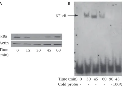
In order to relate the kinetics of infection to the stabil-ity of IκBa in human osteoblasts, human osteoblasts were infected with S. aureus at an MOI of 250 for indicated times. The infected cell lysates showed a quick IκBa degradation after 15 min and with a nearly loss of immunoreactivity at 30 min, and nearly removed the prior levels at 60 min. How-ever, uninfected cells had stable levels of IκBa expression (Figure 1A).
To correlate the degradation of IκBa with the nuclear translocation of NF-κB in human osteoblasts, EMSAs were used to investigate the nuclear translocation of NF-κB. As shown in Figure 1B, nuclear proteins from the cells infected with S. aureus stimulated NF-κB DNA binding activity to rise at 30 min, reached the maximal level at 45 min, nearly loss at 90 min at an MOI of 250, whereas nuclear proteins
Figure 1: Time-dependent activation of NF-kB signaling
pathway by Staphylococcus aureus in human osteoblasts.
Human osteoblasts were either uninfected (0 min) or infected with Staphylococcus aureus at a multiplicity of infections (MOI) of 250 for the indicated times. Immunoblot demonstrated
cytosoic IkBa degradation (A) and EMSA showed nuclear
NF-kB activation (B) in human osteoblasts. The exposed film is
representative of results from three independent experiments.
IkBa
Actin Time (min)
0 15 30 45 60
A B
NF-kB
Time (min) 0 30 45 60 90 45
from uninfected cells nearly failed to display binding ac-tivity to DNA. To confirm the specificity of the DNA pro-tein interaction, a 100-fold excess of cold probes was used as a competitor to inhibit the binding activity of NF-κB at 45 min after infection. As shown in Figure 1B, cold probes completely inhibited the DNA binding activity of NF-κB, in-dicating that the increased binding is specific to the NF-κB-binding sequence. Collectively, these findings suggest that S. aureus can induce activation of NF-κB in human osteoblasts in a time-dependent manner.
S. aureus induced IκB degradation and NF-κB activa-tion in human osteoblasts in a dose-dependent manner to determine the effect of the infectious dose on IκBa degradation, human osteoblats were infected with S. au-reus for 30 min at the indicated MOIs. S. aureus-infected cells demonstrated IκBa degradation substantially in-creased from MOI of 25 to 500 at 30 min (Figure 2A). To further determine the infectious doses on the roles of NF-κB DNA binding activity in human osteoblasts, NF-κB DNA binding activity from cells infected with
S. aureus at 45 min quickly increased from an MOI of 25 to 250 (Figure 2B), but started to decline when the MOI reached to 500. These results indicate that S. aureus can induce activation of NF-κB in human osteoblasts in a dose-dependent manner. Activation of NF-κB signaling pathway by S. aureus regulated the secretion of IL-6 and MCP-1 in human osteoblats.
S. aureus-infected human osteoblasts can significantly augment IL-6 and MCP-1 secretion at 24 h postinfection
in vitro.3,4 Therefore, in our present study, we examined the supernatants of human osteoblasts at 24 h postinfection of
S. aureus to determine if activation of NF-κB signaling path-way by S. aureus regulates the secretion of IL-6 and MCP-1 in human osteoblasts. As shown in Figure 3, uninfected cells (controls) could secrete IL-6 (0.22 ng/mL) and MCP-1 (0.84 ng/mL), respectively. However, the secretion of IL-6 and MCP-1 in the supernatants of human osteoblasts infect-ed with S. aureus at an MOI of 250 for 1 h was significantly
Figure 2: Dose-dependent activation of NF-kB signaling
pathway by Staphylococcus aureus in human osteoblasts.
Human osteoblasts were either uninfected (0 min) or infected
with Staphylococcus aureus at the indicated multiplicity of
infections (MOI). Immunoblot demonstrated cytosoic IkBa
degradation at 30 min postinfection (A) and EMSA showed
nuclear NF-kB activation at 45 min postinfection (B) in human
osteoblasts. The exposed film is representative of results from three independent experiments.
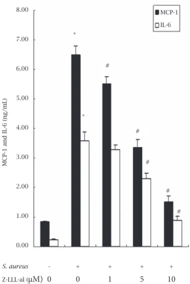
Figure 3: Effect of carbobenzoxyl-l-leucinyl-l-leucinyl-l-leucinal
(Z-LLL-al), an inhibitor of NF-kB, on the secretion of MCP-1
and IL-6 in human osteoblasts infected with Staphylococcus
aureus. Human osteoblasts were uninfected or infected with
Staphylococcus aureus (S. aureus) at a multiplicity of infections (MOI) of 250 for 1 h or pretreated with Z-LLL-al at different
concentrations for 1 h, then infected with S. aureus at an MOI
of 250 for 1 h. Following infection, the cells were washed and grown in the culture medium for 24 h, and then the culture supernatants were collected. IL-6 and MCP-1 in the culture supernatants were measured with enzyme-linked immunosorbent assay (ELISA) kits. Data are presented as mean ± SD from three independent experiments. Statistical analysis comparing groups was performed by ANOVA and SNK test.
* p < 0.05, cells infected with S. aureus versus controls, # p
< 0.05, cells infected with S. aureus versus cells pretreated
with different concentrations of Z-LLL-al prior to infection of
S. aureus.
IkBa
Actin MOI
NF-kB
MOI 0 25 75 250 500
0 25 75 250 500
A B
MCP-1 and IL-6 (ng/mL)
MCP-1 IL-6
* *
#
#
#
#
#
S. aureus - + + + +
Z-LLL-al (µM) 0 0 1 5 10
8.00
7.00
6.00
5.00
4.00
3.00
2.00
1.00
enhanced to 3.58 ± 0.31 ng/mL (p < 0.05) and 6.48 ± 0.30 ng/mL (p < 0.05), compared with controls, respectively. In addition, Figure 3 also shows that the secretion of Il-6 and MCP-1 in the supernatants of human osteoblasts infected with S. aureus were suppressed by the addition of Z-LLL-al (1 to 10 µM) in a dodependent manner, and IL-6 se-cretion was reduced from 3.58 ± 0.31 to 3.27 ± 0.18, 2.30 ± 0.20 (p < 0.05), 0.89 ± 0.13 ng/mL (p < 0.05) and MCP-1 secretion was reduced from 6.48 ± 0.30 to 5.52 ± 0. 22 (p < 0.05), 3.36 ± 0.26 (p < 0.05), 1.52 ± 0.19 ng/mL (p < 0.05) at 1, 5, 10 µM Z-LLL-al, respectively.
DISCUSSION
This study shows that S. aureus can activate NF-κB in hu-man osteoblasts in a time and dose-dependent hu-manner and
S. aureus-activated NF-κB signaling pathway in human os-teoblasts regulates the secretion of IL-6 and MCP-1.
The nuclear translocation of NF-κB is regulated by the cytoplasmic inhibitory species IκBa via its binding to the nuclear localization sequence of p65; degradation of IκBa triggers the activation of NF-κB.8,9 In our current study, activation of NF-κB by S. aureus in human osteoblasts has been demonstrated by two different methods. First, our im-munoblot experiments demonstrated that degradation of cytoplasmic IκBa in S. aureus-infected human osteoblasts occurred in a time and dose-dependent manner (Figures 1A and 2A). Second, correlated IκBa degradation with NF-κB DNA binding activity, our findings by EMSA confirmed that nuclear proteins from S. aureus-infected human osteoblasts could stimulate the DNA binding activity of NF-κB in a time and dose-depenent manner (Figures 1B and 2B).
In addition, we demonstrated that S. aureus-activated NF-κB signaling pathway in human osteoblasts regulates the secretion of IL-6 and MCP-1. As shown in Figure 3, human osteoblats infected with S. aureus at MOI of 250 significant-ly augment IL-6 (p < 0.05) and MCP-1 (p < 0.05) secretion compared with controls, respectively. These results are con-sistent with previous studies suggesting that S. aureus could stimulate osteoblasts to secrete high levels of interleukin-6 and monocyte chemoattractant protein-1.3,4 Furthermore, Figure 3 also demonstrated that the secretion of IL-6 and MCP-1 in the supernatants of S. aureus-infected human oste-oblats were significantly suppressed by the addition of Z-LLL-al, an inhibitor of NF-κB,12 in a dose-dependent manner, sug-gesting that S. aureus-activated NF-κB signaling pathway in human osteoblasts regulates the secretion of IL-6 and MCP-1. These findings are consistent with previous studies suggesting that NF-κB signaling pathway can regulate the secretion of IL-613 and MCP-1.26,27 Indeed, the overall infectious process that human osteoblasts were exposed to S. aureus clearly involved a variety of converging signaling transduction pathway; it is possible that S. aureus may activate another signaling pathway to regulate the secretion of IL-6 and MCP-1.
NF-κB is the predominantly modulated transcription factor functioning in immune and inflammatory response to microbial ligands by mediating signaling from the Toll-like receptors (TLRs).8,28 TLRs can recognize bacterial cell components such as M protein,29 lipoteichoic acid,30 LPS, peptidoglycans and lipopeptides, flagella and bacte-rial DNA.28,31,32 The different TLRs recognize different li-gands in a pathogen-associated molecular pattern. It has been reported that a number of these TLR homologues are present on osteoblasts, such as TLR2,33,34 TLR4,34,35 TLR5,36 and TLR9.37 We are now in the process of exploring if the pathogen associated molecular pattern between TLRs of S. aureus-infected human osteoblasts and S. aureus cell com-ponents is a predominant transcriptional factor that regu-lates the activation of NF-κB.
In conclusion, our results demonstrate for the first time that
S. aureus can activate NF-κB in human osteoblasts in response to S. aureus infection, and activation of NF-κB by S. aureus regu-lates the secretion of IL-6 and MCP-1 in human osteoblasts. The NF-κB transcription factor regulates a number of genes in-volved in a wide variety of biological processes.38 Further investi-gation of the effects of S. aureus on activation of NF-κB in human osteoblast may provide us with more insight into the regulation of the immune mechanisms in osteomyelitis. More understand-ing about the pathogenesis of osteomyelitis may provide new therapeutic targets for the treatment of osteomyelitis.
ACKNOWLEDGEMENTS
We thank Xiaoli Lou for her technical assistance. The re-search was financially supported by the National Natural Science Foundation of China (No. 30470455).
REFERENCES
1. Jorge LS, Chueire AG, Rossit ARB. Osteomyelitis: a current challenge. Braz J Infect Dis 2010; 14:310-5.
2. Lew DP, Waldvogel FA. Osteomyelitis. Lancet 2004; 364:369-79.
3. Bost KL, Ramp WK, Nicholson NC et al. Staphylococcus
au-reus infection of mouse or human osteoblasts induces high levels of interleukin-6 and interleukin-12 production. J Infect Dis 1999; 180:1912-20.
4. Bost KL, Bento JL, Petty CC et al. Monocyte chemoattractant
protein-1 expression by osteoblasts following infection with
Staphylococcus aureus or Salmonella. J Interferon Cytokine
Res 2001; 21:297-304.
5. Marriott I. Osteoblast responses to bacterial pathogens: a pre-viously unappreciated role for bone-forming cells in host de-fense and disease progression. Immunol Res 2004; 30:291-308. 6. Wright KM, Friedland JS. Regulation of chemokine gene ex-pression and secretion in Staphylococcus aureus-infected os-teoblasts. Microbes Infect 2004; 6:844-52.
7. Naumann M, Wessler S, Bartsch C et al.Neisseria gonorrhoeae
8. Carmody RJ, Chen YH. Nuclear factor-kappaB: activation and regulation during toll-like receptor signaling. Cell Mol Immu-nol 2007; 4:31-41.
9. Kriete A, Mayo KL. Atypical pathways of NF-kappaB activa-tion and aging. Exp Gerontol 2009; 44:250-5.
10. Morotti A, Cilloni D, Pautasso M et al. NF-kB inhibition as a
strategy to enhance etoposide-induced apoptosis in K562 cell line. Am J Hematol 2006; 81:938-45.
11. Panzer U, Steinmetz OM, Turner JE et al. Resolution of renal
inflammation: a new role for NF-kappaB1 (p50) in inflam-matory kidney diseases. Am J Physiol Renal Physiol 2009; 297:F429-39.
12. Ohno T, Okahashi N, Morisaki I, Amano A. Signaling path-ways in osteoblast proinflammatory responses to infection by Porphyromonas gingivalis. Oral Microbiol Immunol 2008; 23:96-104.
13. Tsai PJ, Chen YH, Hsueh CH et al.Streptococcus pyogenes
in-duces epithelial inflammatory responses through NF-kappaB/ MAPK signaling pathways. Microbes Infect 2006; 8:1440-9. 14. Medina E, Anders D, Chhatwal GS. Induction of NF-kappaB
nuclear translocation in human respiratory epithelial cells by group A streptococci. Microb Pathog 2002; 33:307-13.
15. Mendez-Samperio P, Perez A, Rivera L. Mycobacterium bovis
Bacillus Calmette-Guerin (BCG)-induced activation of PI3K/
Akt and NF-kB signaling pathways regulates expression of
CXCL10 in epithelial cells. Cell Immunol 2009; 256:12-8. 16. Nishimoto N, Kishimoto T. Inhibition of IL-6 for the
treat-ment of inflammatory diseases. Curr Opin Pharmacol 2004; 4:386-91.
17. Van Snick J. Interleukin-6: an overview. Annu Rev Immunol 1990; 8:253-78.
18. Pasare C, Medzhitov R. Toll pathway-dependent blockade of CD4+CD25+ T cell-mediated suppression by dendritic cells. Science 2003; 299:1033-6.
19. Deshmane SL, Kremlev S, Amini S, Sawaya BE. Monocyte chemoattractant protein-1 (MCP-1): an overview. J Interferon Cytokine Res 2009; 29:313-26.
20. Sakamoto A, Ishibashi-Ueda H, Sugamoto Y et al. Expression
and function of ephrin-B1 and its cognate receptor EphB2 in human atherosclerosis: from an aspect of chemotaxis. Clin Sci (Lond) 2008; 114:643-50.
21. Marriott I, Gray DL, Tranguch SL et al. Osteoblasts express
the inflammatory cytokine interleukin-6 in a murine model of Staphylococcus aureus osteomyelitis and infected human bone tissue. Am J Pathol 2004; 164:1399-406.
22. Marriott I, Gray DL, Rati DM et al. Osteoblasts produce
mono-cyte chemoattractant protein-1 in a murine model of Staphy-lococcus aureus osteomyelitis and infected human bone tissue. Bone 2005; 37:504-12.
23. Somayaji SN, Ritchie S, Sahraei M et al. Staphylococcus aureus
induces expression of receptor activator of NF-kappaB ligand and prostaglandin E2 in infected murine osteoblasts. Infect Immun 2008; 76:5120-6.
24. Zhu J, Zhang X, Wang C, Peng X. Periprosthetic strain mag-nitude-dependent upregulation of type I collagen synthesis in human osteoblasts through an ERK1/2 pathway. Int Orthop 2009; 33:1455-60.
25. Andela VB, Rosier RN. The proteosome inhibitor MG132 at-tenuates retinoic acid receptor trans-activation and enhances trans-repression of nuclear factor kappaB. Potential relevance to chemo-preventive interventions with retinoids. Mol Cancer 2004; 3:8.
26. Fantuzzi L, Spadaro F, Purificato C et al.
Phosphatidylcholine-specific phospholipase C activation is required for
CCR5-de-pendent, NF-kB-driven CCL2 secretion elicited in response to
HIV-1 gp120 in human primary macrophages. Blood 2008; 111:3355-63.
27. Mitchell D, Olive C. Regulation of Toll-like receptor-induced chemokine production in murine dendritic cells by mitogen-activated protein kinases. Mol Immunol 2010; 47:2065-73. 28. Akira S, Uematsu S, Takeuchi O. Pathogen recognition and
in-nate immunity. Cell 2006; 124:783-801.
29. Bekeredjian-Ding I, Inamura S, Giese T et al. Staphylococcus
aureus protein A triggers T cell-independent B cell prolifera-tion by sensitizing B cells for TLR2 ligands. J Immunol 2007; 178:2803-12.
30. Dessing MC, Schouten M, Draing C et al. Role played by
Toll-like receptors 2 and 4 in lipoteichoic acid-induced lung in-flammation and coagulation. J Infect Dis 2008; 197:245-52. 31. Miyake K. Innate recognition of lipopolysaccharide by
Toll-like receptor 4-MD-2. Trends Microbiol 2004; 12:186-92. 32. Takeuchi O, Akira S. MyD88 as a bottle neck in Toll/IL-1
sign-aling. Curr Top Microbiol Immunol 2002; 270:155-67.
33. Kikuchi T, Matsuguchi T, Tsuboi N et al. Gene expression of
osteoclast differentiation factor is induced by lipopolysaccha-ride in mouse osteoblasts via Toll-like receptors. J Immunol 2001; 166:3574-9.
34. Varoga D, Wruck CJ, Tohidnezhad M et al. Osteoblasts
partici-pate in the innate immunity of the bone by producing human beta defensin-3. Histochem Cell Biol 2009; 131:207-18.
35. Gasper NA, Petty CC, Schrum LW et al. Bacterium-induced
CXCL10 secretion by osteoblasts can be mediated in part through toll-like receptor 4. Infect Immun 2002; 70:4075-82. 36. Madrazo DR, Tranguch SL, Marriott I. Signaling via Toll-like
receptor 5 can initiate inflammatory mediator production by murine osteoblasts. Infect Immun 2003; 71:5418-21.
37. Zou W, Amcheslavsky A, Bar-Shavit Z. CpG oligodeoxynu-cleotides modulate the osteoclastogenic activity of osteoblasts via Toll-like receptor 9. J Biol Chem 2003; 278:16732-40. 38. Liou HC. Regulation of the immune system by NF-kappaB