Ana Maria Rodrigues Jorge
Dissert at ion present ed t o obt ain t he Ph.D degree in Biology
Instituto de Tecnologia Quím ica e Biológica | Universidade Nova de Lisboa
Oeiras
April, 2012
Ana Maria Rodrigues Jorge
Dissertation presented to obtain the Ph.D degree in Biology
Instituto de Tecnologia Química e Biológica | Universidade Nova de LisboaOeiras, April 2012
When I committed myself in this PhD to study the cell wall machinery of Staphylococcus aureus, I thought I would be able to purify the whole complex and even, why not, obtain a crystal structure of it! That would have been amazing! For those who are not familiar with scientific methods, essentially, what I aimed to do is still an utopia nowadays. Thus, it was not long after that I realized my ideal aim would be quite difficult to achieve… Science can be hard. Yet, patience, a lot of persistence and a good lab environment builds up our motivation and small steps in science are then achieved! The work conducting
this thesis, mainly done at the Bacterial Cell Biology Laboratory, at ITQB-UNL, was only possible with the strong support of several people.
My first acknowledgment is to my supervisor, Dr. Mariana Pinho, to whom I am sincerely grateful for accepting me in her recent lab, in 2005. Now I recognize the privilege of being taught by her at the bench, during the first years. It is an honor and a responsibility being her first PhD student. Thank you for all the helpful discussions and wise guidance that motivated me to go on. I would like to express a special acknowledgment for helping me turn ideas and many different results into this thesis.
I am deeply thankful to Dr. Dirk-Jan Scheffers, my supervisor at the Vrije University, in Holland. You were the best tutor in Science I ever had! Your excitement in the results, your support and scientific knowledge were important in my work and taught me to be more effective doing experiments.
To Dr. Sérgio Filipe, from the BCSP Laboratory, at ITQB and James Yates, who were always close by and accompanied my project during all these years, thank you for all the helpful discussions.
To Dr. João Paulo Gomes, who received me in his Laboratory at INSRJ, and helped me validating my results. I am convinced: RT-PCR is powerful! Thank you for your time, help, dedication and valuable discussions.
To the hardest-working Post-Doc I ever met, Trish. Science should give us more successful days, right? Thank you for your friendship, support, for your guidance, for putting order in the lab, for reviewing papers, and now, this thesis.
a prosperous future.
To João Monteiro, an extraordinary bench neighbor and friend, thanks for being so tolerant! To Helena, I will never forget those pleasant years; Mafalda and Tatiana, for
creating an excellent environment (the exceptional ones who also like to start working early and have lunch always at 12h!). To Vanessa, your joy is incredible; you should have come more times to our lab! To all current members of our lab, Raquel, Pedro F., Teresa and to the other members of the BCSP Lab Filipa, Joana and Maria João, for creating an essential good environment.
To Dr. Ana Coelho and Dr. Peter Boross, from the MS Lab at ITQB for all the dedication and help, in the Mass Spectrometry analysis.
To the neighbor Lab members, particularly, Mónica S. (the precious Post-Doc, expert in spores, but not only! Thanks for all the discussions and for inspiring me!), Claúdia (always present and helpful either on day-time, after-hours, on weekends… I wish you the best luck in your life!), Fátima (let’s run a marathon, next?), Bruno and Ivo (ideas come faster while running, isn’t it?), José A. (a sprinter runner and a scientist always ready to help with RNA and radioactivity. Thank you!).
I loved the short but intensely lived three months in Amsterdam, for all the learning and especially for the good friends I made. Ana Sauri and Ovi, I miss you so much! Latin-like cultures make a lab environment so much better!
To my Biochemists’ friends from FCUL, Catarina and Irina (it is wonderful seeing our lives going on, isn’t it?), Bruno (I miss our long talks and diners), Pedro, Tiago, Filipe, Raquel, João, Nuno L. and Nuno C., we are represented in almost all parts of the world!
longas e animadas jantaradas. Obrigada pela vossa vivacidade!
Ao meu querido Tio, Professor Virgolino Jorge, que me transmitiu o fascínio da investigação desde pequena. O seu rigor e sabedoria continuam a contribuir para a minha aprendizagem. À minha querida prima Susana, designer de louvor, que se prontificou na execução da paginação e do design gráfico desta tese. Agradeço-te o precioso tempo de extrema dedicação, a aprendizagem e todo o companheirismo!
E a quem dedico esta tese. Aos meus pais pelo apoio incondicional, pela força, e pelos valores que me transmitiram. É sempre bom voltar a casa… To my dearest brother, who continuously pushes me forward, even being far, far away… Your positive mind is precious and motivating for me. To Antje and my niece Kaia, whom I miss and wish we could be just a few Km closer.
To FCT, FEBS and EMBN-Train, MPSi and E-MeP-Lab for financial support that allowed me to undergo my studies and participate in valuable courses that contributed to my formation.
Dr. Mariana Gomes de Pinho, Principal Investigator and Head of the Bacterial Cell Biology Laboratory, at ITQB-UNL, Oeiras, Portugal.
Jury
Professor Simon Foster, Director of the Krebs Institute, Department of Molecular Biol -ogy and Biotechnol-ogy, University of Sheffield, United Kingdom;
Professor Andrèa Dessen, Head of the Bacterial Pathogenesis Group, Institut de Biolo
-gie Structurale, France;
Dr. João Paulo Gomes, Head of the Research Unit, Department of Infectious Diseases,
National Institute of Health, Lisbon, Portugal;
Jorge, A. M., E. Hoiczyk, J. P. Gomes & M. G. Pinho, (2011) EzrA contributes to the regula -tion of cell size in Staphylococcus aureus. PLoS One6: e27542.
Additional Publications
Gohring, N., I. Fedtke, G. Xia, A. M. Jorge, M. G. Pinho, U. Bertsche & A. Peschel, (2011)
New role of the disulfide stress effector YjbH in Staphylococcus aureus β-lactam suscep -tibility. Antimicrob Agents Chemother.
Veiga, H., A. M. Jorge & M. G. Pinho, (2011) Absence of nucleoid occlusion effector Noc
impairs formation of orthogonal FtsZ rings during Staphylococcus aureus cell division. Mol Microbiol80: 1366-1380.
Reed, P., H. Veiga, A. M. Jorge, M. Terrak & M. G. Pinho, (2011) Monofunctional
transglycosylases are not essential for Staphylococcus aureus cell wall synthesis. J Bacteriol193: 2549-2556.
Pereira, P. M., H. Veiga, A. M. Jorge & M. G. Pinho, (2010) Fluorescent reporters for studies
of cellular localization of proteins in Staphylococcus aureus. Appl Environ Microbiol76:
in Staphylococcus aureus
COVER IMAGE
Staphylococcus aureus mutant cells labeled with BocillinFL (green). scale bar 1μm
SECOND EDITION April 2012
AUTHOR
Ana Maria Rodrigues Jorge
DESIGN Susana Marques
All rights reserved
Abbreviations and Acronyms
Abstract
Resumo
Thesis outline
Chapter I
Chapter II
Chapter III
Chapter IV
Chapter V
13
15
19
22
27
87
131
175
209
Abbreviations and Acronyms
2D SDS-PAGE
ASPRE Second dimension denaturing gelActive-site serine penicillin recognizing enzymes
ACA Aminocaproic acid
Amp Ampicillin
BN-PAGE Blue-native polyacrylamide gel electrophoresis
bp Base-pair
BTH Bacterial Two Hybrid
Cm Chloramphenicol
CN-PAGE High resolution clear native polyacrylamide gel electrophoresis
CW Cell wall
dcw Division and cell wall cluster
DDM Dodecyl-β-D-maltoside
DOC Sodium deoxycholate
DTT Dithiothreitol
EMRSA Epidemic MRSA
Ery Erythromycin
GTase Glycosyltransferase
HMW High-molecular weight
IPTG Isopropyl-β-D-thiogalactopyranoside
Kan Kanamycin
LA Luria-Bertani agar
LB Luria–Bertani broth
LMW Low-molecular weight
MGT Monofunctional glycosyltransferase
MIC Minimal inhibitory concentration
MRSA Methicillin resistant S. aureus
MS Mass spectrometry
MSSA Methicillin susceptible S. aureus
MW Molecular weight
NAG N-acetyl-glucosamine
NAM N- acetyl-muramic acid
NAMOS Native antibody-based mobility-shift
ONPG o-nitrophenol-β-galactoside
PAGE Polyacrylamide gel electrophoresis
PAPs Population analysis profiles
PBP Penicillin Binding Protein
PBS Phosphate Buffered Saline
PCR Polymerase Chain Reaction
PG Peptidoglycan
PVDF Hybond-P polyvinylidene difluoride
SDS-PAGE Sodium dodecyl sulfate polyacrylamide gel electrophoresis
TC Tetra-cysteine
Tet Tetracycline
TM Transmembrane
TPase Transpeptidase
TSA Tryptic soy agar
TSB Tryptic soy broth
WT Wild type
Abstract
Staphylococcus aureus is a gram-positive bacterial pathogen that besides persistently colonizing healthy individuals, is responsible for a large number of hospital-associated bacterial infections. The extraordinary capacity of S. aureus to acquire resistance to antibiotics led to the emergence of highly resistant strains, mainly methicillin-resistant
S. aureus (MRSA) strains, that are a major cause of soft skin and tissue infections and
bacteremia. In one third of European countries, including Portugal, more than 25% of S. aureus infections are caused by MRSA strains.
The capacity of MRSA strains to resist β-lactam antibiotics (such as penicillin) is mainly due to the acquisition of an extra-species penicillin-binding protein (PBP), PBP2A. PBPs are bacterial enzymes involved in the synthesis of the cell wall polymer peptidoglycan. Besides PBP2A, which is present only in MRSA strains, S. aureus has 4
native PBPs (PBP1-4), which catalyze the polymerization (transglycosylation) and the cross-linking (transpeptidation) of glycan chains, forming a strong yet flexible structure that protects the cell from the high internal osmotic pressure. Peptidoglycan is unique to the bacterial kingdom and its biosynthesis is the target of a vast number of clinically
important antibiotics such as β-lactams and glycopeptides. β-lactam antibiotics target
the transpeptidase domain of PBPs, halting peptidoglycan synthesis and eventually leading to cell lysis. However, in MRSA strains the existence of PBP2A, which has a low affinity for β-lactams, enables cell wall synthesis to continue even in the presence of these antibiotics. Under these conditions, the transpeptidase domain of PBP2A functionally cooperates with the transglycosylase domain of the unique bifunctional PBP, PBP2, to ensure continued cell wall synthesis and cell survival.
PBP2A without these four residues had a decreased level of resistance to β-lactams. We propose that the N-terminal of PBP2A is necessary for interactions with either cytoplasmic proteins or cell wall synthetic enzymes required, in addition to PBP2A, for the full expression of resistance, such as the fem or aux factors, which have been shown to be important for resistance.
PBPs are believed to work within a multi-enzyme complex, which includes cell
wall synthetic enzymes and hydrolases, in order to coordinate the different reactions of peptidoglycan synthesis, that occur at the septum. This cell wall synthetic complex works in coordination with cell division proteins, ensuring proper synthesis of the septum during cell division. In the third chapter of this thesis we aimed to identify the components of the cell wall synthetic machinery of S. aureus. Using biochemical
approaches, mainly 1D high resolution clear native-polyacrylamide gel electrophoresis (CN-PAGE) and 2D SDS-PAGE of purified membranes solubilized with a mild detergent, we found that PBPs were indeed present in protein complexes. The most stable of these complexes, with an approximate molecular weight of 242 kDa, was analyzed by Mass Spectrometry and shown to contain PBP2. A second protein was identified together with PBP2 and shown to be the cell division protein EzrA, characterized in detail in the fourth chapter of this thesis.
To further support the data obtained with the native PAGE, strains expressing a Histidine tag fused to either the N-terminal or the C-terminal of PBP2 were constructed in order to purify PBP2-containing complexes. Surprisingly, it was noted that strains expressing an N-terminally tagged PBP2 were susceptible to β-lactams and had decreased PBP2 expression levels. The role of the N-terminal domain of this protein was therefore investigated by constructing strains with a PBP2 protein lacking the
N-terminal cytoplasmic amino acids or lacking the native transmembrane anchor, which
was exchanged for that of B. subtilis PBP2c. Indeed, the minimal inhibitory concentration
of both strains to oxacillin was drastically reduced. We suggest that protein interactions of PBP2 that occur via the N-terminal domain must be important for retaining PBP2 and/or other cell wall synthetic proteins in place when native PBPs are acylated.
As the cell division protein ErzA had not been previously characterized in S. aureus, strains lacking this protein were generated in order to study its role. In B. subtilis, EzrA
is involved in the regulation of FtsZ assembly, by negatively affecting the polymerization of FtsZ, as well as in the localization of PBP1. We showed that EzrA is not essential in
S. aureus and that the major phenotype found in S. aureus cells depleted of EzrA was
mainly in the larger cells. Based on this observation, we proposed a role for EzrA in the regulation of cell size in S. aureus in which EzrA coordinates cell growth and division
through the regulation of FtsZ polymerization, ensuring that a complete Z-ring is formed only when the cell reaches the appropriate size. After the formation of the Z-ring, EzrA may also have a role linking cell division and cell wall synthetic proteins, acting as a scaffold for those proteins, as recently suggested.
Resumo
Staphylococcus aureus é uma bactéria gram-positiva, patogénica, que para além de
colonizar persistentemente indivíduos saudáveis, é também responsável por um grande número de infecções hospitalares. A capacidade extraordinária de S. aureus adquirir resistência aos antibióticos levou ao aparecimento de estirpes altamente resistentes,
principalmente S. aureus resistentes à meticilina (MRSA), que são a maior causa de
infecções da pele e tecidos moles, assim como de bacteriemia. Em cerca de um terço dos países Europeus, incluindo Portugal, mais de 25% das infecções causadas por S. aureus
correspondem a estirpes MRSA.
A capacidade de resistência das estirpes MRSA aos antibióticos β-lactâmicos (como a penicilina), deve-se principalmente à aquisição de uma proteina de ligação à penicilina (PBP), PBP2A, de origem exógena a S. aureus. As PBPs são enzimas envolvidos na síntese
do peptidoglicano, o principal constituinte da parede celular. Para além da PBP2A, que apenas está presente nas estirpes MRSA, S. aureus tem 4 PBPs nativas (PBP1-4), que
catalizam a polimerização (transglicosilação) e a ligação peptídica (transpeptidação) entre as cadeias de glicanos, formando uma estrutura forte mas flexível, que protege a célula da elevada pressão osmótica interna. O peptidoglicano é exclusivo do reino bacteriano e a sua biosíntese é alvo de um vasto número de antibióticos clinicamente relevantes, como é o caso dos β-lactâmicos e dos glicopéptidos. Os antibióticos β-lactâmicos têm como alvo o domínio de transpeptidação das PBPs, inibindo a síntese do peptidoglicano e tendo como consequência eventual a lise celular. No entanto, nas estirpes MRSA, a existência da PBP2A, que tem baixa afinidade para os β-lactâmicos, permite a continuação da síntese da parede celular, mesmo na presença desses antibióticos. Nestas condições, o domínio de transpeptidação da PBP2A coopera funcionalmente com o domínio de transglicosilação da única PBP bifuncional, a PBP2, garantindo a síntese contínua da parede celular e a sobrevivência da célula na presença dos antibióticos.
que expressam derivados fluorescentes da PBP2A, mostraram uma diminuição da resistência aos β-lactâmicos, o que sugere que fusões a N-terminal da PBP2A interferem com a expressão da resistência. Por este motivo, investigamos a importância dos quatro aminoacidos citoplasmáticos localizados na extremidade N-terminal, adjacentes ao domínio transmembranar da PBP2A. Estirpes que expressam uma forma truncada da PBP2A, sem os quatro resíduos iniciais, apresentaram um nível inferior de resistência aos β-lactâmicos. Assim, propomos que o N-terminal da PBP2A é necessário para interacções com proteinas citoplasmáticas ou enzimas envolvidos na síntese da parede celular, como é o caso dos factores fem ou aux, que são necessários, em conjunto com a
PBP2A, para a expressão de resistência.
Foi proposto há cerca de uma década que as PBPs fazem parte de um complexo multi-enzimático, que incluiria tanto enzimas envolvidos na síntese como na degradação do peptidoglicano, de modo a garantir a coordenação entre as diferentes reacções necessárias à síntese da parede celular, que ocorre no septo. Este complexo multi-enzimático, deverá também funcionar em coordenação com proteínas envolvidas na divisão celular, assegurando uma síntese eficaz do septo durante a divisão celular. No terceiro capítulo desta tese, tivemos como objectivo a identificação dos componentes da maquinaria de síntese da parede celular de S. aureus. Com esse intuito, recorremos
ao uso de técnicas bioquímicas, principalmente electroforese 1D em gel “claro” nativo de poliacrilamida de alta resolução (CN-PAGE) e electroforese em gel de poliacrilamida desnaturante (SDS-PAGE) 2D para separação de complexos membranares isolados de membranas purificadas e solubilizadas. Usando estas metodologias descobrimos que, de facto, as PBPs estavam presentes em complexos proteicos. O complexo mais estável, com um peso molecular de cerca de 242kDa, foi analizado por Espectrometria de Massa que levou à identificação da PBP2. Uma segunda proteina foi identificada juntamente com a PBP2, a proteina EzrA, envolvida na divisão celular, a qual foi caracterizada em detalhe no capítulo quatro desta tese.
transmembranar, foi substituída pelo domínio correspondente da proteína PBP2c de
Bacillus subtilis. De facto, a resistência de ambas as estirpes à oxacilina foi drásticamente
reduzida. Esta observação levou-nos a sugerir que interacções da PBP2 que ocorram via o domínio N-terminal devem ser importantes para manter a PBP2 e/ou outras proteínas envolvidas na síntese da parede celular no local apropriado, quando as PBPs nativas se encontram aciladas com antibiótico.
Como a proteina de divisão celular, EzrA, identificada nos geis nativos, não tinha sido previamente caracterizada em S. aureus, foram construídas estirpes que não expressam
esta proteína de modo a podermos estudar a sua função. Em B. subtilis, o EzrA está
envolvido na regulação da formação de filamentos da proteina de divisão FtsZ, afectando negativamente a sua polimerização, e contribui para a localização da PBP1. Neste estudo mostrámos que o EzrA não é essencial em S. aureus e que o fenótipo mais relevante observado em células sem EzrA foi o aumento generalizado do tamanho dessas células, com uma concomitante deslocalização do FtsZ e das PBPs, principalmente nas células de maiores dimensões.
Baseados nesta observação, propusemos que a principal função do EzrA consiste na regulação do tamanho das células em S. aureus, onde o EzrA poderá coordenar o
crescimento celular com a divisão através da regulação da polimerização do FtsZ. Desta forma, é assegurada a formação de um anel completo de FtsZ (Z-ring) apenas quando a célula atinge um tamanho apropriado. Após a formação do anel de FtsZ, o EzrA pode também ter um papel na ligação da divisão celular com as proteínas de síntese da parede celular, actuando como uma estrutura/suporte para estas proteínas, como foi sugerido recentemente.
Em conclusão, esta tese contribui para o melhor conhecimento dos mecanismos envolvidos na acção coordenada entre as proteínas de divisão celular e os enzimas de síntese da parede celular, numa das bactérias de maior relevância patogénica para o ser humano. Os nossos resultados sugerem que o divisoma de S. aureus é altamente
Thesis outline
Chapter Aim
I. Introduction General introduction focusing on S. aureus
epidemiology and factors of β-lactam resistance. State of the art of bacterial PBPs, CW architecture, peptidoglycan biosynthesis, regulation of FtsZ-ring formation and assembly of cell division and CW synthetic
machineries.
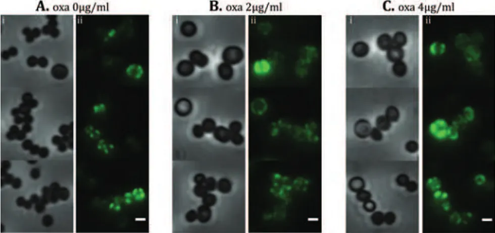
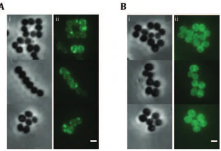
II. PBP2A localization studies. Importance of its N-terminal cytoplasmic domain for resistance.
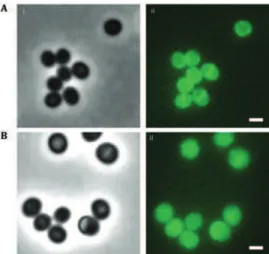
Localization of PBP2A in MSSA and MRSA
strains. Genetic manipulation of Fluorescence microscopy.
Immunofluorescence.
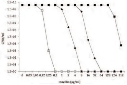
Antibiotic susceptibility tests: PAPs and microdilution assay.
SDS-PAGE, western blot, GFP fluorescence.
PBP2A localizes all around the membrane both in MSSA and MRSA strains.
Tagged-PBP2A is not fully functional. Four residues at the N-terminal of PBP2A are
in MRSA strains.
III. Identification of components of the
cell division and cell wall synthesis machinery in S. aureus.
Unravel the interacting partners of PBPs within the multy-enzyme complex of CW synthesis.
Cloning, expression and purification of recombinant His-PBP2.
Purification and solubilization of membranes.
1D CN-PAGE/2D SDS-PAGE.
. MS and MS/MS.
Immunoprecipitation of PBP2. BTH assays.
Antibiotic susceptibility tests: PAPs and microdilution assay.
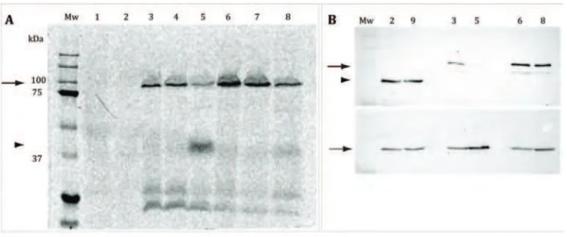
PBP2 and PBP2A form independent and stable complexes in 1D CN-PAGE with ~242
PBP1 and PBP3 are in complexes that have ~300 kDa.
Acylated PBP2 likely undergoes conformational changes.
Methods Conclusions
General introduction focusing on epidemiology and factors of β-lactam resistance. State of the art of bacterial PBPs,
regulation of FtsZ-ring formation and
.
Localization of PBP2A in MSSA and MRSA
strains. Genetic manipulation of
S. aureus.
Fluorescence microscopy. Immunofluorescence.
Antibiotic susceptibility tests: PAPs and microdilution assay.
SDS-PAGE, western blot, in gel detection of GFP fluorescence.
PBP2A localizes all around the membrane both in MSSA and MRSA strains.
Tagged-PBP2A is not fully functional. Four residues at the N-terminal of PBP2A are important for the full expression of resistance in MRSA strains.
Identification of components of the
synthesis.
Cloning, expression and purification of recombinant His-PBP2.
Purification and solubilization of S. aureus
membranes.
1D CN-PAGE/2D SDS-PAGE.
Western blot, detection of bocillin-labeled PBPs, in gel.
MS and MS/MS.
Immunoprecipitation of PBP2. BTH assays.
Antibiotic susceptibility tests: PAPs and microdilution assay.
PBP2 and PBP2A form independent and stable complexes in 1D CN-PAGE with ~242 kDa
PBP1 and PBP3 are in complexes that have ~300 kDa.
Acylated PBP2 likely undergoes conformational changes.
Chapter Aim
IV. EzrA contributes to the regulation of cell size in S. aureus
Characterization of the role of the cell division protein EzrA, in S. aureus.
Genetic manipulation of .
RNA and Quantitative Real-Time PCR.
Fluorescence microscopy. BTH assays.
Electron Microscopy.
EzrA is not essential in .
Cells increase in size in the absence of EzrA. FtsZ and PBPs mislocalize in larger cells. EzrA interacts with PBPs.
V. General discussion and future perspectives
A general overview of all the work involved in this thesis is given. An integration of the results obtained in all the chapters is discussed. Further experiments are suggested in order to complement the work presented.
Abbreviations: BTH - bacterial two hybrid; CN - High resolution Clear Native; CW - cell wall; GFP - green fluorescent protein; MRSA - methicillin resistant S. aureus; MS - Mass spectrometry; MSSA - methicillin
susceptible S. aureus; PAPs - Population analysis profiles; PCR - Polymerase Chain Reaction; SDS-PAGE -
sodium dodecyl sulfate polyacrylamide gel electrophoresis; TM - transmembrane
Methods Conclusions
protein EzrA, in . Genetic manipulation of
S. aureus.
Extraction of S. aureus RNA and Quantitative
Real-Time PCR.
Fluorescence microscopy. BTH assays.
Electron Microscopy.
EzrA is not essential in S. aureus.
Cells increase in size in the absence of EzrA. FtsZ and PBPs mislocalize in larger ezrA- cells.
EzrA interacts with PBPs.
A general overview of all the work involved in this thesis is given. An integration of
Contents
1 Staphylococcus aureus 1.1 Challenging microbes
1.2 Staphylococcus aureus overview 1.3 Staphylococcus aureus epidemiology
2 Genetic basis for β-lactam resistance in S. aureus 2.1 Penicillin-binding protein 2A
2.2 Other factors required for β-lactam resistance
3 Bacterial cell wall
3.1 Peptidoglycan chemical composition 3.2 Cell wall architecture
3.3 Biochemical pathway of PG synthesis 3.4 Penicillin Binding Proteins
3.4.1 Glycosyltransferase and transpeptidation reactions 3.5 S. aureus penicillin-binding proteins
4 Cell division and the divisome 4.1 Cytokinesis
4.1.1 Spatial regulation of cytokinesis 4.2 Z-ring assembly
4.2.1 Structure and spatial organization of FtsZ 4.2.2 Regulation of FtsZ assembly
4.3 Divisome assembly
4.4 Cell wall machinery assembly 4.5 Constriction
5 Final remarks
6 References
Introduction
1. Staphylococcus aureus
1.1 Challenging microbes“Microbes have lived for a long time without animals. At no point have animals been free of microbes”. Abigail Salyers
Bacteria colonize different areas of the human body, such as the airways, skin,
gastrointestinal tract, oral cavity and the urinary tract, living in equilibrium with our organism. Bacterial communities can be commensals1 or even beneficial to several functions of the host (Blaser, 2010, Fujimura et al., 2010) and this relationship is essential
for our life. The concern about co-habiting with microorganisms arrives when bacteria become pathogenic and life threatening. Over the years, the world assisted to several epidemic diseases, many of them caused by bacteria (Cholera by Vibrio cholerae, Typhoid Fever by Salmonella enterica, Bacillary Dysentery by Shigella dysenteriae, Tuberculosis by
Mycobacterium tuberculosis, Plague by Yersinia pestis, Syphilis by Treponema pallidum).
In the last decades, life expectancy has increased abruptly, mainly in the so-called developed countries. Healthcare improvement was perhaps the most important factor mainly through control of diseases with the production of vaccines and the discovery of antibiotics, arguably the most successful chemotherapy agents ever found. After Alexander Fleming’s discovery of penicillin in 1928 (Fleming, 1929) this compound turned out to be crucial to treat infections, becoming essential to guaranty the life of patients which included many soldiers in the following wars. Soon, the entire world recognized the importance of antibiotics and its use was accordingly spread to treat the majority of infections in clinical practices. However, despite the hope in the eradication of virtually all bacterial infections, physicians reported the emergence of resistant strains shortly after antibiotics were introduced. The misuse of antibiotics was one of the factors responsible for the selection of resistant bacteria. Despite the relative small size of the bacterial genome, it contains enough information and plasticity so that
bacteria can adapt and survive not only in the presence of antibiotics but also in a wide
variety of environments (extreme temperatures, pressure, salt, oxygenation, lack of nutrients, and others). As a consequence, the search for new antibiotics and the study of the molecular mechanisms underlying bacterial resistance became an urgent need.
As a matter of fact, nowadays we continue to struggle with the emergence of new resistant strains of pathogenic bacteria. Nosocomial infections, which are transmitted within the healthcare systems, have been a major concern. It is difficult to estimate and compare the incidence of bacterial infections worldwide, but in the US, the Center for Disease Control and Prevention reported that, per year, 10% of hospital patients become infected by all types of bacteria combined (www.cdc.gov). In Europe, the European Centre for Disease Control and Infection reported that approximately 4million patients are estimated to acquire a healthcare-associated infection in the EU every year (www.ecdc.europa.eu).
During the recent years, Staphylococcus aureus, Streptococcus pneumoniae,
Enterococcus spp., Escherichia coli, Klebsiella pneumoniae, Pseudomonas aeruginosa, and
Acinetobacter spp. became the species mostly associated with healthcare-associated
infections and prone to multidrug resistance (European Centre for Disease Prevention and Control, 2010).In particular, S. aureus, the focus of this thesis,is a major human pathogen (Lowy, 1998). In the early 1940s, prior to the introduction of penicillin for the treatment of S. aureus infections, the mortality rate of individuals with serious S. aureus infection was about 80% (Deurenberg & Stobberingh, 2008). Nowadays, 2%
of all newly admitted patients acquire a hospital infection by S. aureus that results in disease (Jones, 2003).
1.2 Staphylococcus aureus overview
The word Staphylococcus is derived from the Greek words “staphyle” meaning bunch of grapes plus the word “coccus” as for their round shape, and was adopted due to its characteristic growth in clusters, an observation first reported by Alexander Ogston in 1881 (Ogston, 1881). Staphylococci are gram-positive bacteria and belong to the phylum Firmicutes of low G+C content bacteria, to the class Bacilli and order Bacillales. Staphylococci are facultative anaerobes that grow by aerobic respiration or by fermentation that yields mainly lactic acid. Three years after the discovery of Staphylococcus, Rosenbach identified the species S. aureus and proposed the Latin word “aureus” meaning gold, due to the yellow color of the pigmented colonies growing on agar. S. aureus can grow in a temperature range from 15 to 45 degrees Celsius and
characteristic for species identification (Sonenshein, 1993). Strikingly, S. aureus has a strong dessication tolerance with an estimated ability to survive on dry plastic surfaces for more than one thousand days (Chaibenjawong & Foster, 2011).
1.3 Staphylococcus aureus epidemiology
S. aureus is a commensal organism that persistently colonizes 20 to 30% of the
human population (European Centre for Disease Prevention and Control, 2010) and intermittently colonizes a further 60% throughout their lifetime (Edwards & Massey, 2011). Our innate immunity is the primary defense against S. aureus. Although nearly
everyone has antibodies against Staphylococcus, these are generally not efficient in
preventing an infection (Foster, 2005).
In fact, S. aureus can cause disease if it leaves the asymptomatic colonization site to reach the blood, for example due to a breach in the skin caused by a surgical wound or an intravenous catheter. In most cases, S. aureus causes skin and soft tissue infections (SSTI) (furuncles, abscesses) being responsible for 71% of all SSTI, in Europe (Sader
et al., 2010). However, it can pass to the bloodstream and disseminate throughout the
body causing bacteremia2, which can cause death as a result of a septic shock. If S. aureus leaves the bloodstream and enters the surrounding tissues, it can lead to secondary (metastatic) infection foci causing endocarditis, abscesses in organs, septic arthritis, vertebral osteomyelitis or meningitis (Edwards & Massey, 2011). S. aureus is therefore an extremely successful and versatile pathogen. This is largely due to the number of virulence factors expressed and the ability to resist a larger number of antibiotics.
Two major groups of virulence factors play a key role in S. aureus infections: proteins
expressed on the surface of the bacterial cell and secreted proteins. These proteins are required for a pathway of infection that involves bacterial attachment to the host tissues (mucosa), colonization, invasion or penetration, and evasion of host immunity.
Perhaps even more surprising than S. aureus virulence is it capacity to acquire resistance to antibiotics. Within 5 years after penicillin wide use as antimicrobial agent, 50% of all S. aureus strains became resistant (Rammelkamp, 1942) by expressing β-lactamases enzymes,
which hydrolyse the β-lactam ring of penicillin. In 1960 the first semi-synthetic penicillin derivative, methicillin, was introduced into clinical use for the treatment of infections caused
by penicillin-resistant S. aureus. However, within one year, the first isolate of
resistant S. aureus (MRSA) was reported in the UK (Jevons et al., 1963). Methicillin is no
longer in use but the acronym MRSA continues to be applied to S. aureus strains resistant to β-lactams. The occurrence of MRSA strains, particularly clone ST250, was reported in several countries in the following years, but by unknown reasons, this clone subsequently declined from European Hospitals during the late 1970s. In the early 1980s epidemic strains (so-called EMRSA, mostly represented by the Iberian, Brazilian, NW/Japan, Pedriatic, EMRSA-16 and Berlin clones) emerged and are still present nowadays (Chambers & Deleo, 2009). Until the early 1990s MRSA was only associated with nosocomial infections, however, by that time, MRSA strains started to appear in communities outside the healthcare environment (CA-MRSA), generally being highly virulent (Udo et al., 1993, Johnson, 2011).
Treatment of the most severe cases of MRSA infections is frequently done with the glycopeptide vancomycin which was introduced in the late 1960s. Nonetheless, its broad use caused an intensive selective pressure and in the late 1990s vancomycin-intermediate S. aureus (VISA) strains were reported (Hiramatsu et al., 1997), followed
by the emergence of vancomycin-resistant S. aureus (VRSA) in 2002 (Tiwari & Sen,
2006). Those occurrences were often associated with prolonged glycopeptide therapy and fortunately VISA and VRSA strains have not yet spread widely presumably due to the high fitness cost of vancomycin resistance in S. aureus.
MRSA-associated infections are still widely identified in hospitals worldwide (European Centre for Disease Prevention and Control, 2010) constituting an important financial burden to the healthcare systems. In the US, MRSA infections represent above 60% of the S. aureus isolates. The number of MRSA infection-associated deaths in the
United States is about 19,000 annually which is similar to the number of deaths due to AIDS, tuberculosis, and viral hepatitis combined (Bucher, 2008). In Europe, although the proportion of S. aureus bacteremia due to MRSA is declining in many countries (from
2006 to 2009), in 10 out of 28 European countries, MRSA proportion is still above 25% of all S. aureus infections identified. In Portugal, rates of MRSA (almost 50% of all S. aureus
isolates) are among the highest of European countries, representing an important public health burden (European Centre for Disease Prevention and Control, 2010).
2 Genetic basis for β-lactam resistance in S. aureus
vary extensively among different strains and about 22% of each genome comprises dispensable regions which may include virulence factors or proteins mediating antibiotic
resistance and various mobile genetic elements, such as chromosomal cassettes, insertion sequences, transposons, bacteriophages, plasmids, “pathogenicity islands” and genomic islands (Fitzgerald et al., 2001) (Gill et al., 2005). Together, all the mobile genetic elements
constitute approximately 7% of the S. aureus COL genome (Gill et al., 2005).
In 1984, Hartman and Tomasz identified the genetic marker responsible for methicillin resistance, the mecA gene. β-lactams target Penicillin-binding proteins
(PBPs), involved in the last stages of cell wall (CW) synthesis. The mecA gene encodes an extra PBP, PBP2A, which is absent in susceptible strains and has low affinity for most semi-synthetic penicillins (e.g., methicillin, nafcillin and oxacillin) (Hartman & Tomasz, 1984). Thus, despite the presence of otherwise inhibitory concentrations of β-lactam antibiotics, MRSA can continue CW synthesis due to the uninhibited activity of PBP2A (Matthews & Tomasz, 1990).
2.1 Penicillin-binding protein 2A
The PBP2A-encoding mecA gene is located on a mobile genetic element of 21-67kb,
known as the Staphylococcal Cassette Chromosome mec (SCCmec) (Ito et al., 1999)
(Katayama et al., 2000). SCCs are excellent and sophisticated vehicles for genetic
exchange among staphylococcal species. The mec complex contains the mecA gene, regulatory genes (mecR1 and mecI both upstream of the mecA gene) and associated
insertion sequences downstream of the mecA gene. Besides the mec complex, the SCCmec encodes additional genes which promote its integration and excision, necessary for the recombination in the staphylococcal chromosome, as well as insertion sequences and different resistance or virulence determinants. Particularly, specific recombinases of the invertase/resolvase family (ccr) are encoded within the SCCmec which allow its movement between different strains. Integration occurs always at a specific site, the
attBSCC (bacterial chromosomal attachment site), near the origin of replication and at
the 3’end of an open reading frame (ORF) of unknown function, orfX (Ito et al., 2001).
Up to this date, eleven SCCmec types (named from I to XI) have been described and
are currently updated at the International Working Group on the Staphylococcal Cassette Chromosome elements website (www.SCCmec.org). The classification of the different SCCmec types is based on the combination of ccr genes and mec complexes. Although the
from the same common ancestor and that the mecA gene might have been acquired from
Staphylococcus sciuri and moved by horizontal transfer among staphylococcal species (Couto et al., 1996). Recently, Tsubakishita et al reported S. fleurettii as a probable candidate for the origin of the mecA gene which was found not associated to a SCCmec
within the chromosome, but rather incorporated among the essential genes of S. fleurettii (Tsubakishita et al., 2010). The same authors propose that the SCCmec might have been formed through the acquisition of two distinguished regions: i) mecA gene complex from
S. fleurettii and ii) an SCC element with no mecA gene.
Although the major genetic factor for methicillin resistance, the mecA gene, is common to virtually all MRSA strains, MRSA isolates exhibit a broad range of resistance to β-lactams, having minimum inhibitory concentrations (MIC3) that range from 1 to 800μg/ml of methicillin and resistance profiles that vary from homogeneous to heterogeneous or borderline. Heterogeneously resistant isolates show a majority of the population susceptible to low concentrations of β-lactam antibiotics (e.g. 1 to 5 μg/ml of methicillin) while a small subpopulation is able to grow in the presence of higher methicillin concentrations. When these strains are exposed to selective antibiotic pressure, they may alter their
resistance phenotype due to selection of highly resistant mutants that arise as a result of
mutational events in the chromosome, in a loci different from the SCCmec (Ryffel et al., 1994). The selected strains became homogeneous and able to grow in the presence of high concentrations of β-lactam antibiotics (Chambers, 1997). Generally, these highly resistant clones keep their resistance even in the absence of antibiotic pressure (Berger-Bachi & Rohrer, 2002). Clinical isolates showing constitutive expression of homogeneous resistance are rare, with the strain COL being the most extensively studied (Gill et al., 2005). Borderline resistance is characterized by low β-lactam MICs, generally 4 to 8 μg/ml. Interestingly, some of these borderline isolates do not carry the mecA gene. In these clones other factors, such as
modified expression of native PBPs (Tomasz et al., 1989) (Henze & Berger-Bachi, 1996) and
modified acylation rates due to point mutations in the penicillin binding domain of native PBP1 and PBP2 (Chambers et al., 1994), play an important role in resistance.
2.2 Other factors required for β-lactam resistance
The level of methicillin resistance, defined by its MIC, depends on several factors besides the presence of PBP2A. It is known that factors such as pH, temperature, osmolarity, the
availability of divalent cations and the composition of the growth medium, influence methicillin resistance levels (Matthews, 1984). Furthermore, the genetic background also affects the stability of mecA in S. aureus (Katayama et al., 2005). In addition, several
genetic factors were identified as being necessary for the full expression of resistance. Initially, those factors were found by transposon insertional mutagenesis on MRSA strains, which rendered them susceptible to β-lactams antibiotics (Berger-Bachi, 1983) (de Lencastre & Tomasz, 1994). These genetic determinants, named fem and aux genes (for factor/auxiliary essential for methicillin resistance), are mostly housekeeping genes found both in susceptible and resistant strains. In recent years, an increasing number of factors have been identified and a list can be found below in Table 1.
Table 1 – Genes involved in β-lactam resistance
Gene Known function Reference
fmhB (femX) Interpeptide bridge formation in muropeptides; addition of the
first glycine to the stem peptide. (Rohrer
et al., 1999)
femA Interpeptide bridge formation in muropeptides; addition of the 2nd and 3rd glycine to the stem peptide. FemA and FemB are the
first described members of the class of non-ribosomal peptidyl transferases.
(Stranden et al., 1997)
(Berger-Bachi et al.,
1989)
femB Interpeptide bridge formation in muropeptides; addition of the
4th and 5th glycine to the stem peptide.
(Henze et al., 1993) femC (glnR) Glutamine synthetase repressor; inactivation reduces amidation
of the D-glutamate of the stem peptide. (Gustafson
et al.,
1994)
femD (femR315) (glmM)
Phosphoglucosamine mutase; catalyzes the interconversion
of glucosamine-6-phosphate to the cytoplasmic PG precursor glucosamine-1-phosphate.
(Jolly et al., 1997)
femE Function unknown; inactivation slightly reduces methicillin
resistance. (de Lencastre & Tomasz, 1994)
femF (murE) UDP-N-acetylmuramyl tripeptide synthetase; catalyzes the addition of the L-lysine residue to the UDP-linked muramyl
dipeptide CW precursor.
(Ornelas-Soares et al.,
1994)(Gardete et al.,
2004)
murF Attachment of the D-Ala-D-Ala to the UDP-NAM-tripeptide,
completing the formation of the PG building block, the UDP-NAM-pentapeptide.
(Sobral et al., 2003)
fmtA CW associated membrane protein containing two of the three conserved motifs found in PBPs and penicillinases; inactivation
decreases cross-linking and amidation of PG; it is proposed to participate in PG biosynthesis under β-lactam induced CW stress conditions.
(Komatsuzawa et al.,
Gene Known function Reference
fmtB (mrp) Cell surface protein; probably involved with CW biosynthesis; inactivation reduces pentaglycyl-substituted monomer of the CW fraction while increasing the amount of unsubstituted
pentapeptide.
(Komatsuzawa et al.,
2000)
fmtC (mprF) Membrane-associated protein; inactivation reduces
modification of phosphatidyl-glycerol with L-lysine, and
consequently leads to an increased negative charge of the
membrane surface.
(Komatsuzawa et al.,
2001) (Peschel et al.,
2001)
llm (tagO) (tarO)
First enzyme of the wall teichoic acids biosynthesis pathway;
inactivation increases Triton-X-100-induced autolysis and PBP4 delocalization.
(Maki et al., 1994,
Campbell et al., 2011) pbp2 catalyses the transpeptidation and glycosyltransferase reactions
of the PG; a functional glycosyltransferase domain of PBP2 is needed for methicillin resistance.
(Pinho et al., 2001a)
sigB Alternative transcription factor; inactivation reduces both
methicillin and glycopeptides-intermediate resistance.
(Wu et al., 1996) dlt operon Transfer of D-alanine into teichoic acids; inactivation increases
methicillin resistance.
(Nakao et al., 2000) agr and sar Control of the synthesis of CW-associated and excreted proteins
depending on the growth rate.
(Piriz Duran et al.,
1996)
lytH Encodes a putative lytic enzyme with homology to a
N-acetylmuramyl-L-alanine amidase. (Fujimura & Murakami, 1997)
vraSR Sensor and response regulator of a two component system. VraS
inactivation leads to a massive reduction of the resistance to
β-lactams and vancomycin antibiotics.
(Gardete et al., 2006)
spoVG Potential downstream regulators of σB. Inactivation impedes
capsule formation in capsular polysaccharide-producing strain
Newman and decreases resistance in MRSA and GISA strains.
(Schulthess et al.,
2009)
hmrA Encodes a putative aminohydrolase. Upregulation leads to
increased levels of resistance. (Kondo
et al., 2001)
SA1665 A putative helix-turn-helix DNA-binding protein, which binds
to the mec operator region, modulates resistance by decreasing
methicillin resistance levels in a strain dependent manner.
(Ender et al., 2009)
secDF SecDF is an accessory factor of the conserved Sec protein translocation machinery and belongs to the
resistance-nodulation-cell division family of multidrug exporters.
(Quiblier et al., 2011)
Abbreviations: PG - peptidoglycan; CW - cell wall; NAM - N-acetylmuramic acid
PBP2A, with the exception of PBP2 (Pinho et al., 2001a). It has been suggested that, in the presence of β-lactams, PBP2A transpeptidase (TPase) domain cooperates with the glycosyltransferase (GTase) activity of PBP2 (whose TPase active site becomes inactivated by the presence of β-lactams) so that CW synthesis can occur and consequently the cell continues dividing (Pinho et al., 2001a).
Therefore, as a result of the role of the majority of the factors described above, expression of full methicillin resistance requires an optimal rate of PG precursor formation as well as a correct structure of the PBP2A substrate. Interestingly, PBP2A has a strict requirement for the presence of the pentaglycine brigde characteristic of S. aureus stem peptide PG precursor to mediate resistance (Berger-Bachi & Tschierske, 1998) and of a normal stem peptide configuration (Ludovice et al., 1998) (de Jonge et al., 1996). Autolytic
activity further contributes to the full expression of resistance. Moreover, the regulation of all these factors might be dependent on the control of global regulators.
3. Bacterial cell wall
The CW of bacteria is an essential cell component required for protection of the cell membrane against high internal osmotic pressure and for maintenance of cell morphology. It functions as a sieve that allows diffusion of large macromolecules and as an attachment site for surface proteins and CW polymers essential for adhesion, colonization and infection. Inhibition of the CW biosynthesis machinery can lead to bacterial lysis and consequent death. Although the existence of a CW is common to the majority of bacteria, its composition differs according to their gram-type. Accordingly, gram-positive bacteria CWs are composed of a thick (20-80nm) multilayered PG sheath that includes proteins and embedded teichoic acids. The later anionic polymers can account for over 60% of the mass of the gram-positive CW (Silhavy et al., 2010) and can be either covalently linked to the PG (wall teichoic acids)
or anchored to the outside of the bacterial membrane (lipoteichoic acids) (Xia et al., 2010).
On the other hand, gram-negative CWs include an outer membrane surrounding a thin layer of PG (1-7nm) within a periplasmic space that includes lipoproteins (Silhavy et al., 2010).
3.1 Peptidoglycan chemical composition
and adhesins, and as a scaffold for anchoring and displaying other CW components on the staphylococcal surface, namely proteins, teichoic acids and carbohydrates. The PG chemical structure is well studied and its presence is widely conserved among species with
different shapes, except for Mycoplasma and other rare species with no CW. It is a polymer
formed of long glycan strands made of the aminosugars N-acetylglucosamine (NAG) and N-acetylmuramic acid (NAM), which are alternately linked together by β-1,4 glycosidic
bonds and further cross-linked by short peptide flexible bridges (stem peptides) that are attached to the lactyl group of NAM (Figure 1) (Vollmer et al., 2008). The composition of the
stem peptide vary with species, with a major difference in the third amino acid which can be a diaminopimelic-acid (most gram-negative bacteria, Mycobacteria, Bacilli) or L-Lysine (most gram-positive bacteria) (Figure 1). The association between stem peptides generally occurs between the D-Ala at position 4 and the amino acid at position 3 of two stem peptides. Furthermore, this association can be either direct or through an interpeptide bridge.
Figure 1 – Stem peptides and interpeptide bridge of (a) Escherichia coli (direct 3-4 cross-link) and (b) Staphylococcus aureus (3-4 cross-link through a pentaglycine bridge). G – NAG; M – NAM. Adapted from
(Vollmer et al., 2008)
In addition to the variability of the composition of the stem peptides, the degree of cross-linkage also differs among species, corresponding to 20% in E. coli (Glauner,
1988) and over 93% in S. aureus (Snowden & Perkins, 1990).
Glycan chains vary in length among different bacteria and, interestingly, their length is not related to their gram-type or the thickness of the CW. In E. coli the average chain length is 30 disaccharide units and this may vary to some extent depending on the strain and growth conditions (Harz et al., 1990). Glycan chains of S. aureus are much shorter, with 85-90% having an average length of about 6 disaccharide units (Tipper et al., 1967, Ward,
with about 50% of glycan chains having more than 50 disaccharide units and 14% with more than 100 disaccharide units (Ward, 1973, Wheeler et al., 2011). Cryo-transmission electron microscopy (Cryo-TEM4) and atomic force microscopy (AFM5) of E. coli cells have shown that the PG layer is on average 6.0 nm thick (Matias et al., 2003, Yao et al., 1999). Cryo-TEM applied to gram-positive bacteria revealed the existence of a bipartite organization of the CW, composed of a low density zone or periplasmic space adjacent to the cell membrane, with a thickness of 16nm in S. aureus and 22nm in B. subtilis, and an outer wall zone. This latter zone likely includes polymeric complexes of PG with attached teichoic acids and surface proteins and was determined to be 19 ± 4.3nm in S. aureus and 33.3 ± 4.7nm in B. subtilis (Matias & Beveridge, 2007, Matias & Beveridge, 2005).
3.2 Cell wall architecture
One of the most challenging paradigms on the structure of the bacteria cell is the organization of the CW components in a tri-dimensional perspective, which includes the
orientation of the glycan strands and peptides with regard to the surface and axis of the cell. Due to their size and complexity, glycan strands and peptides cannot be visualized by conventional microscopy. Nevertheless, two distinct models for the three-dimensional arrangement of PG structure have been proposed (Holtje, 1998) (Dmitriev et al., 2005).
In the classical “layered” model glycan chains run parallel to the cytoplasmic membrane (Koch, 1998, Holtje, 1998, Yao et al., 1999). In contrast, the “scaffold model” predicts that the glycan chains run perpendicular to the plasma membrane (Dmitriev et al., 2005).
Lately, Cryo-TEM and AFM have shed some light into the field of the architecture of the PG for gram-negative (Gan et al., 2008) and gram-positive bacteria (Hayhurst et al., 2008, Turner et al., 2010, Wheeler et al., 2011). Currently, based on the results obtained
with those techniques, a “classical” layered model is favored for gram-negative bacteria in which both the glycan strands and the peptide cross-links run parallel to the cytoplasmic membrane (Gan et al., 2008). A more elaborate PG architecture has been revealed for
the gram-positive bacteria analyzed so far, although the specific architecture varies with species (Wheeler et al., 2011, Turner et al., 2010, Hayhurst et al., 2008). For B. subtilis it
4 Cryo-TEM: A microscopy technique that allows the visualization of biological molecules in a near-native state. Unfixed samples are flash frozen, held at cryogenic temperature and visualized by transmission electron microscopy.
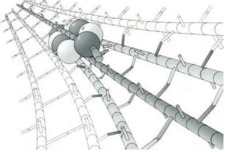
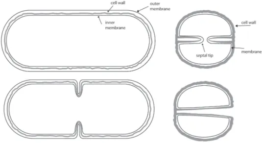
was suggested that the PG is organized in coiled cables running around the cell cylinder, providing strength and yet permitting growth (Hayhurst et al., 2008). The gram-positive cocci S. aureus has a very different PG architecture, probably as a result of its different
shape, mode of growth and division. In a model proposed in 1987, septal PG is synthesized in a spiral running in a plane from the periphery to the center of the cell. Expansion of newly synthesized PG at the septum, to a hemisphere, would then mainly stretch the flexible peptides but not the rigid glycan strands (Seligman & Pincus, 1987) (Seligman, 1987). Electron microscopy and AFM applied to S. aureus cells and purified sacculi allowed
the visualization of concentric rings surrounding a central depression in CWs that had just undergone cell separation (Touhami et al., 2004). At the old poles, however, the cell surface
is similar to a network of fibers with large empty spaces (Giesbrecht et al., 1998, Francius et al., 2008, Touhami et al., 2004, Turner et al., 2010). AFM applied to purified S. aureus
PG revealed that, at the division site, the PG is a dynamic structure that remodels itself by continuous autolysis. These modifications lead to the lateral expansion of PG which, as a result, matures into a knobble architecture (Turner et al., 2010). Moreover, the same study identified the presence of PG features with the capability of demarking the previous division. It was suggested that those features function as epigenetic factors important for division site localization, therefore allowing division of S. aureus in 3 sequential planes.
Thus, according to this model, PG carries the necessary information across the generations to allow the sequential orthogonal division to occur with fidelity (Turner et al., 2010).
3.3 Biochemical pathway of PG synthesis
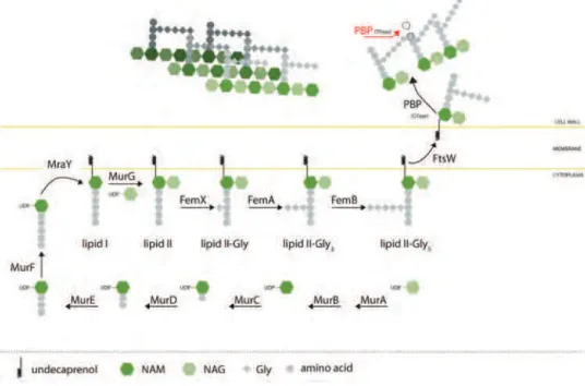
The biochemical pathway of PG synthesis takes place in three cellular compartments: cytoplasm, cytoplasmic membrane, and periplasm (or outside) (Figure 2).
CW synthesis is initiated in the bacterial cytoplasm where the first precursor of the CW pathway, fructose-6-P is transformed in uridine diphosphate (UDP)-NAG by the action of GlmSMU enzymes and using acetylCoA and uridine triphosphate (UTP).
Then, PG biosynthesis pathway continues with the action of the Mur enzymes (named MurA to MurF), which catalyze the formation of UDP-NAM from a UDP-NAG precursor (MurA and MurB), to which five amino acids are subsequently added onto the D-lactoyl group of UDP-NAM (MurC to MurF) (Figure 2). The peptide sequence of the five amino acids may vary according to the bacterial species, but it ends with a D-alanyl-D-alanine moiety (except in certain vancomycin-resistant enterococcal strains, which
Figure 2 - Biochemical pathway of S. aureus peptidoglycan biosynthesis.
This soluble molecule, UDP-NAM-pentapeptide, becomes associated to the bacterial membrane through the action of MraY, a ten transmembrane helix protein, which catalyzes its linkage to a 55-carbon-long phospholipid, harbouring a membrane embedded undecaprenyl diphosphate group (van Heijenoort, 2001) (Bouhss et al., 2004). The resulting molecule, called lipid I, is subsequently transformed by MurG, an N-acetylglucosaminyl transferase, which adds a soluble UDP-NAG group to lipid I. This reaction generates lipid II, the disaccharidic precursor for PG biosynthesis and substrate for PBPs for the majority of the bacterial species (Goffin & Ghuysen, 2002). At this stage, in Staphylococci, five glycines are sequentially added to the L-Lysine of the stem peptide by the FemX, FemA and FemB enzymes. The amino acids linked to the ε-amino acid of the stem peptide, or their presence, vary among different species.
Little is known about how lipid II translocates across the membrane, as structural and biochemical assays of putative proteins are difficult to obtain due to its membrane topology. However, putative candidates included FtsW and RodA (Holtje, 1998). Recently, biochemical experiments on E. coli vesicles gave strong evidences that FtsW is, indeed, the flippase (Mohammadi et al., 2011). Interestingly, composition of the membrane
phospholipid synthesis, results in cessation of PG synthesis (Ehlert & Holtje, 1996), suggesting that lipid II translocation depends on ongoing phospholipid synthesis.
Once on the outer side of the membrane, the PG muropeptide is polymerized and cross-linked by PBPs, that catalyze the polymerization of the glycan strands (glycosyltransferase reaction) and the cross-linking of the stem peptides between glycan chains (transpeptidation).
3.4 Penicillin Binding Proteins
PBPs belong to the superfamily of “active-site serine penicillin recognizing enzymes” (ASPRE) which includes transpeptidases, carboxypeptidases and β-lactamases. This group of enzymes is characterized by the presence of a penicillin-binding domain with variable affinity for β-lactams, which constitutes the transpeptidation catalytic domain (Ghuysen, 1991). The penicillin-binding module possesses three conserved active-site motifs: SXXK, S(Y)XN(V) and KT(S)G, where the serine of the first motif is the catalytic residue (Goffin & Ghuysen, 1998).
PBPs are classified in two main groups: high molecular weight (HMW) and low molecular weight (LMW). The first group is further subdivided in Class A and Class B depending on the reactions that PBPs are able to catalyze, as predicted by the presence of the catalytic domains. Class A PBPs are bifunctional (with TPase and GTase domains) and Class B are monofunctional (with only TPase domain). Some PBPs can remove the last D-alanine of the stem peptides (DD-carboxypeptidation) or hydrolyze a D-alanine peptide bond of two adjacent chains, connecting two glycan strands (endopeptidation) (Goffin & Ghuysen, 1998) (Sauvage et al., 2008).
In addition to PBPs, monofunctional glycosyltransferases (MGTs), which have only GTase activity, can also be found in many bacteria (Spratt et al., 1996). However, it is
surprising to observe that these enzymes do not exist in all bacteria (Kunst et al., 1997)
and when present may be dispensable for growth (Schiffer & Holtje, 1999, Reed et al., 2011). S. aureus has two MGTs in its genome, MGT and SgtA, which are not required for CW
synthesis. Interestingly, although these proteins are not essential, in the absence of PBP2 GTase activity, MGT but not SgtA becomes essential for cell viability (Reed et al., 2011).
HMW PBPs contain an N-terminal cytoplasmic tail, a transmembrane anchor, and two domains joined by a short β-rich linker and located at the outer surface of the cytoplasmic membrane (Lovering et al., 2007). The penicillin binding domain with TPase activity is
domain is highly conserved within ASPRE enzymes, being composed of a central five stranded β-sheet surrounded by α-helices (Macheboeuf et al., 2006). The active site of the
bifunctional PBP1b from S. pneumoniae showed closed and open conformations in the absence or presence of substrates or antibiotics, revealing that PBPs might exist in inactive
and active conformations, which can have a regulatory role in PBPs catalytic activity
(Macheboeuf et al., 2005). The GTase domain structure of S. aureus PBP2 and Aquifex aeolicus PBP1a contains almost only α-helices and is composed of a globular head and a
flexible, small jaw lobe separated by an extended cleft that contains the active site (Figure 3) (Lovering et al., 2007, Yuan et al., 2007). The GTase domains share five conserved motifs of which motif 1 (EDxxFxxHxG) and motif 3 (RKxxE) contain the two conserved catalytic glutamic acids. Interestingly, comparisons of the apo and moenomycin-bound S. aureus
PBP2 structures show that antibiotic or substrate binding induces a conformational change in the small subdomain and that some flexibility occurs around the linker region connecting the GTase and the TPase domains (Lovering et al., 2008).
Figure 3 – Overall structure of PBP2. The predicted N-terminal TM anchor, whose structure is not known, is
represented by a vertical blue rectangle. The GTase domain (in blue) is connected to the TPase domain (in green) by a short linker region (in yellow). The active-site residues of both domains, the GTase (lower, E171; upper, E114) and TPase (S398) are represented as red spheres. Adapted from (Lovering et al., 2007).
In Class B PBPs, the N-terminal domain is believed to play a structural role for the interaction with other proteins (Holtje, 1998) or to serve as a pedestal/connector, placing the catalytic region of the protein away from the cell membrane and towards the PG (Macheboeuf et al., 2006).
TPase domain and the transmembrane anchor or amphipathic helix is accomplished by a long β-rich region that is proposed to act as pedestal which could position the catalytic region of LMW PBPs towards the PG. The existence of an amphipathic helix could permit the enzymes to skid on the membrane surface without being associated within it, while the pedestal could situate the catalytic domain at the specific level for substrate recognition (Morlot et al., 2005).
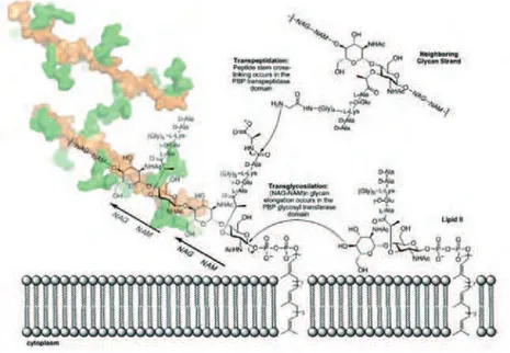
3.4.1 Glycosyltransferase and transpeptidation reactions
Figure 4 – Schematic representation of glycosyltransferase and transpeptidation reactions during synthesis of S. aureus CW. Both reactions assemble the PG on the surface of the cytoplasmic membrane. The 3D structure
of the PG is depicted as described in (Meroueh et al., 2006) with the sugar backbone (NAG-NAM polymer) in
orange and the peptides in green. Two strands of PG are shown with cross-linking through a peptide bridge shared between the two. NHAc, N-acetyl group. Adapted from (Llarrull et al., 2009)
The GTase reaction results in the association of the NAM and NAG groups of the lipid II and the growing chain of the PG (Lovering et al., 2007) (Figure 4). The mechanism of glycan chain elongation proceeds by successive attacks of the growing chain (donor) at the reducing end by lipid II (acceptor). The reaction is catalyzed by deprotonation of the 4-OH nucleophile of NAG by the active site (a glutamate) of the enzyme GTase domain. Concomitantly, stabilization of the leaving diphosphoundecaprenyl group is achieved by a second glutamate probably requiring the presence of a divalent metal (Schwartz et al., 2002)
exception as it does not depend on metal ions (Barrett et al., 2005). The glycolipid antibiotic
moenomycin inhibits the TGase reaction by binding to the active site of the enzymes due to a structural analogy to the lipid-linked PG precursor. Nonetheless, the toxicity of moenomycin for human cells prevents its use in clinical practice (Yuan et al., 2008).
The TPase reaction consists in the cross-linking between glycan chains through peptide bridges, and generally requires that the enzyme recognizes the terminal D-Ala-D-Ala of the muropeptides substrate at its active site (Figure 4). The attachment of the carbonyl group of the penultimate D-Ala of the PG muropeptide to the Serine present in the active site of the TPase domain, promotes the cleavage of the D-Ala-D-Ala, with concomitant formation of a Ser-D-Ala ester bond and release of the last D-Ala residue. This acyl-enzyme intermediate reacts with the side chain of the terminal amino group of the pentaglycine bridge (in S. aureus) forming a cross-link peptide amide bond. In other bacteria where
a pentaglycine bridge does not exist, the acceptor amino group is the ε-amino group of lysine or diaminopimelic acid present at the stem peptide (Macheboeuf et al., 2006). In some cases, the acceptor in the TPase reaction is a water molecule, which will result in a D-Ala-D-Ala (or D, D-) carboxypeptidation reaction, catalyzed by LMW PBPs, with the consequent removal of the last D-Ala of the muropeptide, therefore generating a product which can no longer be cross-linked to other PG molecules.
Due to the structural analogy between penicillin and the substrate of PBPs, namely the terminal D-Ala–D-Ala, PBPs can also react with penicillin and cleave the β-lactam bond, forming a covalently linked penicilloyl-enzyme intermediate with consequent opening of the β-lactam ring (Tipper & Strominger, 1965). The resulting acyl-enzyme can only be hydrolyzed at a very slow rate. Thus, penicillin functions as a suicide substrate in this reaction, by freezing the reaction at the penicilloyl-enzyme intermediate.
3.5 S. aureus penicillin-binding proteins
PBPs were named after the discovery that they were the targets of penicillin, although the sensitivity to β-lactams, with which they form a stable acyl-enzyme complex, vary among different PBPs. The numbering of PBPs is associated with their migration in sodium dodecyl sulfate-polyacrylamide gel electrophoresis (SDS-PAGE).
S. aureus has 4 native PBPs, described below, three of which are HMW PBPs. As stated
above, MRSA strains have acquired an additional HMW Class B PBP, PBP2A, from an extra-species source.
PBP1: The pbp1 gene is located within the division and CW (dcw) cluster together