Receptor
b
Deficiency
Yung-Hsiang Chen1,2, Chao-Jung Chen1,2, Shuyuan Yeh3, Yu-Ning Lin1, Yang-Chang Wu1, Wen-Tsong Hsieh1, Bor-Tsang Wu1, Wen-Lung Ma2, Wen-Chi Chen1,2, Chawnshang Chang2,3*, Huey-Yi Chen1,2*
1Graduate Institute of Integrated Medicine, College of Chinese Medicine, School of Pharmacy, College of Pharmacy, Department of Pharmacology, Department of Physical Therapy, Graduate Institute of Rehabilitation Science, China Medical University, Taichung, Taiwan,2Departments of Medical Research, Urology, and Obstetrics and Gynecology, Sex Hormone Research Center, China Medical University Hospital, Taichung, Taiwan,3Department of Urology, George H Whipple Laboratory for Cancer Research, Wilmot Cancer Center, University of Rochester Medical Center, Rochester, New York, United States of America
Abstract
Estrogen has various regulatory functions in the growth, development, and differentiation of the female urogenital system. This study investigated the roles of ERb in stress urinary incontinence (SUI). Wild-type (ERb+/+) and knockout (ERb2/2) female mice were generated (aged 6–8 weeks, n = 6) and urethral function and protein expression were measured. Leak point pressures (LPP) and maximum urethral closure pressure (MUCP) were assessed in mice under urethane anesthesia. After the measurements, the urethras were removed for proteomic analysis using label-free quantitative proteomics by nano-liquid chromatography–mass spectrometry (LC-MS/MS) analysis. The interaction between these proteins was further analysed using MetaCore. Lastly, Western blot was used to confirm the candidate proteins. Compared with the ERb+/+
group, the LPP and MUCP values of the ERb2/2 group were significantly decreased. Additionally, we identified 85 differentially expressed proteins in the urethra of ERb2/2female mice; 57 proteins were up-regulated and 28 were down-regulated. The majority of the ERbknockout-modified proteins were involved in cell-matrix adhesion, metabolism, immune response, signal transduction, nuclear receptor translational regelation, and muscle contraction and development. Western blot confirmed the up-regulation of myosin and collagen in urethra. By contrast, elastin was down-regulated in the ERb2/2 mice. This study is the first study to estimate protein expression changes in urethras from ERb2/2 female mice. These changes could be related to the molecular mechanism of ERbin SUI.
Citation:Chen Y-H, Chen C-J, Yeh S, Lin Y-N, Wu Y-C, et al. (2014) Urethral Dysfunction in Female Mice with Estrogen ReceptorbDeficiency. PLoS ONE 9(10): e109058. doi:10.1371/journal.pone.0109058
Editor:Andrew Wolfe, John Hopkins University School of Medicine, United States of America
ReceivedMay 29, 2014;AcceptedAugust 28, 2014;PublishedOctober 2, 2014
Copyright:ß2014 Chen et al. This is an open-access article distributed under the terms of the Creative Commons Attribution License, which permits
unrestricted use, distribution, and reproduction in any medium, provided the original author and source are credited.
Data Availability:The authors confirm that all data underlying the findings are fully available without restriction. All relevant data are within the paper.
Funding:This work was supported in part by Taiwan National Science Council (NSC101-2314-B-039-018 and NSC102-2320-B-039-025), China Medical University Hospital (DMR-103-063), and Taiwan Department of Health Clinical Trial and Research Center of Excellence (DOH102-TD-B-111-004). The funders had no role in study design, data collection and analysis, decision to publish, or preparation of the manuscript.
Competing Interests:The authors have declared that no competing interests exist. * Email: d888208@ms45.hinet.net (HYC); chang@urmc.rochester.edu (CC)
Introduction
Stress urinary incontinence (SUI) is defined as the involuntary leakage of urine under stress conditions such as coughing and sneezing [1–3]. The effects of birth trauma, menopause, and aging may contribute to the development of SUI [4]. Although improvement has been made in SUI treatment [5], our comprehension of the molecular mechanisms underlying this condition is inadequate.
Estrogen exerts a variety of regulatory functions on growth, development, and differentiation in the female urogenital system [6]. Estrogen actions are mediated by estrogen receptors (ERs) [7], encoded by two distinct genes, ERaand ERb. Due to the female predominance of autoimmune diseases, the role of gender and sex hormones in the immune system is of interest. The primary effects of estrogen are mediated via ERs that are expressed on most immune cells. ERs are nuclear hormone receptors that can either directly bind to estrogen response elements in gene promoters or serve as cofactors with other transcription factors. ERs have
prominent effects on immune function in both the innate and adaptive immune responses [8]. The discovery of ERb in 1996 stimulated great interest in the physiological roles and molecular mechanisms of its action. ERb plays a major role in mediating estrogen action in several tissues and organ systems, including the immune system [9]. Genetic deficiency of ERbhad minimal to no effect in autoimmune models [8].
[13]. Therefore, the specific roles of estrogen and ERb in SUI remained elusive.
Because of the limited availability of human tissue for study, animal models are an important adjunct in improving our understanding of SUI [14]. Over the last decade, animal models of SUI have increasingly been used to understand the pathogenesis of SUI [15]. Vaginal distension (VD) [16] and pudendal nerve transaction [17] have been used for creation of SUI in rats, as evidenced by lowered leak point pressures (LPP) on urodynamic testing. The use of mice in various lines of translational research has made available transgenic and knockout technologies for conducting mechanistic studies of varied target diseases [18,19]. The C57BL/6 mouse, for example, has been widely used for genetic manipulation in previous studies concerning urinary and pelvic disorders [20]. Interestingly, the decrease of ER in the pelvic floor tissues in pelvic organ prolapse (POP) patients may be closely related to the occurrence of SUI [4].
Proteomics approaches to identify and quantify the entire protein content (proteome) of a tissue at a given time may provide insights into the mechanisms of diseases [21]. Our aim was to understand the molecular mechanism of ERbin SUI and in this study using label free quantitative proteomics by nanoLC-MS/MS (liquid chromatography–mass spectrometry) analysis we identified candidate target proteins in urethra from ERbdeficiency female mice.
Results
Decreased LPP and maximum urethral closure pressure (MUCP) in ERb2/2 mice
ERbgenotyping was based on genomic sequence of ERbexon 3. We used the primer sequence as below to identify PstI site insert on ERbexon 3 transgene animal. The size of fragments for wild-type and knockout allele was about 150 and 350 b.p., respectively (Figure 1). Female C57BL/6 ERb+/+
mice, aged 6–8 weeks, were used as control.
LPP and MUCP values were slightly decreased in the ERb+
/-group without statistical significance. By contrast, LPP and MUCP
values were significantly decreased in the ERb2/2 group compared with the ERb+/+
group (Figures 2A and 2B).
Protein expression profile from proteomic analysis
We identified 85 urethra proteins differentially expressed with statistical significance in ERb+/+
and ERb2/2 female mice.
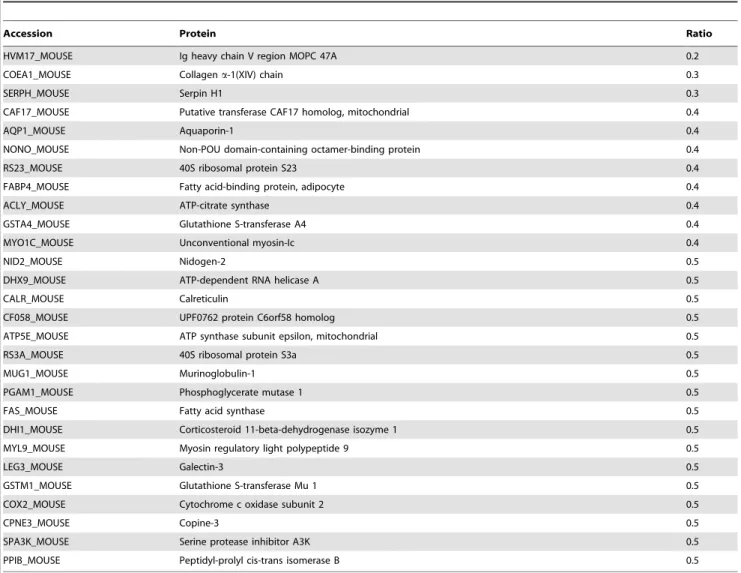
Additionally, 57 proteins were up-regulated (Table 1) and 28 were down-regulated (Table 2). The majority of the ERb
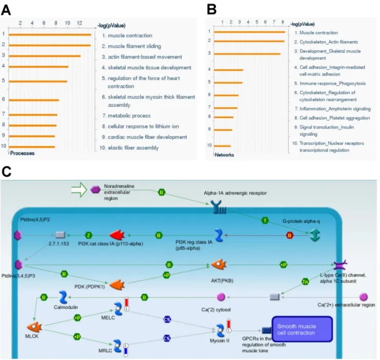
knockout-modified proteins were involved in cell-matrix adhesion, metabolism, immune response, signal transduction, nuclear receptor translational regelation, and muscle contraction and development (Figures 3A, 3B, and 3C).
Figure 1. Genotyping of different ERbmutant mice.Lane 1, ERb+/+
mice; Lane 2, ERb+
/-mice; Lane 3, ERb-/-mice. doi:10.1371/journal.pone.0109058.g001
Figure 2. Decreased urodynamic testing in ERb2/2mice.(A) LPP
and (B) MUCP values in the different groups. Each bar represents the mean6standard deviation of six individual mice. *P,0.05 different from the value in the control group. **P,0.01 different from the value in the control group.
Table 1.Up-regulated proteins in ERb-/-mouse urethra.
Accession Protein Ratio
A1AT1_MOUSE a-1-antitrypsin 1-1 1.5
KAD1_MOUSE Adenylate kinase isoenzyme 1 1.5
MYH1_MOUSE Myosin-1 1.5
RAC3_MOUSE Ras-related C3 botulinum toxin substrate 3 1.5
TRFE_MOUSE Serotransferrin 1.5
B3AT_MOUSE Band 3 anion transport protein 1.5
H3C_MOUSE Histone H3.3C 1.5
RS9_MOUSE 40S ribosomal protein S9 1.6
GPX3_MOUSE Glutathione peroxidase 3 1.6
GSTO1_MOUSE Glutathione S-transferaseV-1 1.6
A1AT2_MOUSE a-1-antitrypsin 1–2 1.6
RD23B_MOUSE UV excision repair protein RAD23 homolog B 1.6
FIBG_MOUSE Fibrinogencchain 1.6
FBN1_MOUSE Fibrillin-1 1.6
A1AT4_MOUSE a-1-antitrypsin 1–4 1.6
RS14_MOUSE 40S ribosomal protein S14 1.6
DHB11_MOUSE Estradiol 17-b-dehydrogenase 11 1.7
PEBP1_MOUSE Phosphatidylethanolamine-binding protein 1 1.7
CASQ1_MOUSE Calsequestrin-1 1.7
MYH4_MOUSE Myosin-4 1.7
SPG16_MOUSE Sperm-associated antigen 16 protein 1.7
K1C10_MOUSE Keratin, type I cytoskeletal 10 1.7
PGS2_MOUSE Decorin 1.7
KCRM_MOUSE Creatine kinase M-type 1.7
MYP0_MOUSE Myelin protein P0 1.7
WFS1_MOUSE Wolframin 1.7
ITIH4_MOUSE Intera-trypsin inhibitor, heavy chain 4 1.7
HBB1_MOUSE Hemoglobin subunitb-1 1.8
CO6A1_MOUSE Collagena-1(VI) chain 1.8
H14_MOUSE Histone H1.4 1.8
APOE_MOUSE Apolipoprotein E 1.8
LUM_MOUSE Lumican 1.9
CLUS_MOUSE Clusterin 1.9
CO6A2_MOUSE Collagena-2(VI) chain 1.9
MIME_MOUSE Mimecan 1.9
HBA_MOUSE Hemoglobin subunita 1.9
MYH8_MOUSE Myosin-8 2.0
PURA1_MOUSE Adenylosuccinate synthetase isozyme 1 2.1
TNNT3_MOUSE Troponin T, fast skeletal muscle 2.2
HMGB1_MOUSE High mobility group protein B1 2.2
MPC2_MOUSE Mitochondrial pyruvate carrier 2 2.3
MYG_MOUSE Myoglobin 2.3
CAH2_MOUSE Carbonic anhydrase 2 2.3
ILEUA_MOUSE Leukocyte elastase inhibitor A 2.5
A1BG_MOUSE a-1B-glycoprotein 2.6
MYL3_MOUSE Myosin light chain 3 2.6
COPD_MOUSE Coatomer subunitd 3.0
IQGA1_MOUSE Ras GTPase-activating-like protein IQGAP1 3.2
MK01_MOUSE Mitogen-activated protein kinase 1 3.2
Myosin, collagen, and elastin expressions in the urethra of ERb2/2mice
We further focused on urethral dysfunction-related proteins including myosin, collagen, and elastin and confirmed their expressions by Western blot analysis. There is a contradiction between the different subtypes of urethral dysfunction-related protein expressions. For example, four types of myosin (myosin-1,
4, 8, and myosin light chain 3) that were overexpressed in ERb2/2
female mice, whereas other two types of myosin (unconventional myosin-Ic and myosin regulatory light polypeptide 9) were decreased. Thus, the common commercial available antibodies, including anti-myosin heavy chain (clone A4.1025), anti-collagen
a-1(III) (FH-7A), and anti-elastin (BA-4), were chosen for the subsequent Western blot analysis.
Table 1.Cont.
Accession Protein Ratio
KV3A3_MOUSE Igkchain V-III region MOPC 70 3.8
IGHM_MOUSE Igmchain C region secreted form 4.2
NID1_MOUSE Nidogen-1 4.3
ACDSB_MOUSE Short/branched chain specific acyl-CoA dehydrogenase 4.5
IGKC_MOUSE Igkchain C region 4.8
IGG2B_MOUSE Igc-2B chain C region 5.3
GCAB_MOUSE Igc-2A chain C region secreted form 13.4
doi:10.1371/journal.pone.0109058.t001
Table 2.Down-regulated proteins in ERb-/-mouse urethra.
Accession Protein Ratio
HVM17_MOUSE Ig heavy chain V region MOPC 47A 0.2
COEA1_MOUSE Collagena-1(XIV) chain 0.3
SERPH_MOUSE Serpin H1 0.3
CAF17_MOUSE Putative transferase CAF17 homolog, mitochondrial 0.4
AQP1_MOUSE Aquaporin-1 0.4
NONO_MOUSE Non-POU domain-containing octamer-binding protein 0.4
RS23_MOUSE 40S ribosomal protein S23 0.4
FABP4_MOUSE Fatty acid-binding protein, adipocyte 0.4
ACLY_MOUSE ATP-citrate synthase 0.4
GSTA4_MOUSE Glutathione S-transferase A4 0.4
MYO1C_MOUSE Unconventional myosin-Ic 0.4
NID2_MOUSE Nidogen-2 0.5
DHX9_MOUSE ATP-dependent RNA helicase A 0.5
CALR_MOUSE Calreticulin 0.5
CF058_MOUSE UPF0762 protein C6orf58 homolog 0.5
ATP5E_MOUSE ATP synthase subunit epsilon, mitochondrial 0.5
RS3A_MOUSE 40S ribosomal protein S3a 0.5
MUG1_MOUSE Murinoglobulin-1 0.5
PGAM1_MOUSE Phosphoglycerate mutase 1 0.5
FAS_MOUSE Fatty acid synthase 0.5
DHI1_MOUSE Corticosteroid 11-beta-dehydrogenase isozyme 1 0.5
MYL9_MOUSE Myosin regulatory light polypeptide 9 0.5
LEG3_MOUSE Galectin-3 0.5
GSTM1_MOUSE Glutathione S-transferase Mu 1 0.5
COX2_MOUSE Cytochrome c oxidase subunit 2 0.5
CPNE3_MOUSE Copine-3 0.5
SPA3K_MOUSE Serine protease inhibitor A3K 0.5
PPIB_MOUSE Peptidyl-prolyl cis-trans isomerase B 0.5
Myosin (Figure 4A) and collagen (Figure 4B) expressions in the urethra was significantly increased in the ERb2/2 group as
compared with the ERb+/+
group. By contrast, elastin ( Fig-ure 4C) expression in the urethra was significantly decreased in the ERb2/2group.
Discussion
Estrogen actions mediated by ERs are known to modulate lower urinary tract (LUT) trophicity [22]. In the present study, LPP and MUCP values were significantly decreased in the ERb2/2group
compared with the ERb+/+
group, indicating an important role of ERbin SUI.
To the best of our knowledge, this is the first study that estimates the changes in protein expression related to ERbin SUI. We used nanoLC-MS/MS analysis to evaluate the proteomic profile of urethral samples collected from ERb+/+
and ERb2/2mice. We
found 85 differentially expressed proteins between ERb+/+ and ERb2/2 female mice. The majority of the identified ERb
knockout-modified proteins were involved in cell-matrix adhesion, metabolism, immune response, signal transduction, nuclear receptor translational regulation, and muscle contraction and development. We further focused on urethral dysfunction-related proteins including myosin, collagen, and elastin and confirmed their expressions by Western blot analyses.
Figure 3. Protein expression profile from proteomic analysis.In term of (A) Gene Ontology and (B) biological networks databases, the
differentially expressed proteins of urethra from ERb-/- female mice were divided into different categories. (C) Biological network analysis for differentially expressed proteins of urethra from ERb-/-female mice using MetaCore mapping tool. The network was generated using shortest path algorithm to map interaction between the proteins.
Four of the proteins that exhibited overexpression in ERb2/2
female mice were related to myosin; specifically, Myosin-1, 4, 8, and myosin light chain 3 were overexpressed in ERb2/2female mice, whereas other two types of myosin (unconventional myosin-Ic and myosin regulatory light polypeptide 9) were decreased. Myosin proteins are composed of both heavy and light chains and are essential components of muscles. Myosin heavy chains help to determine the speed of muscle contraction; in contrast, the role of myosin regulatory light polypeptide 9 (whose expression was 50% decreased in ERb2/2 female mice) is unclear. Since myosin is
responsible for muscle contraction, this overexpression of myosin heavy chains may be a mechanism to counteract the loss of normal urethral function in SUI. This finding is consistent with previous works showing overexpression myosin genes in the pubococcygeus muscle of women with POP [23,24].
The phenotype of the ERb2/2 mice with respect to collagen biosynthesis appears to be more complex. Collagen biosynthesis and deposition is a multiphase process, which is tightly regulated to maintain proper tissue homeostasis. Collagen production is regulated by a variety of molecules, including growth factors, cytokines, and hormones. However, the factors and pathways involved in this process are not fully defined [25]. In the present study, type III collagen (a fibrillar collagen that is found in extensible connective tissues such as skin, lung, and the vascular system, frequently in association with type I collagen) expression in the urethra was significantly increased in ERb2/2 group as
compared with that in the ERb+/+
group, indicating an increased synthesis of collagen or a decreased proteolysis in the urethra. Several factors may have contributed to the high deposition of extracellular matrix (ECM) in ERb2/2mice, including elevated
collagen synthesis by fibroblasts [26,27], and a decreased expression of matrix metalloproteinases in mouse tissue. For example, ER retained their responsiveness to estradiol with respect to collagen biosynthesis. There is evidence that ERb knockout plays a role in the development of SUI. In addition, ERbdeletion in mice has been described to lead to fibrosis in various tissues [28]. For example, Pedram et al. showed that in the hearts of ovariectomized female mice, cardiac hypertrophy and fibrosis were prevented by estradiol administration to wild type but not ERb knockout rodents. Their results established the cardiac
fibroblast as an important target for hypertrophic/fibrosis-inducing peptides the actions of which were mitigated by estrogen/ERb acting in these stromal cells [29]. This supports the findings that collagen increases in the urethra following deletion of ERb.
Certain alterations in connective tissue metabolism are known to be modified in postmenopausal women with genuine stress incontinence. Jacksonet al. showed that treatment with oestrogen has profound effects upon pelvic collagen metabolism, stimulating collagen degradation via increased proteinase activity. While aged collagen is being lost, new collagen is synthesized as witnessed by the increase in the immature cross-links and the decrease in both mature cross-links and advanced glycation end-products. In the present study, collagen deposition contradicts previous reports; perhaps aged collagen degradation is merely an early response to estrogen stimulation [30]. Further studies within this field are indeed needed.
By contrast, elastin was down-regulated in ERb2/2mice. Our previous study [31] suggested that some ECM remodelling enzymes, including lysyl oxidase, are required for the oxidative deamination of amino acid residues in collagen and elastin molecules a step that is required for fibre cross-linking and are therefore essential for the stabilization of collagen fibrils and for the integrity and elasticity of mature elastin [32]. The assembly and cross-linking of elastin/collagen fibres is crucial for the recovery of tissue elasticity and urethral support. The loss of elasticity and resiliency might be attributed to an imbalanced cross-linking of collagen to elastin. When these urethral charac-teristics manifest in a severe form, descriptive terms such as low-pressure urethra, lead-pipe urethra (urethra and bladder neck areas are open at rest) [33,34], or patulous urethra have been used. Because the bladder and urethra are composed of both active (smooth muscle) and passive elements (collagen and elastin) [35,36], our study model characterizes a long-term phase of ERbknockout, representing lower urinary dysfunction in meno-pause-related development of SUI.
Our study has certain limitations. First, the pelvic floor structure of the mouse, which is a quadruped and has a lax abdominal wall, is different from that of a human female; therefore, the results of this study need to be carefully applied to human subjects. Second,
Figure 4. Urethral dysfunction-related proteins expressions.Alterations of (A) myosin, (B) collagen, and (C) elastin expressions in urethra as
indicated by Western blot analyses. The values are calculated by intensity of each band (ratio of target protein/GAPDH) and expressed as mean6
urodynamic studies were conducted under anaesthesia; fortunate-ly, none of our subjects manifested any evidence of bladder instability, implying that detrusor overactivity was not present, and giving credence to our interpretation of fluid expulsion in the absence of increased bladder pressure as evidence of SUI. Third, since a fairly large number of the protein expression profiles were found from the proteomic study, it is difficult to confirm all the expression levels of potential target proteins by Western blot analysis. Fourth, the experimental conditions and the sample preparation technique could have affected the detection of some proteins. In label-free analysis (as in any other proteomics method), not all proteins can be resolved simultaneously; this is due to differences in their properties (such as hydrophobicity, charge, and solubility properties). The specific protocol used in our study is optimal for the solubility of cytosolic proteins [24]. Changes in the protein extraction protocol are needed for easier detection of ECM proteins.
In conclusion, our results suggest a role for ERbin SUI. This pilot study is the first one to estimate protein expression changes in urethras from ERb+/+
and ERb2/2female mice. These changes
could be related to the molecular mechanism of ERbin SUI. A further study confirming the expression levels of potential target proteins in urethras from ERb+/+
and ERb2/2female mice with
Western blot analysis and changing the protein extraction protocol for proteomic analysis is needed. These findings may also have therapeutic implications [37] and perhaps provide pharmacolog-ical strategy for the treatment of urethral dysfunction with ERb
agonists [38,39].
Materials and Methods
Ethics statement
All animals were housed and handled in accordance with criteria outlined in the National Institutes of Health ‘‘Guide for Care and Use of Laboratory Animals’’. The study was approved by the Institutional Animal Care and Use Committee of China Medical University (Reference number: 101-201-N). All efforts were made to ameliorate animal suffering. Animal sacrifice was performed by CO2asphyxiation followed by cervical dislocation.
Animals
ERb knockout mice were generated as previously described [40,41]. ERbgenotyping was based on genomic sequence of ERb
exon 3. We used the primer sequence as below to identify PstI site insert on ERbexon 3 transgene animal. 59-exon 3 (3145): GTT GTG CCA GCC CTG TTA CT (AY028415), 39-exon 3 (8140): GGG CCA GCT CAT TCC ACT C 8161 (X76683). The size of fragments for wild-type and knockout allele was about 150 and 350 b.p., respectively. Female C57BL/6 ERb+/+
mice, aged 6–8 weeks, were used as control (n = 6).
The mice underwent suprapubic bladder tubing (SPT) place-ment [31,42]. LPP and MUCP were assessed in these mice under urethane (1 g/kg, i.p.) anesthesia. After measurements, the animals were sacrificed, and the urethras were removed for proteomic and further analyses.
Suprapubic tube implantation
The surgical procedure was carried out under 1.5% isoflurane anesthesia according to previous methods [31,42]. An SPT (PE-10 tubing, Clay Adams, Parsippany, NJ) was implanted in the bladder. Key points of the operation were as follows: (1) a midline longitudinal abdominal incision was made, 0.5 cm above the urethral meatus; (2) a small incision was made in the bladder wall, and PE-10 tubing with a flared tip was implanted in the bladder
dome; and (3) a purse-string suture with 8-0 silk was tightened around the catheter, which was tunneled subcutaneously to the neck, where it exited the skin.
LPP measurement
Two days after implanting the bladder catheter, the LPP was assessed in these mice under urethane anesthesia. The bladder catheter was connected to both a syringe pump and a pressure transducer. Pressure and force transducer signals were amplified and digitized for computer data collection at 10 samples per second (PowerLabs, AD Instruments, Bella Vista, Australia). The mice were placed supine at the level of zero pressure while bladders were filled with room temperature saline at 1 ml/h through the bladder catheter. If a mouse voided, the bladder was emptied manually using Crede’s maneuver. The average bladder capacity of each mouse was determined after 3–5 voiding cycles. Subsequently, the LPP was measured in the following manner [31,42]. When half-bladder capacity was reached, gentle pressure with one finger was applied to the mouse’s abdomen. Pressure was gently increased until urine leaked, at which time the externally applied pressure was quickly removed. The peak bladder pressure was taken as the LPP. At least three LPPs were obtained for each animal, and the mean LPP was calculated [43,44].
Urethral pressure profile
Urethral pressure profile (UPP) was assessed in these mice under urethane (1 g/kg, i.p.) anesthesia. The bladder catheter (PE-10 tubing, Clay Adams, Parsippany, NJ) was connected to a syringe pump with room temperature saline at 1 ml/hr. The urethral catheter (PE-10 tubing, Clay Adams, Parsippany, NJ) was connected to a pressure transducer. A withdrawal speed of 10mm per minute was used. Pressure and force transducer signals were amplified and digitized for computer data collection at 10 samples per second (PowerLabs, ADInstruments, Bella Vista, Australia). Three successive profiles were obtained in the supine position. The urethral closure pressure (Pclose) is the difference between the urethral pressure (Pure) and the bladder pressure (Pves): Pclose = Pure – Pves [45]. Maximum urethral pressure and MUCP were determined from the UPP measurements taken. The mice were sacrificed immediately after completing the measurements of LPP and MUCP, and the urethras were harvested [31,42].
Protein preparation
Frozen pieces of urethra were weighed and then pulverized with a liquid nitrogen-chilled mortar and pestle. Tissue powder was then homogenized in buffer (16 mmol/l potassium phosphate, pH 7.8, 0.12 mol/l NaCl, 1 mmol/l ethylenediaminetetraacetic acid) containing a protease inhibitor cocktail (Complete Mini, product number 11836153001, Roche Diagnostics, Penzberg, Germany), and then centrifuged at 10,0006g. The supernatant
Label free quantitative proteomics by nanoLC-MS/MS analysis
The nanoLC-MS/MS was performed with a nanoflow UPLC system (UltiMate 3000 RSLCnano system, Dionex, Amsterdam, Netherlands) coupled with a captive spray ion source and hybrid Q-TOF mass spectrometer (maXis impact, Bruker). The sample was injected into a tunnel-frit trap column (C18, 5mm, 100 A˚ , packed length of 2 cm, 375mm od6180mm id) with a flow rate of 8ml/min and a duration of 5 min. The trapped analyses were separated by a commercial analytical column (Acclaim PepMap C18, 2mm 100 A˚ , 75mm6250 mm, Thermo Scientific, USA)
with a flow rate of 300 nl/min. An acetonitrile/water gradient of 1%–40% within 90 min was used for peptide separation. For MS/ MS detection, peptides with charge 2+, 3+or 4+and the intensity greater than 20 counts were selected for data dependent acquisition, which was set to one full MS scan (400–2000 m/z) with 1 Hz and switched to ten product ion scans (100–2000 m/z) with ten Hz.
The LC-MS/MS spectra were deisotoped, centroided, and converted to xml files using DataAnalysis (version 4.1, Bruker). The xml files were searched against the Swissport (release 51.0) database using the MASCOT search algorithm (version 2.2.07). The search parameters for MASCOT for peptide and MS/MS mass tolerance were 50 ppm and 0.07 Da, respectively. Search parameters were selected as Taxonomy – mus; enzyme–trypsin; fixed modifications – carbamidomethyl (C); variable modifications – oxidation (M). Peptides were considered as identified if their MASCOT individual ion score was higher than 25 (P,0.01).
Label free quantitative proteomics was achieved by LC-MS replicated runs (n = 4) of different groups. After LC-MS runs finished, LC-MS/MS runs of each group was performed for protein identification. LC-MS results were processed to have molecular features with DataAnalysis 4.1 (Bruker Daltonics, Germany), which were then loaded into ProfileAnalysis software 2.0 (Bruker Daltonics) fort-test comparison between two groups. Thet-test results among different groups were further transferred to ProteinScape 3.0 (Bruker Daltonics) and combined with protein identification results of each group for the integration of quantified peptide information into each protein [46].
Networks analysis using MetaCore
MetaCore (GeneGo, St. Joseph, MI) was used to map the differentially expressed proteins into biological networks. It is an integrated software suited for functional analysis of protein– protein, protein–DNA and protein compound interactions, metabolic and signaling pathways, and the effects of bioactive molecules [47]. Differentially expressed proteins were converted into gene symbols and uploaded into MetaCore for analysis. The biological process enrichment was analyzed based on Gene
Ontology processes. For network analysis, three algorithms were used: (1) the direct interaction algorithm to map direct protein-protein interactions; (2) the shortest path algorithm to map shortest path for interaction between differentially expressed proteins; and (3) the analyze network algorithm to deduce top scoring processes that are regulated by differentially expressed proteins [42].
Western blot analysis
Urethral tissue were prepared by homogenization of cells in a lysis buffer containing 1% IGEPAL CA-630, 0.5% sodium deoxycholate, 0.1% sodium dodecyl sulfate, aprotinin (10 mg/ mL), leupeptin (10 mg/mL), and phosphate-buffered saline (PBS). Cell lysates containing 100mg of protein were subjected to sodium dodecyl sulfate polyacrylamide gel electrophoresis and then transferred to a polyvinylidene fluoride membrane (Millipore Corp, Bedford, MA, USA). The membrane was stained with Ponceau S to verify the integrity of the transferred proteins and to monitor the unbiased transfer of all protein samples [48,49]. Detection of myosin and collagen on the membranes was performed with an electrochemiluminescence kit (Amersham Life Sciences Inc, Arlington Heights, IL, USA) with the use of the antibody derived from rabbit (anti-myosin heavy chain (clone A4.1025), 1:500 dilution, Millipore, MA, USA; anti-collagen a -1(III) (FH-7A) antibody, 1:500 dilution, Abcam, Cambridge, UK; anti-elastin (BA-4) antibody, 1:500 dilution, Abcam, Cambridge, UK). The intensity of each band was quantified using a densitometer (Molecular Dynamics, Sunnyvale, CA, USA) [50,51].
Statistical analyses
The changes of the target expressions were compared by Student’s t-test or analysis of variance (ANOVA). One-way ANOVA andpost-hoc test (Bonferroni correction) were given for more than two groups are being compared [52].P-value less than 0.05 was considered statistically significant. All calculations were performed using the Statistical Package for Social Sciences (SPSS for Windows, SPSS Inc, Chicago, IL, USA).
Acknowledgments
We thank Professor Shuyuan Yeh to provide ERbknockout mice for this study.
Author Contributions
Conceived and designed the experiments: CC HYC. Performed the experiments: CJC YNL WCC. Analyzed the data: YHC CJC YNL HYC. Contributed reagents/materials/analysis tools: CJC SY YCW WTH BTW WLM CC HYC. Wrote the paper: YHC HYC.
References
1. Wu CY, Hu HY, Huang N, Fang YT, Chou YJ, et al. (2014) Determinants of long-term care services among the elderly: a population-based study in Taiwan. PLoS One 9: e89213.
2. Martinez-Gonzalez NA, Tandjung R, Djalali S, Huber-Geismann F, Markun S, et al. (2014) Effects of physician-nurse substitution on clinical parameters: a systematic review and meta-analysis. PLoS One 9: e89181.
3. Gunetti M, Tomasi S, Giammo A, Boido M, Rustichelli D, et al. (2012) Myogenic potential of whole bone marrow mesenchymal stem cells in vitro and in vivo for usage in urinary incontinence. PLoS One 7: e45538.
4. Zhu L, Lang J, Feng R, Chen J, Wong F (2004) Estrogen receptor in pelvic floor tissues in patients with stress urinary incontinence. Int Urogynecol J Pelvic Floor Dysfunct 15: 340–343.
5. Feifer A, Corcos J (2007) The use of synthetic sub-urethral slings in the treatment of female stress urinary incontinence. Int Urogynecol J Pelvic Floor Dysfunct 18: 1087–1095.
6. Asada H, Yamagata Y, Taketani T, Matsuoka A, Tamura H, et al. (2008) Potential link between estrogen receptor-alpha gene hypomethylation and uterine fibroid formation. Mol Hum Reprod 14: 539–545.
7. Paech K, Webb P, Kuiper GG, Nilsson S, Gustafsson J, et al. (1997) Differential ligand activation of estrogen receptors ERalpha and ERbeta at AP1 sites. Science 277: 1508–1510.
8. Cunningham M, Gilkeson G (2011) Estrogen receptors in immunity and autoimmunity. Clin Rev Allergy Immunol 40: 66–73.
9. Deroo BJ, Buensuceso AV (2010) Minireview: Estrogen receptor-beta: mechanistic insights from recent studies. Mol Endocrinol 24: 1703–1714. 10. Lindberg MK, Alatalo SL, Halleen JM, Mohan S, Gustafsson JA, et al. (2001)
Estrogen receptor specificity in the regulation of the skeleton in female mice. J Endocrinol 171: 229–236.
12. Kaur J, Thakur MK (1991) Effect of age on physico-chemical properties of the uterine nuclear estrogen receptors of albino rats. Mech Ageing Dev 57: 111–123. 13. Hirai K, Tsuda H (2009) Estrogen and urinary incontinence. Int J Urol 16:
45–48.
14. Sievert KD, Emre Bakircioglu M, Tsai T, Dahms SE, Nunes L, et al. (2001) The effect of simulated birth trauma and/or ovariectomy on rodent continence mechanism. Part I: functional and structural change. J Urol 166: 311–317. 15. Hijaz A, Daneshgari F, Sievert KD, Damaser MS (2008) Animal models of
female stress urinary incontinence. J Urol 179: 2103–2110.
16. Cannon TW, Wojcik EM, Ferguson CL, Saraga S, Thomas C, et al. (2002) Effects of vaginal distension on urethral anatomy and function. BJU Int 90: 403– 407.
17. Hijaz A, Daneshgari F, Huang X, Bena J, Liu G, et al. (2005) Role of sling integrity in the restoration of leak point pressure in the rat vaginal sling model. J Urol 174: 771–775.
18. Ma WL, Jeng LB, Yeh CC, Chang C (2012) Androgen and androgen receptor signals jamming monocyte/macrophage functions in premalignant phase of livers. Biomedicine 2: 155–159.
19. Lin DY, Tsai FJ, Tsai CH, Huang CY (2011) Mechanisms governing the protective effect of 17b-estradiol and estrogen receptors against cardiomyocyte injury. BioMedicine 1: 21–28.
20. Drewes PG, Yanagisawa H, Starcher B, Hornstra I, Csiszar K, et al. (2007) Pelvic organ prolapse in fibulin-5 knockout mice: pregnancy-induced changes in elastic fiber homeostasis in mouse vagina. Am J Pathol 170: 578–589. 21. Hammack BN, Fung KY, Hunsucker SW, Duncan MW, Burgoon MP, et al.
(2004) Proteomic analysis of multiple sclerosis cerebrospinal fluid. Mult Scler 10: 245–260.
22. Game X, Allard J, Escourrou G, Gourdy P, Tack I, et al. (2008) Estradiol increases urethral tone through the local inhibition of neuronal nitric oxide synthase expression. Am J Physiol Regul Integr Comp Physiol 294: R851–857. 23. Visco AG, Yuan L (2003) Differential gene expression in pubococcygeus muscle from patients with pelvic organ prolapse. Am J Obstet Gynecol 189: 102–112. 24. Athanasiou S, Lymberopoulos E, Kanellopoulou S, Rodolakis A, Vlachos G, et al. (2010) Proteomic analysis of pubocervical fascia in women with and without pelvic organ prolapse and urodynamic stress incontinence. Int Urogynecol J 21: 1377–1384.
25. Markiewicz M, Znoyko S, Stawski L, Ghatnekar A, Gilkeson G, et al. (2013) A role for estrogen receptor-alpha and estrogen receptor-beta in collagen biosynthesis in mouse skin. J Invest Dermatol 133: 120–127.
26. Liu PL, Tsai JR, Hwang JJ, Chou SH, Cheng YJ, et al. (2010) High-mobility group box 1-mediated matrix metalloproteinase-9 expression in non-small cell lung cancer contributes to tumor cell invasiveness. Am J Respir Cell Mol Biol 43: 530–538.
27. Chen HY, Lin WY, Chen YH, Chen WC, Tsai FJ, et al. (2010) Matrix metalloproteinase-9 polymorphism and risk of pelvic organ prolapse in Taiwanese women. Eur J Obstet Gynecol Reprod Biol 149: 222–224. 28. Wang XX, Jiang T, Levi M (2010) Nuclear hormone receptors in diabetic
nephropathy. Nat Rev Nephrol 6: 342–351.
29. Pedram A, Razandi M, O’Mahony F, Lubahn D, Levin ER (2010) Estrogen receptor-beta prevents cardiac fibrosis. Mol Endocrinol 24: 2152–2165. 30. Jackson S, James M, Abrams P (2002) The effect of oestradiol on vaginal
collagen metabolism in postmenopausal women with genuine stress inconti-nence. BJOG 109: 339–344.
31. Chen HY, Lin YN, Chen YH, Chen WC (2012) Stress urinary incontinence following vaginal trauma involves remodeling of urethral connective tissue in female mice. Eur J Obstet Gynecol Reprod Biol 163: 224–229.
32. Klutke J, Stanczyk FZ, Ji Q, Campeau JD, Klutke CG (2010) Suppression of lysyl oxidase gene expression by methylation in pelvic organ prolapse. Int Urogynecol J 21: 869–872.
33. Crivellaro S, Smith JJ 3rd (2009) Minimally invasive therapies for female stress urinary incontinence: the current status of bioinjectables/new devices (adjustable continence therapy, urethral submucosal collagen denaturation by radiofre-quency). ScientificWorldJournal 9: 466–478.
34. Chew SY (1989) Investigation and treatment of female urinary incontinence. Singapore Med J 30: 396–399.
35. Levin RM, Horan P, Liu SP (1999) Metabolic aspects of urinary bladder filling. Scand J Urol Nephrol Suppl 201: 59–66; discussion 76–99.
36. McLennan MT, Leong FC, Steele AC (2007) Evaluation of urinary incontinence and voiding dysfunction in women. Mo Med 104: 77–81.
37. Liao WL, Tsai FJ (2013) Personalized medicine: A paradigm shift in healthcare. Biomedicine 3: 66–72.
38. Skala CE, Petry IB, Albrich S, Puhl A, Naumann G, et al. (2011) The effect of genital and lower urinary tract symptoms on steroid receptor expression in women with genital prolapse. Int Urogynecol J 22: 705–712.
39. Cheng CL, de Groat WC (2014) Effects of agonists for estrogen receptor alpha and beta on ovariectomy-induced lower urinary tract dysfunction in the rat. Am J Physiol Renal Physiol 306: F181–187.
40. Krege JH, Hodgin JB, Couse JF, Enmark E, Warner M, et al. (1998) Generation and reproductive phenotypes of mice lacking estrogen receptor beta. Proc Natl Acad Sci U S A 95: 15677–15682.
41. Hsu I, Chuang KL, Slavin S, Da J, Lim WX, et al. (2014) Suppression of ERbeta signaling via ERbeta knockout or antagonist protects against bladder cancer development. Carcinogenesis 35: 651–661.
42. Chen HY, Chen CJ, Lin YN, Chen YH, Chen WC, et al. (2013) Proteomic analysis related to stress urinary incontinence following vaginal trauma in female mice. Eur J Obstet Gynecol Reprod Biol 171: 171–179.
43. Chen YH, Lin YN, Chen WC, Hsieh WT, Chen HY (2014) Treatment of stress urinary incontinence by ginsenoside rh2. Am J Chin Med 42: 817–831. 44. Chen YH, Lin YN, Chen WC, Hsieh WT, Chen HY (2014) Treatment of stress
urinary incontinence by cinnamaldehyde, the major constituent of the chinese medicinal herb ramulus cinnamomi. Evid Based Complement Alternat Med 2014: 280204.
45. Hilton P, Stanton SL (1983) Urethral pressure measurement by microtransdu-cer: the results in symptom-free women and in those with genuine stress incontinence. Br J Obstet Gynaecol 90: 919–933.
46. Chen CJ, Chen WY, Tseng MC, Chen YR (2012) Tunnel frit: a nonmetallic in-capillary frit for nanoflow ultra high-performance liquid chromatography-mass spectrometryapplications. Anal Chem 84: 297–303.
47. Li CC, Lo HY, Hsiang CY, Ho TY (2012) DNA microarray analysis as a tool to investigate the therapeutic mechanisms and drug development of Chinese medicinal herbs. BioMedicine 2: 10–16.
48. Yin WH, Chen YH, Wei J, Jen HL, Huang WP, et al. (2011) Associations between endothelin-1 and adiponectin in chronic heart failure. Cardiology 118: 207–216.
49. Lin FY, Lin YW, Huang CY, Chang YJ, Tsao NW, et al. (2011) GroEL1, a heat shock protein 60 of Chlamydia pneumoniae, induces lectin-like oxidized low-density lipoprotein receptor 1 expression in endothelial cells and enhances atherogenesis in hypercholesterolemic rabbits. J Immunol 186: 4405–4414. 50. Liu PL, Tsai JR, Chiu CC, Hwang JJ, Chou SH, et al. (2010) Decreased
expression of thrombomodulin is correlated with tumor cell invasiveness and poor prognosis in nonsmall cell lung cancer. Mol Carcinog 49: 874–881. 51. Yang TL, Lin FY, Chen YH, Chiu JJ, Shiao MS, et al. (2011) Salvianolic acid B
inhibits low-density lipoprotein oxidation and neointimal hyperplasia in endothelium-denuded hypercholesterolaemic rabbits. J Sci Food Agric 91: 134–141.


