Specific Deletion of AMP-Activated Protein
Kinase (
α
1AMPK) in Murine Oocytes Alters
Junctional Protein Expression and
Mitochondrial Physiology
Michael J. Bertoldo1,5, Edith Guibert1, Melanie Faure1, Christelle Ramé1, Marc Foretz2,3,4, Benoit Viollet2,3,4, Joëlle Dupont1, Pascal Froment1*
1UMR 7247 INRA CNRS Université de Tours Haras Nationaux Physiologie de la Reproduction et des Comportements, 37380, Nouzilly, France,2INSERM, U1016, Institut Cochin, Paris, France,3CNRS, UMR8104, Paris, France,4Université Paris Descartes, Sorbonne Paris Cité, Paris, France,5School of Women’s and Children’s Health, Discipline of Obstetrics and Gynaecology, University of New South Wales, Sydney, NSW, Australia
Abstract
Oogenesis and folliculogenesis are dynamic processes that are regulated by endocrine, paracrine and autocrine signals. These signals are exchanged between the oocyte and the somatic cells of the follicle. Here we analyzed the role of AMP-activated protein kinase (AMPK), an important regulator of cellular energy homeostasis, by using transgenic mice deficient inα1AMPK specifically in the oocyte. We found a decrease of 27% in litter size was observed in ZP3-α1AMPK-/-(ZP3-KO) female mice. Followingin vitrofertilization, where conditions are stressful for the oocyte and embryo, ZP3-KO oocytes were 68% less likely to pass the 2-cell stage.In vivoand in cumulus-oocyte complexes, several proteins in-volved in junctional communication, such as connexin37 and N-cadherin were down-regulated in the absence ofα1AMPK. While the two signalling pathways (PKA and MAPK) involved in the junctional communication between the cumulus/granulosa cells and the oo-cyte were stimulated in control oooo-cytes, ZP3-KO oooo-cytes exhibited only low phosphorylation of MAPK or CREB proteins. In addition, MII oocytes deficient inα1AMPK had a 3-fold lower ATP concentration, an increase in abnormal mitochondria, and a decrease in cytochrome C and PGC1αlevels, suggesting perturbed energy production by mitochondria. The absence ofα1AMPK also induced a reduction in histone deacetylase activity, which was associated with an increase in histone H3 acetylation (K9/K14 residues). Together, the results of the present study suggest that absence of AMPK, modifies oocyte quality through energy pro-cesses and oocyte/somatic cell communication. The limited effect observedin vivocould be partly due to a favourable follicle microenvironment where nutrients, growth factors, and ad-equate cell interaction were present. Whereas in a challenging environment such as that of in vitroculture following IVF, the phenotype is revealed.
OPEN ACCESS
Citation:Bertoldo MJ, Guibert E, Faure M, Ramé C, Foretz M, Viollet B, et al. (2015) Specific Deletion of AMP-Activated Protein Kinase (α1AMPK) in Murine
Oocytes Alters Junctional Protein Expression and Mitochondrial Physiology. PLoS ONE 10(3): e0119680. doi:10.1371/journal.pone.0119680
Academic Editor:Qing-Yuan Sun, Institute of Zoology, Chinese Academy of Sciences, CHINA
Received:September 21, 2014
Accepted:January 15, 2015
Published:March 13, 2015
Copyright:© 2015 Bertoldo et al. This is an open access article distributed under the terms of the
Creative Commons Attribution License, which permits unrestricted use, distribution, and reproduction in any medium, provided the original author and source are credited.
Data Availability Statement:All relevant data are within the paper and its Supporting Information files.
Funding:Melanie Faure was supported by the Region Centre and Institut National de la Recherche Agronomique (Ph.D Grant). The national FERTiNERGY programme funded by the French National Research Agency (ANR). The funders had no role in study design, data collection and analysis, decision to publish, or preparation of the manuscript.
Introduction
Numerous studies have emphasized the importance of adequate nutritional status in maintain-ing reproductive function. The challenge is to understand the means of communication be-tween the existing nutritional status, energy metabolism and the reproductive system. Ratchfordet al. have hypothesized that abnormalities in oocyte metabolism, such as that ob-served in diabetes, could potentially preprogramme the oocyte for poor outcomes after fertili-zation [1]. Furthermore, Wanget al. [2] concluded that maternal diabetes results in various oocyte defects. During oogenesis and folliculogenesis, several dynamic processes that are regu-lated by endocrine, paracrine and autocrine signals have been shown to be linked with energet-ic status. For example glucose metabolism is necessary for successful oocyte maturation and the resumption of meiosis [3]. Mitochondria can influence the developmental competence of the oocyte [4]. Indeed, mitochondria play a key role in cellular energy generation, the control of cell death [5] and the dynamic process of meiosis including DNA reorganization [2]. In the case of diabetes, mitochondria are abnormally distributed around the spindle or in the oocyte cytoplasm [2]. For these crucial activities in oocyte maturation, mitochondrial redistribution, activity or dysfunction have been suggested as markers of oocyte quality and are strongly relat-ed to fertilization rates and embryo development [2,6].
A protein kinase called AMP-activated protein kinase (AMPK), plays an important role in cellular energy homeostasis and mitochondrial function. AMPK is sensitive to energy content and responds both by stimulating energy production, including glucose and lipid catabolism, and by inhibiting energy-consuming processes such as protein, fatty acid and cholesterol syn-thesis [7]. AMPK is a heterodimer composed of a catalyticα-subunit bound withβ- andγ -regulatory subunits. It is sensitive to the AMP/ATP ratio and is activated by allosteric regula-tion of increased AMP concentraregula-tion and by the phosphorylaregula-tion of theα-subunit at threo-nine 172 by the upstream kinases; liver kinase B1 (LKB1), Ca2+calmodulin-dependent protein kinase 1 or 2 (CaMKK1 or CaMKK2), TGF-β-activated kinase (TAK1) and possibly kinase suppressor of RAS (KSR2). Apart from its classical functions as a cellular energy sensor, AMPK has a role in the formation and maintenance of cellular junctional complexes and cyto-skeleton dynamics [8–10].
In mammals and birds, AMPK has been identified in the different cell types of the ovary (oocyte cumulus, granulosa cells and theca) and in the corpus luteum [11,12]. Its role has been studied in detail in granulosa cell cultures and during oocyte maturation by using phar-macological agents [13–15]. AMPK activators inhibit the secretion of progesterone and / or estradiol by granulosa cells in mammals [12,16]. AMPK improves resumption of oocyte meio-sis in mice [13,17–19], whereas pharmacological activation of AMPK blocks nuclear oocyte maturation in pigs and cattle [14,15]. Although AMPK has been shown to be involved in sev-eral ovarian functions, no study has yet described the consequences ofα1AMPK ablation from the oocyte.
Materials and Methods
Ethics Statement
All animal procedures were carried out in accordance with european legislation for animal ex-perimentation (Directive 86/609/EEC) and with french legislation on animal research. The procedures using oocyteα1AMPK-deficient mice were approved by the ethics committee of Val de Loire (CEEA VdL, Comité d'Ethique pour l'Expérimentation Animale du Val de Loire, n°2012–12–11).
Animals
Oocyteα1AMPK-deficient mice were obtained by crossing ZP3-Cre mice (C57BL/6-TgN (Zp3-Cre)93Knw, The Jackson Laboratory; Charles River Laboratories, l’arbresle, France) [22] with mice containing floxedα1AMPK subunits on a C57BL/6 background [23,24] in the ani-mal facilities (EU0028, UEPAO, 1297). The ZP3-Cre mice have previously displayed the same fertility characteristics as wild-type mice [22]. Wild-type and mutant mice were maintained under standard conditions of light (12h light, 12h darkness) and temperature withad libitum access to food and water.
The fertility assessment was performed by crossing a 3 month-old wild-type male mouse with two transgenic female mice at the same age for 1 week in the same cage. Two alternative transgenic females were then rotated through a male’s cage. The same male was crossed with the 3 following genotypes of females: 1/ AMPKα1lox/loxmice (control females noted WT, n = 14), 2/ female ZP3-Cre AMPKα1+/-mice (n = 12) and 3/ female ZP3-Cre AMPKα1-/-mice (noted ZP3-KO, n = 20). When a vaginal plug was detected, the mated female was moved in another cage. The litter size was counted at birth and 5 days after birth, to detect neonatal death or cannibalism. Results were presented as number of litters obtained per female at birth, because no neonatal death was observed. We measured 3 litter sizes per female.
To study responsiveness of ovaries to gonadotropins and to recover oocytes and cumulus cell-oocyte complexes (COCs), mice were injected intraperitoneal with 5 IU of equine chorion-ic gonadotropin (eCG). 46 hours later, 5 IU of human chorionchorion-ic gonadotropin was adminis-trated (hCG, Intervet, Boxmeer, Holland), A further 12 hours later, oviducal COCs were retrieved in warmed M2 medium (Sigma) or fixed in 4% paraformaldehyde. Animals were killed by cervical dislocation. Ovaries were fixed in bouin solution for histological studies or di-rectly stored at -80°C for biology molecular analysis.
Oocyte recovery and
In vitro
fertilisation (IVF)
Cumulus oocyte complexes (COCs) were collected from oviducal ampullae in warmed M2 me-dium (Sigma); then the COCs were washed three times in Embryomax human tubal meme-dium (HTF Medium, Millipore, St Quentin en Yvelines, France). The area of the COC (cumulus ex-pansion) was measured using an image analyser (ImageJ, NIH, USA). Zona pellucida thickness, volume of the cytoplasm and volume of perivitelline space were measured. Four points on the zona were used to calculate the zona thickness per oocyte as described by Jenningset al. 2011 [25]. Measurements were performed with 47 ZP3-KO oocytes and 48 WT oocytes retrieved from 6–7 different animals.
COCs were randomly placed into 4-well dishes containing 200μl of HTF medium per well
forin vitrofertilization or incubated in hyaluronidase 0.1% for 2 min at 37°C to obtain denud-ed oocytes. Oocytes were then frozen at -80°C or fixdenud-ed with 4% paraformaldehyde for 15min.
dish containing the mature COCs for 5 hours. After co-incubation, presumptive zygotes were washed three times to remove cumulus cells and excess sperm and placed into 20μl drops of
HTF medium under mineral oil. Embryos were cultured at 37°C in a humidified atmosphere of 5% O2and 6% CO2in air. Embryo assessments were made every 24 h until the 4-cell stage as
detailed in Bertoldoet al. 2014 [26]. The experiment was repeated 3 times, with a total of at least 80 oocytes inseminated per condition.
Immunohistochemistry, histological analysis of ovaries and
immunofluorescence of oocytes
For immunohistochemistry or immunofluorescence, procedures were described in Tosca et al. 2005 and 2006 [11,12]. Sections were incubated overnight at 4°C with 1% Bovine Serum Albumin (BSA)—PBS containing the following primary antibodies at 1:100 dilution: antibody against Cre was purchased from Merck-Novagen (Darmstadt, Germany); phospho-acetyl CoA carboxylase (Ser79) from Upstate Biotechnology Inc., (Lake Placid, NY, USA);α-tubulin, cytochrome C, phosphorylated glycogen synthase kinase 3 beta (phospho-GSK-3β(Ser9)) and Sirt 1 from Cell Signalling (Beverly, MA, USA);β-catenin from Santa Cruz (Santa Cruz, CA, USA); connexin 37 from Alpha Diagnostic (San Antonio TX, USA); N-cadherin and occludin from Sigma (St Louis, MO, USA). Negative controls were rabbit or mouse IgG (Sigma, St Louis, MO, USA). The following day, after two PBS baths for 5 min, sections were incubated for immunofluorescence with secondary antibodies (Alexa fluor 488 goat anti-rabbit IgG or Alexa fluor 488 rabbit anti-mouse IgG (Invitrogen) for 4h at room temperature. Slides were then washed twice for 5 min in PBS and incubated for 10 min with 4’,6’ -diamino-2-phenylindole (DAPI; 10μg/ml, Invitrogen) and mounted with fluorescent mounting medium
(Sigma). Immunostaining was performed on ovarian sections from 6 different animals per ge-notype. Immunofluorescence of denuded oocytes (mitochondria and MII spindle) were per-formed on at least 42 oocytes retrieved from 6–7 different animals. Mitochondrial staining of live denuded oocytes were realized in M2 medium (Sigma) containing 200 nM Mitotracker ((MitoTracker Orange CM-H2TMRos, M7511, Invitrogen) for 30 min at 37°C. Images were captured using a fluorescent microscope (Zeiss Axioplan 2, Zeiss Gruppe, Jena, Germany) or for oocyte analysis, fluorescent images were captured on a laser scanning confocal microscope (Zeiss Gruppe, Jena, Germany) with a ×63 objective and analysed with ZEN software (Zeiss Gruppe, Jena, Germany). Spindles were analysed according to one- and two-dimensional characteristics in X, Y or Z planes and measurements were calculated regardless of spindle orientation.
Transmission electron microscopy
Ovaries (n = 4 mice in each genotype) were fixed in 4% glutaraldehyde, 0.1M sodium cacody-late buffer pH 7.4 (Sigma, St Louis, MO, USA) for 24 h at 4°C, post-fixed in 1% osmium tetrox-ide, and embedded in eponaraldite resin as described in [27]. For ultrastructure analysis, samples were serially sectioned at 70 nm slice thickness, and sections were examined on a CM10 electron microscope (CM 10 Philips, Eindhoven, the Netherlands). Analysis software was used for image acquisition (Soft Imaging System, Olympus, Münster, Germany). Zona pel-lucida was observed in oocytes of ovarian follicle from 4 different animals per genotype. More than 500 mitochondria per genotype (4 animals/genotype) were analysed and classified as nor-mal or exhibiting altered cristae and considered as abnornor-mal mitochondria. The diameter of mitochondria (μm), distance between the outer and inner mitochondrial membrane (nm), and
Western immunoblotting
Oocytes were prepared as detailed in“Oocyte recovery”section, incubated for 2 min at 37°C with 0.1% hyaluronidase to remove cumulus cells, then denuded oocytes were washed two times in PBS and frozen at -80°C. Cumulus cells were retrieved, centrifuged and frozen at -80°C. Lysates were prepared from a group of 40–50 oocytes (or cumulus cells from 40–50 COCs) retrieved from 6–7 different animals. Denuded oocytes and cumulus cells were lysed and exposed to 3 repeated freeze/thaw cycles in lysis buffer. The protein concentration in the supernatant was determined using a calorimetric assay kit (DC assay kit; Uptima Interchim, Montmuçon, France) and equal amounts of proteins were submitted to electrophoresis on SDS-PAGE under reducing conditions and then transferred onto nitrocellulose membranes (Schleicher & Schuell, Ecquevilly, France), as described in Fromentet al. 2004 [27]. Thereafter, membranes were incubated overnight at 4°C with the following rabbit or mouse antibodies:
α1AMPK was purchased from Upstate Biotechnology Inc (Lake, Placid, NY, USA); 3β hydro-xysteroid dehydrogenase (3βHSD) andβ-catenin from Santa Cruz (Santa Cruz, CA, USA); cy-tochrome C, extracellular signal-regulated kinase 2 (ERK2), phosphorylated ERK1/2 (pERK1/ 2), cAMP responsive element-binding protein (CREB), phospho-CREB (Ser 133), histone H3 (H3), peroxisome proliferator-activated receptor-γcoactivator1α(PGC1α) and p53 from Cell Signalling (Beverly, MA, USA); N-cadherin, vinculin from Sigma (St Louis, MO, USA); acety-lated H3 (H3 K9/K14) and mdm2 (2A10) from Millipore (Molsheim, France); connexin 37 from Alpha Diagnostic (San Antonio TX, USA). All antibodies were used at 1:1000 dilution in western-blotting, except phospho-CREB antibodies diluted at 1:500. The band densities were quantified using image analysis software (ImageJ, NIH, USA). The results are expressed as the intensity signal in arbitrary units, after normalization by an internal standard (total protein or vinculin) and correspond to the mean of three separate experiments.
RNA isolation and RT-qPCR
Total RNA was extracted from pools of 50 collected denuded oocytes using NucleoSpin RNA XS (Macherey Nagel, Hoerdt, France) according to the manufacturer’s recommendations. RNA concentrations were measured by spectrophotometry. Following a DNAse treatment (Ambion, Clinisciences, Montrouge, France), 2μg of total RNA were reverse-transcribed using
Super Script II reverse transcriptase (Invitrogen, Cergy Pontoise, France) in the presence of random hexamer primers (Promega, Charbonnieres-les-Bains, France). Real-time quantitative PCRs (Q-PCRs) were performed on Light Cycler 480 (Roche, Meylan, France) as previously described [28]. Briefly, the PCR Master Mix (10μl) was composed of 4μl cDNA diluted (5X),
0.2μl of each primer (200 nM), 2μl of SsoAdvanced SYBR Green Supermix (Biorad,
Marne-la-Coquette, France).
The primer sequences were: forward: 5’AAACTTGCTAGCGGTCCTCA 3’and reverse 5’
TGGCTGGTGCCAGTAAGAG 3’designed and validated for mouse-PGC-1alpha
ATP, cAMP and histone deacetylase activity (HDAC) analysis
Four groups of 30–35 denuded oocytes retrieved from 6–7 different animals were lysed by ex-posing oocytes to 3 repeated freeze/thaw cycles before the commencement of the assays. The protein concentration in the lysate was determined using a calorimetric assay kit (DC assay kit; Uptima Interchim, Montluçon, France). HDAC (NAD+-dependent histone deacetylase) activi-ty was assessed using the HDAC-Glo kit according to the manufacturer’s instructions (Pro-mega, Madison, USA). cAMP and ATP concentrations were measured by using the cAMP-Glo Assay (Promega) and the Cell-Titer-Glo Assay (Promega) according to the manufacturer’s in-structions. Each standard and sample was assessed in duplicate. The results of each assay were normalized with the protein concentration in each sample.
Statistical analysis
All data are presented as means ± SEM. A Student’s t test was used to compare means between each genotype. An ANOVA test was performed for QPCR analysis, the means were compared by Fisher’s test. Probability values0.05 were considered significant.
Results
1- Reduced fertility in oocyte-specific
α
1AMPK
-/-mutant female mice
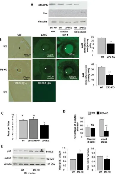
In ZP3-Cre AMPKα1-/-mice (ZP3-KO), Cre recombinase was expressed only in oocytes and was associated with a decrease in phosphorylated Acetyl-CoA carboxylase and Sirt1 expression, two substrates of AMPK (Fig. 1A, B). SIRT1, a nutrient sensor with deacetylase activity, is known to have a reduced downstream signalling in the absence of AMPK [30,31]. Absence of
α1AMPK expression was confirmed in metaphase II (MII) oocytes retrieved after superovula-tion (Fig. 1A, B). Control females (α1AMPKloxlox, WT), heterozygous (ZP3-Cre AMPKα1 +/-mice), and homozygous ZP3-Cre female mice were mated with wild-type males. The mean lit-ter size of ZP3-KO female mice (5.5 ± 0.3 pups/litlit-ter) was reduced significantly (P<0.05), by 27%, compared to that of WT or heterozygous female (7.5 ± 0.4 pups/litter) (Fig. 1C). These re-sults were supported by a lower number (P<0.05) of oocytes recovered in ZP3-KO animals (19.1 ± 1.1) compared to WT animals (23.6 ± 1.1) after superovulation.
Following IVF, there were no differences in the ability of oocytes to cleave between ZP3-KO and WT oocytes. However, there was a 68% reduction in the ability of oocyte-specific ZP3-KO embryos to develop to the 4-cell stage (WT mice: 88% vs ZP3-KO: 28%;Fig. 1D). Moreover, in comparison to WT MII oocytes, oocytes recovered from ZP3-KO mice following superovula-tion presented a higher basal level of p53, a master regulator of cell cycle arrest, associated with a decrease in mdm2, a negative regulator of p53 (Fig. 1E).
Morphological analysis of retrieved oocytes after superovulation have shown a slight in-crease in the volume of the cytoplasm (Fig. 2C) and the thickness of the zona pellucida of ZP3-KO oocytes (11.5 ± 0.1μm) versus WT oocytes (10.4 ± 0.2μm,P<0.001), but no difference between genotypes in the area of cumulus expansion was observed (Fig. 2A-E). Electron micro-scopic observations have shown a uniform appearance of the zona pellucida in WT oocytesin vivo, but a lack of uniformity of the zona pellucida in ZP3-KO oocytes (Fig. 2F).
2- Absence of
α
1AMPK perturbs cell-to-cell communication
surrounding cumulus cells.In vivo, in ZP3-KO oocytes, expression of occludin, a component of tight junctions, was less common at the interface and more localized in the cytoplasm of oo-cytes (Fig. 3A).β-catenin and N-cadherin, two components of adherens junctions were local-ized at the oolemma interface of the oocyte and in the zona pellucida/cumulus interface. They were reduced in ZP3-KO mice (Fig. 3A, B). The glycogen synthase kinase-3β(GSK3β), re-ported to mediateβ-catenin/Wnt signalling [33,34], also showed a reduced intensity of label-ling in the cytoplasm of ZP3-KO oocytes (Fig. 3A). In addition, the localization of connexin37, a critical gap junction protein for oocyte/cumulus cell communication, was barely detectable in ZP3-KO oocytes at the zona pellucida, in contrast to wild-type oocytes (Fig. 3A, B). A similar
Fig 1.α1AMPK inactivation in oocyte.(A) Western blot analysis of theα1AMPK subunit and Cre
recombinase in metaphase II (MII) oocytes, cumulus cells and liver retrieved from wild-type and ZP3-KO mice after superovulation (B) Conditional invalidation ofα1AMPK subunit in the oocyte. Cre expression was localised by immunohistochemistry, and phospho-ACC, and Sirt1, two AMPK substrates, by
immunofluorescence in WT and ZP3-KO ovary sections. Arrows show the oocyte (C) Fertility analysis of WT (genotype AMPKα1lox/lox), heterozygous (genotype ZP3-Cre, AMPKα1+/-), and ZP3-KO (genotype ZP3-Cre,
AMPKα1-/-) female mice were mated with wild-type males. (D) Percentage of oocytes cleaved after in vitro
fertilization with wild type semen and percentage of cleaved oocytes to reach a 4-cell stage embryo. (E) Western blot analysis of p53 and mdm2 protein levels in MII oocytes of WT and ZP3-KO mice.*, P<0.05 ***, P<0.001. a, b Values with different letters differ significantly (P<0.05)
reduction in N-cadherin,β-catenin and Connexin37 protein expression in isolated cumulus cells purified from superovulated ZP3-KO COCs was observed (Fig. 3C).
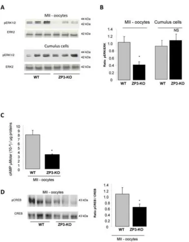
Both PKA and MAPK signal transduction pathways were observed to be involved in the junctional communication between the cumulus/granulosa cells and the oocyte and the somat-ic cells of the follsomat-icle. Communsomat-ication is essential for the growing oocyte and has a role in sup-plying nutrients to the maturing oocyte [35]. Western-blot analyses have revealed a
significantly reduced phosphorylation of a MAP protein, the extracellular signal-regulated ki-nases (ERK1/2) in ZP3-KO MII oocytes when compared with wild-type oocytes (Fig. 4A, B), but not in the ZP3-KO cumulus cells (Fig. 4A, B). Concomitantly, we also observed a reduc-tion in the protein kinase A (PKA) activity as revealed by the diminureduc-tion of CREB phosphory-lation (a target of PKA) and cAMP content (cAMP activates PKA) in ZP3-KO MII oocytes (Fig. 4C, D).
3- Energy production is impaired in the absence of
α
1AMPK in oocyte
When mutant oocytes were placed into a stressful environment, such asin vitroculture, a nota-ble reduction in fertility was observed. This suggested that in the absence ofα1AMPK, energy production within the oocyte was perturbed. Thus, we measured the ATP content in ZP3-KO oocytes, which was about 3-fold lower than in WT oocytes (Fig. 5A). Expression of
Fig 2. Oocyte morphology. (A)Brightfield photomicrographs of the area measured: expanded cumulus 'a'; volume of perivitelline space‘b’; volume of cytoplasm‘c’and zona pellucida thickness‘d’. Method of measurement of zona pellucida, indicating the points on the zona that were used to calculate the zona thickness per oocyte.(B)Analysis of area of cumulus expansion following recovery of COCs 12 h after hCG injection.(C)Volume of cytoplasm.(D)Volume of perivitelline space.(E)Analysis of zona pellucida thickness.(F)Transmission electron microscopic micrographs of the zonae pellucidae of WT and ZP3-KO oocytes. ZP: zona pellucida; GC: Granulosa cells; CO: Cytoplasm oocyte. Arrows show invagination in zona pellucidae.**,P<0.01,***,P<0.001. (n = 40 oocytes/genotypes)
mitochondrial genes showed a decrease in cytochrome B and PGCα1 at the transcript levels (Fig. 5B, C) and a decrease in cytochrome C and PGCα1 at the protein level in ZP3-KO oocytes compared to control oocytes (Fig. 5D). Furthermore when mitochondria were analysed by transmission electron microscopy, ZP3-KO oocytes presented with altered morphology. Mito-chondria displayed a poorly structured matrix, numerous vacuoles, a narrow inter membrane space and rupture of the outer membrane (Fig. 5E). Indeed, in ZP3-KO oocytes, there was a greater proportion (38.1 ± 7.0%) of mitochondria with altered cristae when compared with WT mitochondria (20.6 ± 3.4%;P<0.05) (Fig. 5E). In addition, characteristics associated with swollen mitochondria [36] were observed in ZP3-KO oocytes when compared with mitochon-dria from WT oocytes. These included a larger mitochonmitochon-drial diameter and distance between the outer and inner mitochondrial membrane (Table 1). No difference was observed between genotypes in the intracristal space of mitochondria.
In the growing follicle, two mitochondrial distributions were observed in both genotypes at the germinal vesicle (GV) stage: 1/ the apparent clustering of mitochondria around the GV
Fig 3. Oocyte cell-to-cell communication. (A)Immunofluorescent expression and localization of occludin (a marker for tight junctions), N-cadherin andβ-catenin (markers for adherens junctions), connexin 37 (a marker for gap junctions) and phosphorylated GSK3β(forβ-catenin signalling) in germinal vesicle stage in WT and ZP3-KO oocytes.(B and C)Western blot analysis of N-cadherin,β-catenin and connexin 37 in Metaphase II oocytes and cumulus cells of WT and ZP3-KO mice. Quantification of proteins in both genotypes is shown on the side of the western-blot.*,P<0.05,**,P<0.01.
(Fig. 5F), as reported previously [2]; and 2/ a uniform distribution where the mitochondria were dispersed throughout the cytoplasm. However, with the completion of meiotic matura-tion, we observed clustering and polarized patterns of mitochondrial distribution in WT oo-cytes at the MII stage (Fig. 5F), regarded as a normal distribution pattern [2]. In contrast, ZP3-KO oocytes presented only a clustering pattern of distribution and no polarized distribution (Fig. 5F).
4-
α
1AMPK is not required for spindle formation but reduces HDAC
activity
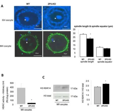
Because pharmacological activators of AMPK lead to resumption or blockage of oocyte meiosis [13,17–19], we analysed spindle formation in MII oocytes. Analysis using confocal microscopy did not detect differences in nuclear morphology at GV stage or in MII oocytes between wild-type or mutant animals. (Fig. 6A). However when spindle length was measured, we observed shorter spindles in ZP3-KO (23.1 ± 1.4μm) oocytes when compared to WT oocyte spindles
(28.8 ± 0.9μm;P<0.05). There were no differences between the size of the spindle equator and chromosomes were well-aligned on the metaphase plate from both genotypes. However
Fig 4. ERK and cAMP content in oocyte. (A)Western blot analysis of phosphorylated ERK1/2 expression in MII oocytes and cumulus cells of WT and ZP3-KO mice and(B)quantification of proteins in both genotypes is shown on the side of the western-blot.(C)Concentration of cAMP in MII WT and ZP3-KO oocytes.(D) Western blot analysis of phospho-CREB expression in MII oocytes of WT and ZP3-KO mice. Total CREB served as a loading control. Quantification of proteins is shown on the side of the western-blot. Results are representative of 3 independent experiments.*,P<0.05.
because AMPK is linked to the deacetylase Sirt, we measured the level of histone deacetylase (HDAC) activity. We observed a decrease in HDAC activity in ZP3-KO MII oocytes (Fig. 6B), and a slight increase in histone H3 acetylation at lysine 9 (H3K9) and lysine 14 (H3K14) resi-dues (Fig. 6C).
Discussion
In the present study, we observed that the absence ofα1AMPK can directly affect the oocyte and its maternally-derived mitochondria. We also demonstrated that the expression of key proteins involved in oocyte/cumulus cell communication, such as connexin 37, is reduced and
Fig 5. Energy production is impaired in the absence ofα1AMPK in oocyte. (A)Concentration of ATP in a MII oocyte of WT and ZP3-KO mice.(B, C)Analysis of mitochondrial gene expression by RT-QPCR
(cytochrome B and PGC1α, normalized by cyclophilin expression) and(D)by western blot for cytochrome C and PGC1αprotein in WT and ZP3-KO oocytes at metaphase II stage.(E)Mitochondrial ultrastructure GV oocytes analysed by transmission electron microscopy. Arrows show crest, intermembrane space and vacuolization in mitochondria. Abnormal mitochondria were quantified.(F)Mitochondrial distribution pattern in GV and MII oocytes. Distribution pattern of mitochondria (see arrow) localized in GV oocytes using immunofluorecence against cytochrome C in WT and ZP3-KO follicle. Two patterns of distribution were observed; clustering and homogeneous. Mitochondrial distribution pattern (arrow) in MII oocytes was visualized after fluorescent Mitotracker labelling. DNA was counterstained with DAPI. Clustering and polarized patterns of distribution were observed in MII WT oocytes whereas only a clustering pattern of distribution was observed in ZP3-KO oocytes*,P<0.05,***,P<0.001.
signalling pathways associated with cell communication are altered (MAPK and PKA) in the absence ofα1AMPK. Moreover, chromosome morphology at the MII stage did not seem to be modified. However spindle length is reduced and an increase in acetylation is associated with the knock-out of theα1AMPK gene. The harmful consequences of absence ofα1AMPK were stronger in oocytes analysedin vitrothanin vivo. Together, these results could explain the re-duction in fertility that was observed in oocyte ZP3-KO animals. However, we cannot exclude the possibility that some of the observed effects are due to the Cre expression in the oocyte.
Table 1. Analysis of mitochondrial ultrastructure.
Genotype percentage of mitochondria with altered cristae
diameter mitochondria (μm)
distance between the outer and inner mitochondrial membrane (nm)
intracristal space (nm)
WT 20.6±3.4 0.55±0.02 25.6±1.8 31.8±1.7
ZP3-KO 36.2±7.0 0.67±0.01 31.1±1.5 35.3±1.8
p = 0.02 P<0.0001 0.02 0.22
* *** * Ns
data are presented as means±SEM. *,P<0.05;
***,P<0.001; NS: non significant.
doi:10.1371/journal.pone.0119680.t001
Fig 6.α1AMPK is not required for spindle formation but reduces HDAC activity. (A)Configuration of DNA in GV oocytes and in ovulated MII oocytes in WT and ZP3-KO mice was analysed on confocal microscope after immunofluorescence. Spindle (green) was stained withα-tubulin antibody and DNA was counter-stained with DAPI (see arrow). Morphometric parameters were measured for each spindle: spindle length and spindle equator (μm). Scale bar: 20μm.(B)Analysis of HDAC activity in MII WT and ZP3-KO oocytes.(C)Western blot analysis of acetylated histone H3 at lysine 9 and 14. Quantification of proteins in both genotypes is shown on the side of the western-blot.*,P<0.05.
Communication between cumulus cells and oocytes is crucial for oocyte meiotic maturation and to acquire full developmental competence. Communication between the oocyte and the surrounding cumulus cells is established by the opening of bidirectional channels. We have lo-calized proteins involved in junction communication between the germinal and somatic com-partments (connexin37, N-cadherin,β-catenin and occludin) [37–39]. A reduction in connexin37 between the zona pelucida in ZP3-KO MII oocytes and their cumulus cells sug-gests a probable reduction in gap junction communication. Connexin37 is known to be a criti-cal gap junction protein in oocytes, as described in the knock-out mouse model for connexin37 [40,41]. The results in ZP3-KO oocytes are not unusual, because AMPK has already been re-ported to regulate ion channels. For example, AMPK down-regulates connexin26 inXenopus oocytes [42] and a potential role for AMPK in the activity of hemichannels was described re-cently [43]. Furthermore in a diabetic mouse model, where oocyte quality is poor, there is de-creased connexin26 and connexin37 expression and oocyte-somatic gap junction
communication [44]. In our model, as in connexin37-/-mice, electron microscopic analysis of oocytes has shown that junctions between granulosa and oocytes were altered or absent [41]. In addition to gap junctions, oocytes interact with granulosa cells through adhesion junctions composed of protein such as E-cadherin and N-cadherin [38,45]. Expression of N-cadherin in-creases throughout maturation, fertilization and early embryogenesis [46]. Thus, N-cadherin mediated cell contact is associated with the maintenance of meiotic arrest [37]. The reduction of N-cadherin early in ZP3-KO mice raises the possibility of a defect during oocyte maturation.
During recent decades, several groups have associated the MAPK and PKA pathways in the regulation of proteins involved in junction communication. Thus, expression of connexin43 in cumulus cells was down-regulated after the LH surge. The use of a treatment with a specific in-hibitor of MAPK kinase inhibits the connexin43 down-regulation [47,48]. Moreover, activa-tion of MAPK in oocytes is important for oocyte maturaactiva-tion induced by FSH and is more closely associated with post-germinal vesicle breakdown events such as meiotic spindle organi-zation [49,50]. In addition, N-cadherin binding could stimulate intra-oocyte cAMP through the GPR3 receptor [51]. High cAMP concentrations in cumulus cells, oocytes, or both lead to a prolonged oocyte-cumulus cell communication and delayed meiotic resumption [52,53]. In-deed, it is well known that a decrease in intra-oocyte cAMP is important for meiosis and for ac-quisition of oocyte competence in numerous species [54–56]. These results suggest that in ZP3-KO oocytes, the alteration of cell communication between the oocyte and cumulus has re-percussions on the MAPK and PKA pathway and potentially in the acquisition of
developmental competence.
suggesting an altered mitochondrial localization as in diabetic mice [2]. Moreover, oocytes from diabetic mice and ZP3-KO mice show an increase in abnormal mitochondria. Similarly to ZP3-KO, diabetic oocytes have also shown low activation of AMPK and its target proteins such as acetyl-CoA carboxylase [1,2,44]. These results are in agreement with Eganet al. [64] where in mouse embryonic fibroblasts, loss of AMPK or ULK1 (an AMPK substrate) resulted in abnormal mitochondria.
While a 27% reduction in litter size was observed in ZP3-KO mice following natural mating, we observed an increase in the number of mutant zygotes arresting at the 2-cell stage following IVF, suggesting that mutant embryos were not adapted to the stresses experienced during cul-ture. It is likely that the normal processes of oocyte maturation were not faithfully completed [65] in an environment where nutrient regulation is not optimal, suggesting that activation of metabolic sensors seems to be crucial for embryo development in stressful conditions. A previ-ous report has reinforced this hypothesis, because the quality of oocytes from diabetic mice, which are metabolically perturbed, can be restored using the AMPK activator AICAR [1]. In addition, under hyperglycaemic conditions, the use of metformin lead to AMPK and SIRT1 ac-tivation and was associated with a decrease in proapoptotic p53 protein abundance [66–68]. In our study, absence ofα1AMPK in the oocyte decreased mdm2 protein level, a strong negative regulator of p53, leading to an increase in the p53 content and probably inducing a cell cycle ar-rest. However, further investigations are needed to learn the exact role of AMPK under stress conditions (i.e.in vitroconditions or under-nutrition before fertilisation and during embryo development).
Consequences for oocyte maturation before fertilization were investigated by showing the spindle formation in metaphase II. Indeed, recently it has been shown that AMPK colocalised withγ-tubulin during metaphase I and II stages, suggesting that AMPK could have a role in spindle function [69]. However, treatment of oocytes with AMPK inhibitor (compound C) did not prevent spindle formation or migration, as observed in theα1AMPK-/-oocyte. An hypoth-esis is that other AMPK related kinases (Salt-inducible Kinase: SIK1, SIK2; Microtubule-Associated protein-Regulating Kinase, MARK) present in the gonad could be allowed to com-pensate for this function in the absence of AMPK. Although we saw a decrease in spindle length, the absence ofα1AMPK does not appear to be necessary for proper spindle function or gross morphology. However,α1AMPK could be involved in chromatin remodelling, because we observed a slight increase in acetylation of H3 histone in oocytes from ZP3-KO mice. These data are correlated with reduction in HDAC activity, and Sirt1 expressionin vivo, a histone deacetylase protein regulated byα1AMPK. Overall, it suggests that AMPK can modify oocyte proteins and histone acetylation status. These observations could be linked to other reports such as those relating to the aorta and heart tissue where a decrease in AMPK and SIRT1 ex-pression is associated with an increased H3 acetylation [70]. Interestingly, acetylation of his-tones H3 and H4 appear to be linked to an overexpression of connexin43 in a prostate cell line [71,72] and PGC1αand p53 can change their availability [66–68]. Moreover, the inadequate histone deacetylation causes changes in gene expression, which can lead to embryopathy in mice [73].
embryo arrest. The relatively non-stressfulin vivoenvironment would explain the discrepan-cies between the number of pups per litter andin vitroembryo development.
In conclusion, this is the first study to describe the absence ofα1AMPK specifically in the oocyte and its effect on fertility. It shows that absence of AMPK, which is a key energy sensor, altered oocyte mitochondrial function, the bidirectional communication between the oocyte and its adjoining cumulus cells, and acetylation status. Together these results suggest a reduc-tion of oocyte developmental competence and raise quesreduc-tions about the oocyte-specific acreduc-tion of AMPK in cases of metabolic disorders such as insulin resistance and polycystic
ovary syndrome.
Acknowledgments
Deborah Crespin, Marine Cirot and Claude Cahier (EU0028, UEPAO, 1297) are thanked for animal care, Bernadette Delaleu and Sophie Picard for the help in electron microscopic prepa-ration and analysis. We thank Alan Scaife for the manuscript editing.
Author Contributions
Conceived and designed the experiments: MB JD PF. Performed the experiments: MB EG CR M. Faure JD PF. Analyzed the data: MB JD PF M. Foretz BV. Contributed reagents/materials/ analysis tools: M. Foretz BV. Wrote the paper: MB PF.
References
1. Ratchford AM, Chang AS, Chi MM-Y, Sheridan R, Moley KH. Maternal diabetes adversely affects AMP-activated protein kinase activity and cellular metabolism in murine oocytes. American Journal of Physiology, Endocrinology and Metabolism 2007; 293: E1198–E1206. PMID:17684106
2. Wang Q, Ratchford AM, Chi MM, Schoeller E, Frolova A, Schedl T, et al. Maternal diabetes causes mi-tochondrial dysfunction and meiotic defects in murine oocytes Molecular Endocrinology 2009; 23 1603–1612. doi:10.1210/me.2009-0033PMID:19574447
3. Downs SM, Mastropolo AM. The participation of energy substrates in the control og meiotic maturation in murine oocytes. Developmental Biology 1994; 162: 154–168. PMID:8125183
4. Thouas GA, Trounson AO, Wolvetang EJ, Jones GM. Mitochondrial dysfunction in mouse oocytes re-sults in preimplantation embryo arrestin vitro. Biology of Reproduction 2004; 71: 1936–1942. PMID:
15286028
5. Perez GI, Trbovich AM, Gosden RG, Tilly JL. Mitochondria and the death of oocytes. Nature 2000; 403: 500–501. PMID:10676949
6. Van Blerkom J. Mitochondria in human oogenesis and preimplantation embryogenesis: engines of me-tabolism, ionic ergulation and developmental competence. Reproduction 2004; 128: 269–280. PMID:
15333778
7. Kahn BB, Alquier T, Carling D, Hardie DG. AMP-activated protein kinase: ancient energu guage pro-vides clues to modern understanding of metabolism. Cell Metabolism 2005; 1: 15–25. PMID:16054041
8. Zheng B, Cantley LC. Regulation of epithelial tight junction assembly and disassembly by AMP-activat-ed protein kinase. PNAS 2007; 104: 819–822. PMID:17204563
9. Zhang L, Young LH, Caplan MJ. AMP-activated protein kinase regulates the assembly of epithelial tight junctions. PNAS 2006; 103: 17272–17277. PMID:17088526
10. Nakano A, Takashima S. LKB1 and AMP-activated protein kinase: regulators of cell polarity. Genes to Cells 2012; 17: 737–747. doi:10.1111/j.1365-2443.2012.01629.xPMID:22892070
11. Tosca L, Crochet S, Ferré P, Foufelle F, Tesseraud S, Dupont J. AMP-activated protein kinase activa-tion modulates progesterone secreactiva-tion in granulosa cells from hen preovulatory follicles. Journal of En-docrinology 2006; 190: 85–97. PMID:16837613
12. Tosca L, Froment P, Solnais P, Foufelle F, Dupont J. Adenosine 5'-monophosphate-activated protein kinase regulates progesterone secretion in rat granulosa cells. Endocrinology 2005; 146: 4500–4513. PMID:16020477
14. Mayes MA, Laforest MF, Guillemette C, Gilchrist RB, Richard FJ. Adenosine 5'-monophosphate ki-nase-activated protein kinase (PRKA) activators delay meiotic resumption in porcine oocytes. Biology of Reproduction 2007; 76: 589–597. PMID:17167165
15. Tosca L, Uzbekova S, Chabrolle C, Dupont J. Possible role of 5' AMP-activated protein kinase in the metformin-mediated arrest of bovine oocytes at the germinal vesicle stage duringin vitromaturation Bi-ology of Reproduction 2007; 77: 452–465. PMID:17567959
16. Reverchon M, Cornuau M, Cloix L, Ramé C, Guerif F, Royère D, et al. Visfatin is expressed in human
granulosa cells: regulation by metformin through AMPK/SIRT1 pathways and its role in steroidogene-sis. Molecular Human Reproduction 2013; 19: 313–326. doi:10.1093/molehr/gat002PMID:23315983
17. Chen J, Downs SM. AMP-activated protein kinase is involved in hormone-induced mouse oocyte mei-otic maturationin vitro. Developmental Biology 2008; 313: 47–57. PMID:18048025
18. Chen J, Hudson E, Chi MM, Chang AS, Moley KH, Hardie DG, et al. AMPK regulation of mouse oocyte meiotic resumptionin vitro. Developmental Biology 2006; 291: 227–238. PMID:16443210
19. LaRosa C, Downs SM. Meiotic inductionb by heat stress in mouse oocytes: Involvement of AMP-activated protein kinase and MAPK family members. Biology of Reproduction 2007; 76: 476–486. PMID:17108331
20. Kidder GM, Vanderhyden BC. Bidirectional communication between oocytes and follicle cells: ensuring oocyte developmental competence. Canadian Journal of Physiology and Pharmacology 2010; 88: 399–413. doi:10.1139/y10-009PMID:20555408
21. Van Blerkom J. Mitochondrial function in the human oocyte and embryo and their role in developmental competence. Mitochondrion 2011; 11: 297–813.
22. de Vries WN, Binns LT, Fancer KS, Dean J, Moore R, Kemler R, et al. Expression of Cre recombinase in mouse oocytes: A means to study maternal effect genes. Genesis 2000; 26: 110–112. PMID:
10686600
23. Fu X, Zhao JX, Zhu MJ, Foretz M, Viollet B, Dodson MV, et al. AMP-activated protein kinase a1 but not a2 catalytic subunit potentiates myogenin expression and myogenesis Molecular and Cellular Biology 2013; 33: 4517–4525. doi:10.1128/MCB.01078-13PMID:24043309
24. Viollet B, Andreelli F, Jorgensen SB, Perrin C, Geloen A, Flamez D, et al. The AMP-activated protein ki-nase alpha2 catalytic subunit controls whole-body insulin sensitivity. Journal of Clinical Investigation 2003; 111: 91–98. PMID:12511592
25. Jennings PC, Merriman JA, Beckett EL, Hansbro PM, Jones KT. Increased zona pellucida thickness and meiotic spindle disruption in oocytes from cigarette smoking mice. Human Reproduction 2011; 26: 878–884. doi:10.1093/humrep/deq393PMID:21233109
26. Bertoldo MJ, Guibert E, Tartarin P, Guillory V, Froment P. Effect of metformin on the fertilizing ability of mouse spermatozoa Cryobiology 2014; 68: 262–268. doi:10.1016/j.cryobiol.2014.02.006PMID:
24556364
27. Froment P, Staub C, Hembert S, Pisselet C, Magistrini M, Delaleu B, et al. Reproductive abnormalities in human insulin-like growth factor-binding protein-1 transgenic male mice. Endocrinology 2004; 145: 2080–2091. PMID:14726451
28. Froment P, Dupont J, Christophe-Marine J. Mdm2 exerts pro-apoptotic activities by antagonizing insu-lin-like growrth factor-1-mediated survivial. Cell Cycle 2008; 7: 3098–3103. PMID:18802403
29. Li L, Pan R, Li R, Niemann B, Aurich AC, Chen Y, et al. Mitochondrial Biogenesis and Peroxisome Pro-liferator–Activated Receptor-γCoactivator-1α(PGC-1α) Deacetylation by Physical Activity. Diabetes 2011; 60: 157–167. doi:10.2337/db10-0331PMID:20929977
30. Canto C, Jiang LQ, Deshmukh AS, Mataki C, Coste A, Lagouge M, et al. Interdependence of AMPK and SIRT1 for metabolic adaptation to fasting and exercise in skeletal muscle. Cell Metabolism 2010; 11: 213–219. doi:10.1016/j.cmet.2010.02.006PMID:20197054
31. Silvestre MF, Viollet B, Caton PW, Leclerc J, Sakakibara I, Foretz M, et al. The AMPK-SIRT signaling network regulates glucose tolerance under calorie restriction conditions. Life Sciences 2014; 100: 55–60. doi:10.1016/j.lfs.2014.01.080PMID:24530742
32. Brower PT, Schultz RM. Intercellular communication between granulosa cells and mouse oocytes: ex-istence and possible nutritional role during oocyte growth. Developmental Biology 1982; 90: 144–153. PMID:7199496
33. Zheng H, Li W, Wang Y, Liu Z, Cai Y, Xie T et al. Glycogen synthase kinase-3 beta regulates Snail and
β-catenin expression during Fas-induced epithelial-mesenchymal transition in gastrointestinal cancer. European Journal of Cancer 2013; 49: 2734–2746. doi:10.1016/j.ejca.2013.03.014PMID:23582741
35. Jorgensen JS. Defining the neighborhoods that escort the oocyte through its early life events and into a functional follicle. Molecular Reproduction and Development 2013; 80: 960–976. doi:10.1002/mrd. 22232PMID:24105719
36. Arismendi-Morillo G. Electron mircroscopy morphology of the mitochondrial network in human cancer. The International Journal of Biochemistry and Cell Biology 2009; 41: 2062–2068. doi:10.1016/j.biocel. 2009.02.002PMID:19703662
37. Peluso JJ. N-cadherin mediated cell contact inhibits germinal vesicle brakdown in mouse oocytes maintainedin vitro. Reproduction 2006; 131: 429–437. PMID:16514186
38. Machell NH, Farookhi R. E- and N-cadherin expression and distribution during luteinization in the rat ovary. Reproduction 2003; 125: 791–800. PMID:12773101
39. Cerda J, Reidenbach S, Pratzel S, Franke WW. Cadherin-catenin complexes during Zebrafish oogenesi: Heterotypic junctions between oocytes and follicle cells. Biology of Reproduction 1999; 61: 692–704. PMID:10456847
40. Gittens JE, Kidder GM. Differential contributions of connexin37 and connexin43 to oogenesis revealed in chimeric reaggregated mouse ovaries. Journal of Cell Science 2005; 118: 5071–5078. PMID:
16254245
41. Simon AM, Goodenough DA, Li E, Paul DL. Female fertility in mice lacking connexin 37. Nature 1997; 385: 525–529. PMID:9020357
42. Alesutan I, Sopjani M, Munoz C, Fraser S, Kemp BE, Föller M, et al. Inhibition of connexin 26 by the AMP-activated protein kinase. Journal of Membrane Biology 2011; 240: 151–158. doi:10.1007/ s00232-011-9353-yPMID:21400101
43. Chi Y, Gao K, Li K, Nakajima S, Kira S, Takeda M, et al. Purinergic control of AMPK activation by ATP released through connexin 43 hemichannels—pivotal roles in hemichannel-mediated cell injury. Jour-nal of Cell Science 2014; 127: 1487–1499. doi:10.1242/jcs.139089PMID:24496445
44. Ratchford AM, Esguerra CR, Moley KH. Decreased oocyte-granulosa cell gap junction communica-tion and connexin expression in a Type 1 diabetic mouse model. Molecular Endocrinology 2008; 22: 2643–2654. doi:10.1210/me.2007-0495PMID:18829945
45. Rufas O, Fisch B, Ziv S, Shalgi R. Expression of cadherin adhesion molecules on human gametes. Mo-lecular Human Reproduction 2000; 6: 163–169. PMID:10655458
46. Ziv S, Rufas O, Shalgi R. Cadherin expression during gamete maturation and fertilzation in the rat. Mo-lecular Reproduction and Development 2002; 62: 547–556. PMID:12112589
47. Edry I, Sela-Abramovich S, Dekel N. Meiotic arrest of oocytes depends on cell-to-cell communication in the ovarian follicle. Molecular and Cellular Endocrinology 2006; 252: 102–106. PMID:16647194
48. Kalma Y, Granot I, Galiani D, Barash A, Dekel N. Luteinizing hormone-induced connexin 43 down-regulation: Inhibition of translation. Endocrinology 2004; 145: 1617–1624. PMID:14684606
49. Araki K, Naito K, Haraguchi S, Suzuki R, Yokoyama M, Inoue M, et al. Meiotic abnormalities of c-mos
knockout mouse oocytes: Activation after first meiosis or entrance into third meoitic metaphase. Biology of Reproduction 1996; 55: 1315–1324. PMID:8949889
50. Choi T, Rulong S, Resau J, Fukasawa K, Matten W, Kuriyama R, et al. Mos/mitogen-activated protein kinase can induce early meiotic phenotypes in the absence of maturation-promoting factor: a novel sys-tem for analyzing spindle formation during meiosis I. PNAS 1996; 93: 4730–4735. PMID:8643471
51. Mehlmann LM, Saeki Y, Tanaka S, Brennan TJ, Evsikov AV, Pendola FL, et al. The Gs-linked receptor GPR3 maintains meiotic arrest in mammalian oocytes. Science 2004; 306: 1947–1950. PMID:
15591206
52. Thomas RE, Armstrong DT, Gilchrist RB. Bovine cumulus cell-oocyte gap junctional communication during in vitro maturation in response to manipulation of cell-specific cyclic adenosine 3',5'-monophoso-phate levels. Biology of Reproduction 2004; 70: 548–556. PMID:14568915
53. Gharibi SH, Hajian M, Ostadhosseini S, Forouzanfar M, Nasr-Esfahani MH. Effect of phosphodiester-ase type 3 inhibitor on nuclear maturation and in vitro development of ovine oocytes. Theriogenology 2013; 80: 302–312. doi:10.1016/j.theriogenology.2013.04.012PMID:23683693
54. Luciano AM, Modina S, Vassena R, Milanesi E, Lauria A, Gandolfi F. Role of intracellular cyclic adeno-sine 3',5'-monophosphate concentration and oocyte-cumulus cells communications on the acquisition of the developmental competence during in vitro maturation of bovine oocyte. Biology of Reproduction 2004; 70: 465–472. PMID:14568913
55. Albuz FK, Sasseville M, Lane M, Armstrong DT, Thompson JG, Gilchrist RB. Simulated physiological oocyte maturation (SPOM): a novelin vitromaturation system that substantially improves embryo yield and pregnancy outcomes. Human Reproduction 2010; 25: 2999–3011. doi:10.1093/humrep/deq246
56. Funahashi H, Cantley TC, Day BN. Synchronization of meiosis in porcine oocytes by exposure to dibu-tyryl cyclic adenosine monophosphate improves developmental competence followingin vitro fertiliza-tion. Biology of Reproduction 1997; 57: 49–53. PMID:9209079
57. Schelbach CJ, Kind KL, Lane M, Thompson JG. Mechanisms contributing to the reduced developmen-tal competence of glucosamine-exposed mouse oocytes. Reproduction, Fertility and Development 2010; 22: 771–779. doi:10.1071/RD09193PMID:20450829
58. Frank LA, Sutton-McDowall ML, Brown HM, Russell DL, Gilchrist RB, Thompson JG. Hyperglycaemic conditions perturb mouse oocytein vitrodevelopmental competence via beta-O-linked glycosylation of heat shock protein 90. Human Reproduction 2014; 29: 1292–1303. doi:10.1093/humrep/deu066
PMID:24713123
59. Moley KH, Vaughn WK, DeCherney AH, Diamond MP. Effect of diabetes mellitus on mouse pre-implantation embryo development. Journal of Reproduction and Fertility 1991; 93: 325–332. PMID:
1787451
60. Quinn P, Wales RG. The relationships betwen the ATP content of preimplantation mouse embryos and their developmentin vitroduring culture. Journal of Reproduction and Fertility 1973; 1973: 301–309. 61. Tartarin P, Guibert E, Touré A, Ouiste C, Leclerc J, Sanz N, et al. Inactivation of AMPKα! induces
asthe-nozoospermia and alters spermatozoa morphology. Endocrinology 2012; 153: 3468–3481. doi:10. 1210/en.2011-1911PMID:22581459
62. Dugan LL, You YH, Ali SS, Diamond-Stanic M, Miyamoto S, DeCleves AE, et al. AMPK dysregulation promotes diabetes-related reduction of superoxide and mitochondrial function Journal of Clinical Inves-tigation 2013; 123: 4888–4899. doi:10.1172/JCI66218PMID:24135141
63. Zong H, Ren JM, Young LH, Pypaert M, Mu J, Birnbaum MJ, et al. AMP Kinase is required for mito-chondrial biogenesis in skeletal muscle in response to chronic energy deprivation PNAS 2012; 99: 15983–15987. PMID:12444247
64. Egan DF, Shackelford DB, Mihaylova MM, Gelino S, Kohnz RA, Mair W, et al. Phosphorylation of ULK1 (hATG1) by AMP-activated protein kinase connects energy sensing to mitophagy. Science 2011; 331: 456–461. doi:10.1126/science.1196371PMID:21205641
65. Bertoldo MJ, Locatelli Y, O'Neill C, Mermillod P. Impacts of and interactions between environmental stress and epigenetic programming during early embryo development. Reproduction, Fertility and De-velopment 2014; In press.
66. Wakeling LA, Ions LJ, Ford D. Could Sirt1-mediated epigenetic effects contribute to the longevity re-sponse to dietary restriction and be mimicked by other dietary interventions? 2009;Age 31: 327–341. doi:10.1007/s11357-009-9104-5PMID:19568959
67. Vaquero A, Scher M, Lee D, Erdjument-Bromage H, Tempst P, Reinberg D. Human SirT1 interacts with histone H1 and promotes formation of facultative heterochromatin. Molecular Cell 2004; 16: 93–105. PMID:15469825
68. Nelson LE, Valentine RJ, Cacicedo JM, Gauthier MS, Ido Y, Ruderman NB. A novel inverse relation-ship between metformin-triggered AMPK-SIRT1 signaling and p53 protein abundance in high glucose-exposed HepG2 cells. American Journal of Physiology Cell Physiology 2012; 303: C4–C13. doi:10. 1152/ajpcell.00296.2011PMID:22378745
69. Ya R, Downs SM. Perturbing microtubule integrity blocks AMP-activated protein kinase-induced meiot-ic resumption in cultured mouse oocytes. Zygote 2014; 22: 91–102. doi:10.1017/S0967199412000457
PMID:23199370
70. Bendale DS, Karpe PA, Chhabra R, Shete SP, Shah H, Tikoo K. 17-βEstradiol prevents cardiovascular dysfunction in post-menopausal metabolic syndrome by involving SIRT1/AMPK/H3 acetylation. British Journal of Pharmacology 2013; 170: 779–795. doi:10.1111/bph.12290PMID:23826814
71. Hernandez M, Shao Q, Yang XJ, Luh SP, Kandouz M, Batist G, et al. A histone deacetylation-dependent mechanism for transcriptional repression of the gap junction gene cx43 in prostate cancer cells. Prostate 2006; 66: 1151–1161. PMID:16652385
72. Ogawa T, Hayashi T, Tokunou M, Nakachi K, Trosko JE, Chang CC, et al. Suberoylanilide hydroxamic acid enhances gap junctional intercellular communication via acetylation of histone containing con-nexin 43 gene locus. Cancer Research 2005; 65: 9771–9778. PMID:16266998
73. Akiyama T, Nagata M, Aoki F. Inadequate histone deacetylation during oocyte meiosis causes aneau-ploidy and embryo death in mice. PNAS 2006; 103: 7339–7344. PMID:16651529
74. Hardie DG. Roles of the AMP-activated/SNF1 protein kinase family in the response to cellular stress. Biochemical Society Symposium 1999; 64: 13–27. PMID:10207618