Journal of Antimicrobials. Photon 128 (2013) 189-197
https://sites.google.com/site/photonfoundationorganization/home/journal-of-antimicrobials Original Research Article. ISJN: 1784-6372
Journal of Antimicrobials
Ph ton
Synergism of Crude Extracts and Essential Oils from Medicinal Plants
with Antimicrobial Drugs
Nathalia Cristina Cirone Silvaa, Lidiane Nunes Barbosaa, Bruna Fernanda Murbach Teles Andradea, Isabella da Silva Probsta, Julio Toshimi Doyamab, Ary Fernandes Juniora*
a
Department of Microbiology and Immunology, Institute of Biosciences, São Paulo State University, UNESP, Botucatu, São Paulo, Brazil
b
Department of chemistry and biochemistry, Institute of Biosciences, São Paulo State University, UNESP, Botucatu, São Paulo, Brazil
Article history:
Received: 27 February, 2013 Accepted: 08 March, 2013 Available online: 28 May, 2013
Keywords:
Antimicrobial activity, essential oils, methanolic extract, medicinal plants, S. aureus, E. coli, P. Aeruginosa
Abbreviations:
Ext.: Extract, EO: Essential oil, MIC: Minimal Inhibitory Concentration, MIC90%: Minimal Inhibitory Concentration 90%, GC-MS: Gas Chromatography-Mass Spectrometry, ATCC: American Type Culture Collection, CLSI/NCCLS: Clinical and Laboratory Standards Institute/ National Committee for Clinical Laboratory Standards.
Corresponding Author:
Junior A.F.* Ph. D
Email: ary@ibb.unesp.br Phone: +551438800412
Silva N.C.C.
Email: natcirone@hotmail.com
Barbosa L.N.
Email: lidianebarbosa@ibb.unesp.br
Andrade B.F.M.T.
Email: brunatura@ibb.unesp.br
Da Silva I.P.
Email: isasprobst@gmail.com
Doyama
Email: julio@ibb.unesp.br
Abstract
The emergence of resistant strains to conventional antimicrobial drugs has been constant as well as research aimed new alternatives of antibacterial agents. Therefore, considering that natural products have been an important potential source of new antimicrobial drugs, aim to verify the synergism by disk and time kill curve method between antimicrobials (extracts-Ext. and essential oils-EO) from four plant and eight antimicrobial drugs against
Staphylococcus aureus and Escherichia coli strains from human specimens. The S. aures strains were highly susceptible with all plant antimicrobials (eg., 1.24 mg/ml with Vernonia polyanthes Ext. and 2.21 mg/ml with Eugenia uniflora EO for the Minimal Inhibitory Concentration-MIC). According disk method, the Bacharis dracunculifolia and V. polyanthes EO had synergism with all eight tested drugs while only Matricaria chamomilla Ext. showed synergism against S. aureus. The synergism was found with V. polyanthes and E. uniflora Ext. while
M. chamomilla Ext. had antagonism against E. coli
strains. By time kill curve, the bacterial growth inhibition was superior when drugs were tested alone and the synergism effect also was verified. The antagonism effect was detected only for E. coli
strains and only with Ext. Results indicated the potential use of these products as coadjutants during treatment of infectious diseases.
Citation:
Silva N.C.C., Barbosa L.N., Andrade B.F.M.T., Da Silva I.P., Doyama J.T., Junior A.F., 2013. Synergism of Crude Extracts and Essential Oils from Medicinal Plants with Antimicrobial Drugs. Journal of Antimicrobials. Photon 128, 189-197.
1. Introduction
The popular tradition of plants and diseases has basic aspects that are essential to modern pharmacology, and several drugs currently used by conventional medicine directly or indirectly originated from medicinal plants. However, some authors reported that such drugs do not often have the same efficacy since they are compounds isolated from plants which were synthesized or purified (Silver and
Bostian, 1993). On the other hand, inventory of biologically active has gained importance in recent years. This involves the process such as extraction, separation purification and characterization (Gomathi et al., 2013).
prove their efficacy. Based on these aspects, phytotherapy has grown in Brazil, becoming an important economic sector due to its popularity as an alternative for health care (Lima et al., 2006).
In the search for new antimicrobials those of plant origin have received interest, and Brazil has been an important center for the study of new antimicrobials since it has the greatest biodiversity in the world; besides, several of its plants have been widely used and tested for hundreds of years for many different purposes. There are also reports indicating that the largest part of the Brazilian population (80%) consumes only 37% of the available medicines, depending almost exclusively on drugs of natural origin (Funari and Ferro, 2005).
Studies on the antimicrobial action of plants have been constant in literature (Candan et al., 2003; Gallucci et al., 2006; Rosato et al., 2007; Aguiar et al., 2008; Costa et al., 2009). (Bertucci et al., 2009) tested the in vitro
antimicrobial action of 3 species from genus
Eugenia and found an important inhibitory
effect against S. aureus, Mycobacterium,
Candida and Aspergillus strains. (Ferronato et
al., 2007) reported that 10µl of B. dracunculifolia essential oil diluted in 15 ml of Mueller Hinton Agar was capable of inhibiting the development of E. coli, S. aureus and P. aeruginosa. (Asolini et al., 2006) reported the
S. aureus growth inhibition with M.
chamomilla, and (Oliveira et al., 2007)
reported that hydroalcoholic extracts of V. polyanthes had significant antimicobacterial activity, decreasing the bacterial growth in 2 log10 CFU/ml in an exposure time from 30 minutes to 1 hour.
Investigations on plant derivatives antimicrobial activities and possible synergisms with conventional antimicrobial drugs have been frequent (Betoni et al., 2006), and the synergistic interaction between antibiotics and medicinal plant extracts against resistant microbial strains may be a new strategy to treat infections, allowing the use of antimicrobial drugs when alone they are not effective against bacterial strains (Kumar et al., 2009). Betoni et al., (2006) found synergism between 8 plant extracts and 13 antimicrobial drugs, of which 2 had synergism with ginger extract and 11 with lemongrass and clove when tested against S. aureus
strains.
Aqueous extract from Cuminum cyminum
(cumin) seeds led to a 35% increase in
rifampicin levels in the plasma of rats, and this activity was attributed to the presence of a flavonoid glycoside in the extract (Sachin, et al., 2007).
2. Objectives of Research
We consider an important advance for the clinical microbiology, the numerous investigations that aim to determine the antimicrobial properties of natural products and its ability to potentiate the effect of drugs that are already in use in the treatment of infectious diseases. Thus, we aimed to study
in vitro antibacterial activities of essential oils (EO) and extracts (Ext.) from samples of M.
chamomilla L, V. polyanthes Less, B.
dracunculifolia D.C. and E. uniflora L and interactions of these natural products with antimicrobial drugs such as chloramphenicol, ciprofloxacin, tetracycline, cefepime, sulfazotrim, rifampicin, cephalothin and gentamicin against S. aureus and E. coli
strains isolated from clinical human specimens.
3. Materials and Methods
Samples were collected from four plants species and their essential oils and methanol extract were prepared and assayed for their antibacterial activities and also found the synergism of these products with antibacterial drugs of conventional use in the treatment of infectious diseases using the disc (Kirby & Bauer) and time kill curve methodology. All experimental was developed in the Laboratory of Bacteriology, Department of Microbiology and Immunology, Institute of Biosciences, São Paulo State University in 2010.
3.1 Plants and chemical characteristics of plant antimicrobials
B. dracunculifolia D.C. and V. polyanthes
the Department of Botany, IBB, UNESP, Botucatu, receiving the deposit numbers: BOTU 25794 (M. chamomilla), BOTU 25795
(B. dracunculifolia D.C.), BOTU 25796 (E.
uniflora) and BOTU 25797 (V. polyanthes
Less).
Dehydrated and powdered plants materials were mixed with 70% methanol solution, followed by 48 hours/±4° C for extraction and filtration using common filter paper during extracts preparation. Then, the solvent (methanol) was evaporated from the filtrate in a rotary evaporator (Brand Phoenix), at ±45° C. One-ml aliquots of the extracts were transferred to containers made of common aluminum foil, previously weighed, and placed over a heated plate for solvent evaporation; the extracts were successively weighed until a constant weight and had their dry weight calculated (mg/ml) (Betoni et al., 2006).
The EO from B. dracunculifolia D.C., V. polyanthes and E. uniflora L were prepared from recently harvested leaves while the M.
chamomilla EO was prepared from dehydrated
flowers and E. uniflora EO was obtained from the producer of essential oils (Fazenda Alpina-Ivo Gregori Ltda). The M. chamomilla, V.
polyanthes and B. dracunculifolia EO were
produced at the Department of Microbiology and Immunology, IBB, UNESP, according to the classic methodology of steam distillation in Clevenger-type equipment (Marconi, model M480) (Souza et al., 2006) while E. uniflora
EO was also produced through steam distillation but in equipment for the commercial production of this oil. The density of oils was determined according to the formula below (Fonseca and Librand, 2008), where P1, obtained in analytical balance, is the weight of the Eppendorf-type container, and P2 is the weight of the Eppendorf-type container with 1 ml (V) of the analyzed essential oil.
ml mg V
P P
D = 2 − 1 =
The qualitative phytochemical analysis of crude extracts included steroids, triterpenes, saponins, fixed strong acids, phenolic compounds, quaternary amines and alkaloids (Matos, 1988). EO chemical analysis was performed using gas chromatography-mass spectrum (GC-MS) (Shimazu, model QP5050A) (Adams, 1989).
3.2 Bacterial strains
Staphylococcus aureus (n=15) and
Escherichia coli (n=15) bacterial strains were isolated from human specimens from patients of the Clinical Hospital of Botucatu Medical
School, UNESP, identified (Konemman et al., 2001) and kept in Nutrient Agar (Difco). We also used two standard ATCC strains (S.
aureus ATCC 33591; E. coli ATCC 25922).
The use of these bacteria was approved by the Ethics Committee on Research at Botucatu Medical School, UNESP, on April 06, 2009, receiving the protocol ECR number 3.152-2009.
3.3 Minimal Inhibitory Concentration (MIC) for
plant products and antimicrobial drugs.
MIC and MIC90% values were found against 32 strains (15- S. aureus, 15- E. coli, 1- S. aureus ATCC 33591, 1-E. coli ATCC 25922) by agar dilution methodology (CLSI/NCCLS, 2005) in Agar Mueller Hinton (MHA) plus 0.2% Tween 80 whose concentrations ranged from 2 to 44 %v/v for extracts and from 0.05 to 3 %v/v for oils. These concentrations values were converted into mg/ml by using the data on the Ext. dry weight and the EO density. Control plates were prepared to observe the normal growth of bacteria and assays were carried out in duplicate (Silva et al., 2009). The strains were inoculated by using a Steer’s replicator with bacterial suspension standardized according to 0.5 Mc Farland scale and diluted to bacterial concentrations around 105-106 CFU/ml and incubation at 37° C/18-24hours. The reading of assays and the determination of MIC values were based on the formation or not of colonies in the plates (NCCLS, 2003).
MIC values for the antimicrobial drugs were obtained through the method of dilution (CLSI/NCCLS, 2005) of salts of the drugs when concentrations between 0.008µg/ml and 1024µg/ml were prepared from a stock solution in a final volume of 2.5 ml Brain Heart Infusion (BHI). The inocula were standardized according to standard 0.5 McFarland scale, and tubes were inoculated with approximately 105CFU/ml. After 37° C /24 hours, assays were read and MIC values for the antimicrobial drugs were determined as those at which there was no turbidity in the medium due to bacterial growth in the culture tube.
3.4 Synergism between plant oils or extracts and antimicrobial drugs by the disk test (Kirby&Bauer method)
Disk diffusion test was employed according to an adaptation used by Fernandes Júnior et al., (2005) and Betoni et al (2006) based on the protocol recommended by NCCLS (2003) (13, 24, 25). Two types of assays were carried out for a total of 10 S. aureus and 10 E. coli
according to the disk diffusion assay (NCCLS, 2003) and another antibiogram treatment, also according to the disk methodology but mixing the respective natural products into the culture medium at concentrations equivalent to ¼ of the MIC90% values previously obtained for each Ext. and EO. The used plates, 150x10mm, contained Mueller Hinton Agar (MHA) culture medium plus Tween 80 (0.2%) (Silva et al., 2009). After 37° C/18-24 hours, inhibition zones (millimeters) were read, and the results for control and treatment antibiograms were compared statistically. The antimicrobial drugs tested against plant products were chloramphenicol (30µg), gentamicin (10µg), cefepime (30µg), tetracycline (30µg), sulfazotrin (25µg), cephalothin (30µg), ciprofloxacin (5µg), and rifampicin (5µg). The antibiogram disks of drugs were all from Laborclin Industries.
3.5 Synergisms by time kill curves method
To confirm only the positive cases of synergism detected through the disk test, assays were done in another step of the study to obtain time kill curves in Mueller Hinton Broth (MHC) plus 0.2% Tween 80 (for one strain of each bacterial species; mixtures of antimicrobial drugs and the respective natural products were used at proportions of ¼ MIC values obtained for the antimicrobial drugs and at proportions of ¼ MIC90% values obtained for the plant products).
These assays were performed in a 24h-incubation period, using initial inocula of approximately 105 CFU/ml, and at the times 0,
1,5, 3, 6, 9 and 12 at 37° C aliquots were collected from the cultures tubes and the subculture (Pour Plate method) in Mueller Hinton Agar medium (MHA Difco) for viable cell count was performed. The values of viable cell count were recorded after 37° C/24 hours and count in a Phoenix digital colony counter. The values of CFU/ml were transformed to log of UFC/ml to establish the bactericidal (when there was a 3-log reduction in the count) or bacteriostatic effects (when the number of viable cells kept close to the initial value of the inoculum). Besides the assays containing the mixtures of antimicrobial products at the defined combinations, curves for natural products and antimicrobial drugs at the respective MIC90% and MIC values were obtained, as well as control growth curve for each bacterial species.
3.6 Statistical analysis
For synergism assays through the disk method, Mann-Whitney Rank Sum Test and T test were adopted, and the result was considered significant if p<0.05 (SAS for Windows version 9.1.3).
4. Results
4.1 Analysis of natural products (dry weight and yield) and Physico-chemical parameters of crude extracts and EO
The values from crude extracts dry weight (mg/ml) and essential oils (mg/ml) density, essential oils yield and Ext. and EO phytochemical analysis are presented in Table 1.
Table 1: Values of extracts (Ext.) dry weight, essential oils (EO) density (mg/ml) and yield (%) and physico-chemical characterization of plant antimicrobials assayed
Vegetal source
Density (mg/ml) (EO)
Dry weight (mg/ml) (Ext.
Yeld (%) (essential oils)
Phytochemical of Ext. GC-MSa of EO
E. uniflora
924.0 145.0 0.19
Phenols, tannins, chalcones, aurones, flavones, catechins, flavonoids, saponins, fixed strong acids, quaternary compounds, free steroids and quinones
Selina 1, 3, 7(11) trien-8-one (30.1%), Selina 1, 3, 7(11) trien-8-one-epoxide (21.89%),
cariofilene (6.51%)
V. polyanthes
856.0 62.5 0.15
Phenols, tannins, chalcones, aurones, flavonoids, fixed strong acids, saponins, free steroids, quinones and flavananois
Germacrene D (27.79%), -Cariofilene (16. 2%), Germacrene B (15.01%)
B.
dracunculifolia 857.0 76.0 0.20
Phenols, tannins, flavones, catechins, flavonoids, saponins, fixed strong acids, quaternary bases and xanthones
M. chamomilla
940.0 100.0 0.17
Phenols, flavones, flavonoids, fixed strong acids, quaternary compounds, quinones, xanthones, free triterpenes
Chamazulene (31.48%)
-bisabolol and Bisabolone oxide (15.71%)
According to mg/ml values of plant products, the Ext. showed around 10% of those obtained in the EO samples and were used for processing the MIC at mg/ml during antibacterial assays. The yields of EO were around 0.15%, which characterizes a very low yield.
4.2 MIC90% values from bacterial strains
MIC90% values for natural products are shown in Table 2, and the MIC values obtained for S. aureus and E. coli strains against the tested antimicrobial drugs are shown in Table 3.
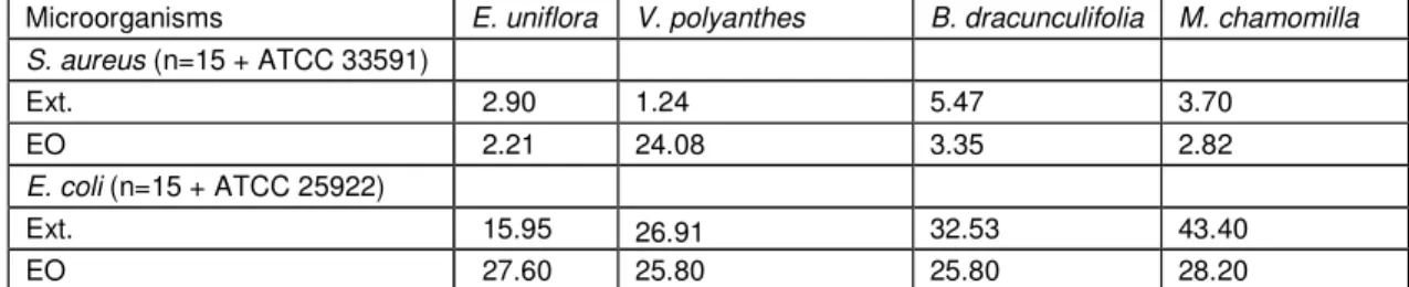
Table 2: Values of Minimal Inhibitory Concentration (MIC - mg/ml) for 90% (MIC90%) of S. aureus and E. coli
strains against extracts (Ext.) and essential oils (EO) from studied plants
Microorganisms E. uniflora V. polyanthes B. dracunculifolia M. chamomilla S. aureus (n=15 + ATCC 33591)
Ext. 2.90 1.24 5.47 3.70
EO 2.21 24.08 3.35 2.82
E. coli (n=15 + ATCC 25922)
Ext. 15.95 26.91 32.53 43.40
EO 27.60 25.80 25.80 28.20
The MIC90% values obtained for the natural products are essential for the assays on interactions between natural products and antimicrobial drugs; although the aim of this study is not to verify the antimicrobial potential
of each of these products, it is possible to infer that all of them were effective to control the growth of the studied bacteria, according to the obtained MIC90% values.
Table 3: Values of minimal inhibitory concentration (MIC - µg/ml) for the tested drugs against S. aureus and E. coli strains
Microorganisms CIP GEN TET CLO RIF CPM SUT
E. coli 1.024 256 0.128 1.024 0.064 64 1024
S. aureus 128 32 128 16 64 0.256 1024
CIP:Ciprofloxacin, GEN: Gentamicin, TET: Tetracycline, CLO: Chloramphenicol, RIF: Rifampicin, CPM: Cefepime, SUT: Sulfazotrin.
4.3 Synergism between antimicrobial products by disk test
In the assays performed to verify synergism between plant products and antimicrobial drugs using the disk methodology, ¼ of the respective MIC90% values were used in the plates that received the plant products, named
treatment, and synergism was established according to the statistical analysis and the comparison of the results obtained for each strain, totaling 10 for S. aureus and 10 for E. coli, in the antibiograms control and treatment. The obtained results are shown in Tables 4 for
S. aureus and E. coli.
Table 4: Results for synergism of essential oils (EO) and extracts (Ext.) with antimicrobial drugs against S. aureus and E. coli strains
Drugs B. dracunculifolia V. polyanthes M. chamomilla E. uniflora
E. coli S. aureus E. coli S. aureus E. coli S. aureus E. coli S. aureus
Chloramphenicol I A S I I I S I I A I I I I I I Rifampicin I I S I I I S I I I S S I I I I Synergism and antagonism when p<0, 05 (S - Synergism, A – Antagonism, I – Indifferent)
There was a noticeable difference for synergism frequency when both bacterial species were considered, whereas the absolute number of synergism cases between the several natural products and the antimicrobial drugs was 24 for S. aureus and 3 for E. coli. It must be emphasized that B.
dracunculifolia and V. polyanthes EO had
synergism with all drugs tested in the assays for S. aureus. On the other hand, there is a superior antagonism frequency (a total of 5 cases) against E. coli, M. chamomilla Ext. presenting a high incidence of antagonism. In addition, the cases of synergism against S. aureus occurred almost exclusively with EO; only two cases of synergism with M. chamomilla Ext. were found. For E. coli, however, there was 100% indifference between EO and antimicrobial drugs.
4.4 Synergism by time kill curve method
The time kill curve assays were performed in order to confirm only the synergism verified in the tested combinations by disk test previously. However, it must be highlighted that although there were cases of synergism with the drug cephalothin, such assays could not be done due to the limitation for obtaining the salt of this drug from suppliers.
For most interactions, the time kill curves showed a similar or even lower profile with the tested drug, and synergism was confirmed for several combinations through the reduction in the bacterial count, including the complete elimination of the bacterium in the culture medium.
According to S. aureus assays, the interactions were detected for sulfazotrim with
V. polyanthes and B. dracunculifolia EO, with a clear synergism capable of more efficiently reducing the microbial growth, compared to the drug tested alone. As shown in Figure 2, ciprofloxacin only had synergistic interaction with M. chamomilla EO, including a bactericidal effect. When combined with rifampicin, both M. chamomilla and V. polyanthes EO showed a synergistic effect. There was also synergism for the combinations of cefepime with V. polyanthes
and B. dracunculifolia EO. In interactions with the drugs chloramphenicol, gentamicin and tetracycline, bacterial growth was greater than that presented by the drugs alone.
When tested against E. coli, the interactions cefepime and V. polyanthes Ext. and cefepime and E. uniflora Ext. yielded a curve profile similar to that of the drugs alone, whereas the curve for the interaction gentamicin and V. polyanthes Ext. showed a bacterial growth rate higher than that of the drug alone.
In general, all curves for the interaction between natural products and drugs showed a bacterial growth profile inferior to those of control curves, i.e. assays without addition of any bacterial growth inhibitor.
5. Discussion
As indicated by Ext.dry weight and EO density (Table 1), there is a great difference of concentration between EO and Ext., the former being more concentrated. This is a possible justification for the higher activity of oils concerning synergism. However, this comparison is difficult since the extracts dry weight represents the solid portion, in solution or in suspension in the solvent, which in the case of extract is basically constituted of water due to the removal of methanol. Conversely, the essential oil represents almost the whole material, which in the case of density is considered all mass in 1 ml volume, for example.
As regards the MIC of natural products, E. uniflora and V. polyanthes Ext. were the most effective against S. aureus, but only M.
chamomilla Ext. had some synergism.
Considering EO, M. chamomilla and E. uniflora had higher inhibitory action against S.
aureus; however, V. polyanthes and B.
dracunculifolia had the highest synergism rate
through disk test, indicating that a higher antimicrobial capacity may not be reflected in a higher synergism rate. For combinations against E. coli, the synergism rate was low, but there were 2 cases of synergism with V. polyanthes Ext. which had high MIC and low antimicrobial action. Betoni et al., (2006) verified a similar result for lemongrass extract, which presented high MIC value; however this extract had the highest synergism rate together with the values obtained for clove extract.
method were promising against S. aureus
strains and the synergism rate for EO was considerable, on other words, 100% for B. dracunculifolia and V. polyanthes. On the other hand, Ext. did not show the same potential and there were only two cases of synergism against S. aureus although no case of antagonism was detected. As regards E. coli, its profile was opposite to that obtained
for S. aureus, since synergism was not
detected for any of the EO tested but was established for V. polyanthes and E. uniflora
Ext. It must be highlighted that M. chamomilla
had the highest antagonism rate (4 events). Thus, the type of plant derivative (EO or Ext.) influences the behavior of antimicrobial drugs, which shows that in such studies, phytochemical characterization is essential to better understand the phenomena observed in the assays of interaction between natural products and drugs.
Another important aspect is the higher sensitivity of Gram-positive bacteria relative to Gram-negative ones, which was somewhat translated into higher synergism rates for S.
aureus, totaling 24 cases of synergism,
compared to 3 cases for E. coli. A natural explanation for this phenomenon is the different structure of the bacterial wall of both bacterial groups. However, for essential oils, which are naturally liposoluble, a higher antimicrobial capacity was expected against E. coli since it has an external membrane rich in lipids, as well as a thin peptidoglycan, which would lead to a lower restriction for its entrance into the bacterial cell. Therefore, according to these observations, a higher efficacy was expected from natural products in the combined therapy with antimicrobials against infections by S. aureus. Studies with other Gram-positive microorganisms are
needed to extend the potential
recommendation of the combined therapy against other Gram-positive bacteria such as Streptococcus, Bacillus, etc.
This study also aimed confirms by the time kill curve the synergism detected previously by disk diffusion assays. Thus, similarly to some cases, the survival curves confirmed the synergism results verified through the disk test (V. polyanthes EO with sulfazotrim, rifampicin and cefepime; M. chamomilla EO with ciprofloxacin and rifampicin; and B.
dracunculifolia EO with sulfazotrim and
cefepime for S. aureus); moreover, in some cases the interactions had a growth profile higher than that of the drug alone, not confirming this type of synergism shown by the
disk test, including for example the interactions with chloramphenicol and tetracycline for which the growth curve of the latter was higher than or equal to that of any other drug. Another example of antagonism through the growth curve and synergism through the disk test is the interaction of rifampicin with M.
chamomilla Ext. and V. polyanthes EO. A
justification for curves that did not show the expected synergism is that previously obtained MIC values were used in the assays, which were different from those established through the Kirby & Bauer disks assays. However, it must be emphasized that even without an evident synergism all tested interactions showed a bacterial growth profile below that of control assays for bacterial growth in absence of any growth inhibitor.
Thus, similarly to reports about the antimicrobial action of natural products, the chosen methodology is essential to obtain conclusive results; besides, the preparation of plant derivatives, the tested microbial strains and the antimicrobial drugs tested in the synergism assays can also influence the obtained results.
Recent studies have also shown synergism between natural products and antimicrobial drugs; Betoni et al., (2006) verified the synergism between 8 plant extracts and 13 antimicrobial drugs and found an interesting incidence of synergism for the drugs acting on the synthesis of bacterial proteins. In the present study, the preference for this type of antimicrobial drug did not occur, and cases of antagonism were more frequent concerning E. coli and the drug chloramphenicol, totalizing two cases of antagonism. Similarly, cases of antagonism between natural products and drugs against the Gram-negative bacterium Klebsiella pneumoniae strains were reported by Van Vuuren et al., (2009). Thus, our results corroborate those obtained for E. coli, demonstrating the more frequent occurrence of cases of antagonism between natural products and antimicrobial drugs besides a higher resistance of Gram-negative bacteria.
Conclusion
bacteria, considering the aspects of similarity among species of this bacterial group. However, it must be highlighted that this study was done in vitro, indicating the need of future studies using in vivo models that will lead to conclusions closer to a real use of these plant products in association with conventional antibacterial drugs to establish potential cytotoxicity of compounds in the plant derivatives during its use as coadjuvant in the treatment of infectious diseases.
Author’s Contribution and Competing Interests
Silva, master's degree student, was responsible for preparing the research project under guidance of professor Fernandes Junior, therefore, responsible for the execution and writing of the manuscript of the study. Barbosa, Andrade and Probst are team members in our laboratory has participated in the implementation of microbiological assays, as well as collaborated in collecting plants and chemical characterization. Doyama, was responsible for carrying out the chemical characterization of essential oils by GC-MS
Ackonowledgments
The authors thank the Prof. Dr. Luciano Barbosa, Department of Biostatistics/ IBB/ UNESP for statistical analysis and Associate Professor Dr. Luiz Claudio Di Stasi for guidance of phytochemical analysis.
References
Adams R.P., 1989. Identification of Essential Oils by Ion Trap Mass Spectroscopy, Academic Press, INC., San Diego, California.
Aguiar J.S., Costa M.C.C.D., Nascimento S.C., Sena K.X.F.R., 2008. Antimicrobial activity of Lippia alba (Mill.) N. E. Brown (Verbenaceae). Brazilian Journal of Pharmacognosy, 18, 436-440.
Asolini F.C., Tedesco A.M., Carpes S.T., Ferraz C., Ferraz S.M., 2006. Antioxidant and antibacterial activities of phenolic compounds from extracts of plants used as tea. Brazilian Journal of Food Technology, 9, 209-215.
Bertucci A., Olivaro C., Silva P.A., Ramos D., Cerdeiras M.P., Vázquez A., 2009. Initial antimicrobial activity studies of plants of the riverside forests of the southern Uruguay River. Brazilian Journal of Pharmacognosy, 19, 20-25.
Betoni J.E.C, Mantovani R.P, Barbosa L.N, Di Stasi L.C, Fernandes Júnior A., 2006. Synergism between plant extract and antimicrobial drugs used
on Staphylococcus aureus diseases. Memórias do Instituto Oswaldo Cruz, 101, 387-390.
Candan F., Unlu M., Tepe B., Daferra D., Polissiou M., Sokmen A., Akpulat H.A., 2003. Antioxidant and antimicrobial activity of the essential oil and methanol extracts of Achillea millefolium subsp. millefolium Afan. (Asteraceae). Journal of Ethnopharmacology, 87, 215-220.
Clinical and Laboratory Standards Institute/National Comitee for clinical Laboratory Standards (CLSI/NCCLS). Performance standards for antimicrobial susceptibility testing; Fifteenth Information Supplement. CLSI/NCCLS document M 100-S15. Wayne, PA, 2005.
Costa A.C., Santos B.H.C., Santos Filho L., Lima E.O., 2009. Antibacterial activity of the essential oil of Origanum vulgare L. (Lamiaceae) against bacterial multiresistant strains isolated from nosocomial patients. Brazilian Journal of Pharmacognosy, 19, 236-241.
Fernandes Júnior A., Balestrin E.C., Betoni J.E.C., Orsi R.O., Cunha M.L.R.S., Montelli A.C., 2005. Propolis: anti-Staphylococcus aureus activity and synergism with antimicrobial drugs. Memórias do Instituto Oswaldo Cruz, 100, 563-566.
Ferronato R., Marchesan E.D., Pezenti E., Bednarski F., Onofre S.B., 2007. Antimicrobial activity of essential oils produced by Baccharis dracunculifolia D.C. and Baccharis uncinella D.C. (Asteraceae). Brazilian Journal of Pharmacognosy, 17, 224-330.
Fonseca P., Librand A.P.L., 2008. Evaluation of physico-chemical and phytochemical characteristics of different tinctures of barbatimão (Stryphnodendron barbatiman). Brazilian Journal of Pharmaceutical Science, 44, 271-277.
Funari C.S., Ferro V.O., 2005. Ethical use of the brazilian biodiversity: necessity and opportunity. Brazilian Journal of Pharmacognosy, 15,178-182.
Gallucci N., Casero C., Oliva M., Zygadlo J., Demo M., 2006. Interactions between terpenes and penicillin on bacterial strains resistant to beta lactam antibiotics. Molecular Medicinal Chemistry, 10, 30-32.
Gomathi S., Ambikapathy V., Panneerselvam A., 2013. Separation of bioactive compounds from
Trichoderma viride isolated from chilli field soil by using Gas Chromatography-Mass Spectrum, FTIR and UV. Journal of Antimicrobials. Photon, 128, 141-146.
Koneman E.W., Allen S.D., Janda N.M., Sherechkenberger P.C., Winn J.R., Atlas C., 2001. textbook of diagnostic microbiology, 5th ed., JB Lippincott, Philadelphia, 1395 p.
Sivakumar T., 2009. Synergistic activity of methanolic extract of Thespesia populnea
(Malvaceae) flowers with oxytetracycline. Bangladesh Journal of Pharmacolog, 4, 13-16.
Lima M.R.F., Ximenez E.C.P.A., Luna J.S., Sant’Ana A.E.G., 2006. The antibiotic activity of some Brazilian medicinal plants. Brazilian Journal of Pharmacognosy, 16, 300-306.
National Committee for clinical Laboratory Standards (NCCLS). Performance standards for antimicrobial disk susceptibility tests. Approved standard-8.ed. NCCLS document M2-A8. Wayne, PA, 2003.
Oliveira D.G., Prince K.A., Higuchi C.T., Santos A.C.B., Lopes L.M.X., 2007. Simões M.J.S., Leite C.Q.F. Antimycobacterial activity of some Brazilian indigenous medicinal drinks. Journal of Basic and Applied Pharmaceutical Sciences, 28, 165-169.
Rosato A., Vitali C., De Laurentis N., Armenise D., Milillo M.A., 2007. Antibacterial effect of some essential oils administered alone or in combination with Norfloxacin, Phytomedicine, 14, 727–732.
Sachin B.S., Sharma S.C., Sethi S., Tasduq S.A., Tikoo M.K., Tikoo A.K., Satti N.K., Gupta B.D., Suri K.A., Johri R.K., Qazi G.N., 2007. Herbal modulation of drug bioavailability: enhancement of rifampicin levels in plasma by herbal products and a flavonoid glycoside derived from Cuminum cyminum. Phytotherapy Research, 21, 157–163.
Silva M.T.N., Ushimaru P.I., Barbosa L.N., Cunha M.L.R.S., Fernandes Júnior A., 2009. Antibacterial activity of plant essential oils against
Staphylococcus aureus and Escherichia coli strains isolated from human specimens. Brazilian Journal of Medicinal Plants, 11, 257-262.
Silver L.L., Bostian K.A., 1993. Discovery and Development of New Antibiotics: the Problem of Antibiotic Resistance. Antimicrobial Agents and Chemotherapy, 37, 377-383.
Souza S.M.C., Pereira M.C., Angélico C.L., Pimenta C.J., 2004. Evaluation of condiments essential oils on micelial growth of fungi associated to bread-making products. Ciência e Agrotecnologia, 28, 685-690.