Prostaglandin E2 and Transforming Growth
Factor-
β
Play a Critical Role in Suppression of
Allergic Airway Inflammation by
Adipose-Derived Stem Cells
Kyu-Sup Cho1☯, Jung-Hoon Lee1☯, Mi-Kyung Park2, Hye-Kyung Park3, Hak-Sun Yu2, Hwan-Jung Roh4*
1Department of Otorhinolaryngology and Biomedical Research Institute, Pusan National University Hospital, Busan, Republic of Korea,2Department of Parasitology, Pusan National University School of Medicine, Yangsan, Republic of Korea,3Department of Internal Medicine, Pusan National University Hospital, Busan, South Korea,4Department of Otorhinolaryngology and Research Institute for Convergence of Biomedical Science and Technology, Pusan National University Yangsan Hospital, Yangsan, Republic of Korea
☯These authors contributed equally to this work. *rohhj@pusan.ac.kr
Abstract
Background
The role of soluble factors in the suppression of allergic airway inflammation by adipose-derived stem cells (ASCs) remains to be elucidated. Moreover, the major soluble factors responsible for the immunomodulatory effects of ASCs in allergic airway diseases have not been well documented. We evaluated the effects of ASCs on allergic inflammation in asth-matic mice treated with a prostaglandin E2 (PGE2) inhibitor or transforming growth factor-β (TGF-β) neutralizing antibodies.
Methods and Findings
Asthmatic mice were injected intraperitoneally with a PGE2 inhibitor or TGF-βneutralizing antibodies at approximately the same time as ASCs injection and were compared with non-treated controls. In asthmatic mice, ASCs significantly reduced airway hyperresponsive-ness, the number of total inflammatory cells and eosinophils in the bronchoalveolar lavage fluid (BALF), eosinophilic inflammation, goblet cell hyperplasia, and serum total and aller-gen-specific IgE and IgG1. ASCs significantly inhibited Th2 cytokines, such as interleukin (IL)-4, IL-5, and IL-13, and enhanced the Th1 cytokine (Interferon-γ) and regulatory cyto-kines (IL-10 and TGF-β) in the BALF and lung draining lymph nodes (LLNs). ASCs engraftment caused significant increases in the regulatory T cell (Treg) and IL-10+T cell populations in LLNs. However, blocking PGE2 or TGF-βeliminated the immunosuppressive effect of ASCs in allergic airway inflammation.
OPEN ACCESS
Citation:Cho K-S, Lee J-H, Park M-K, Park H-K, Yu H-S, Roh H-J (2015) Prostaglandin E2 and Transforming Growth Factor-βPlay a Critical Role in Suppression of Allergic Airway Inflammation by Adipose-Derived Stem Cells. PLoS ONE 10(7): e0131813. doi:10.1371/journal.pone.0131813
Editor:Joao P.B. Viola, National Cancer Institute (INCA), BRAZIL
Received:February 10, 2015
Accepted:June 7, 2015
Published:July 15, 2015
Copyright:© 2015 Cho et al. This is an open access article distributed under the terms of theCreative Commons Attribution License, which permits unrestricted use, distribution, and reproduction in any medium, provided the original author and source are credited.
Data Availability Statement:All relevant data are within the paper.
Funding:This study was supported by a grant of the Korean Health Technology R&D Project, Ministry of Health & Welfare, Republic of Korea (HI12C0315). The funders had no role in study design, data collection and analysis, decision to publish, or preparation of the manuscript.
Conclusions
ASCs are capable of secreting PGE2 and TGF-β, which may play a role in inducing Treg expansion. Furthermore, treatment with a PGE2 inhibitor or TGF-βneutralizing antibodies eliminated the beneficial effect of ASCs treatment in asthmatic mice, suggesting that PGE2 and TGF-βare the major soluble factors responsible for suppressing allergic airway inflammation.
Introduction
Asthma is a chronic inflammatory airway disease affecting more than 300 million people worldwide [1]. It is characterized by Th2-mediated eosinophilic inflammation, mucus hyperse-cretion, and airway hyperresponsiveness (AHR) [1,2]. Excessive activation of Th2 cells is thought to play a major role in the initiation and development of the disease [3]. There is mounting evidence that insufficient suppression of regulatory T cells (Tregs) is responsible for the excessive Th2 response in allergic airway disease [4,5].
Mesenchymal stem cells (MSCs) are ubiquitous multipotent cells abundant in adult bone marrow (BM) and adipose tissue [6,7]. In addition to multi-lineage differentiation potential, MSCs derived from adipose tissue (ASCs) and other MSCs have the unique ability to suppress immune responses and modulate inflammation [8]. Several studies have demonstrated that MSCs can ameliorate allergic airway inflammatory diseases, including asthma [9–11] and aller-gic rhinitis [12–15]. The immunomodulatory effects of MSCs in allergic airway diseases may be mediated by the upregulation of Tregs and increases in several soluble factors such as indo-leamine 2, 3-dioxygenase (IDO), prostaglandin E2 (PGE2), transforming growth factor-β
(TGF-β), and interleukin (IL)-10 [16–19]. However, the role of these soluble factors in the sup-pression of allergic airway inflammation by MSCs remains to be elucidated, and the major sol-uble factors responsible for the immunomodulatory effects of MSCs in allergic airway diseases have not been well documented.
The purpose of this study was to determine whether PGE2 or TGF-βcontributes to the immunomodulatory effects of ASCs in asthmatic mice by evaluating the effects of a PGE2 inhibitor or TGF-β-specific neutralizing antibody (Ab) on allergic inflammation.
Materials and Methods
Animals
Five-week-old female C57BL/6 mice were purchased from Samtako Co. (Osan, Republic of Korea,http://www.samtako.co.kr) and bred in a specific pathogen free animal facility. The ani-mal study protocol was approved by the Institutional Aniani-mal Care and Use Committee of the Pusan National University School of Medicine.
Isolation and culture of ASCs
filtered through a 100-μm nylon mesh to remove cellular debris and then incubated overnight
at 37°C with 5% CO2in control medium (α-MEM, 10% FBS, 100 unit/ml penicillin, 100μg/ml
streptomycin). Following incubation, the plates were washed extensively with PBS to remove residual non-adherent red blood cells. The resulting cell population was maintained at 37°C with 5% CO2in control medium. One week later, once the monolayer of adherent cells had
reached confluence, cells were trypsinized (0.05% trypsin-EDTA; Sigma), resuspended inα -MEM containing 10% FBS, and subcultured at the concentration of 2,000 cells/cm3. For the experiments, third- or fourth-passage ASCs was used.
Flow cytometric analysis was used to characterize the phenotype of ASCs. At least 50,000 cells (in 100μl PBS, 0.5% bovine serum albumin (BSA), 2 mmol/l EDTA) were incubated with
fluorescein isothiocyanate-labeled monoclonal Abs against mouse stem cell antigen-1 (Sca-1), CD44, CD90, CD45, CD117, and CD11b (BD Biosciences Clontech, Palo Alto, CA) or with the respective isotype control. After washing, labeled cells were analyzed by flow cytometry using a FACSCalibur flow cytometer and Cell Quest Pro software (BD Biosciences, San Diego, CA). The expression percentage of each marker on ASCs was determined by the percentage of posi-tive events, as determined by the isotype-matched negaposi-tive control.
ACSs were analyzed for their capacity to differentiate into adipogenic, osteogenic, and chon-drogenic lineages, as described previously [20]. For adipogenic and osteogenic differentiation, cells were seeded in 6-well plates at a density of 20,000 cells/cm2and treated for 3 weeks with adipogenic and osteogenic medium. Adipogenic and osteogenic differentiation was assessed using oil red O staining, as an indicator of intracellular lipid accumulation, and alizarin red S staining, as an indicator of extracellular matrix calcification, respectively. Chondrogenic differ-entiation was induced using the micromass culture technique. Briefly, 10 ml of a concentrated ASC suspension (3 × 10⁵cells/ml) were plated in the center of each well and treated for 3 weeks with chondrogenic medium. Chondrogenesis was confirmed by immunohistochemistry.
Mouse model of allergic airway inflammation
A mouse model of allergic airway inflammation was induced as previously reported with minor modification [16,21]. Briefly, mice were sensitized by intraperitoneal injection of 75μg
of ovalbumin (OVA, Sigma, St. Louis, MO,http://www.sigmaaldrich.com) with 2 mg alumi-num hydroxide (Sigma) in 200μl PBS on days 0, 1, 7, and 8. On days 14, 15, 21, and 22 after
the initial sensitization, the mice were challenged intranasally with 50μg OVA in 50μl PBS
(Fig 1A).
Intravenous transplantation of ASCs and intraperitoneal injection of
PGE2 inhibitor and anti-TGF-
β
Ab
ASCs were washed with PBS and suspended in PBS at a concentration of 1 × 10⁷cells/ml. To evaluate the effect of ASCs, 0.1 ml purified stem cells were injected with a 26-gauge needle via the tail vein of asthmatic mice once a day on days 12, 13, 19, and 20.
PGE2 or TGF-βwere blocked by intraperitoneal injection of a PGE2 inhibitor (1μg/g body
weight in 200μL PBS) (Cayman Chemical, Ann Arbor, MI) (catalog# 10010088; CAY10526;
C12H7BrO3S) or anti-TGF-βAb (10μg/g body weight in 200μL PBS) (R&D Systems,
Minne-apolis, MN) (catalog# MAB1835; TGF-β1, 2, 3 monoclonal Ab (Clone 1D11), mouse IgG1) on days 13, 14, 15, 16, 17, 20, 21, 22, and 23, respectively (Fig 1A).
Mice were divided into five groups with five mice per group: (a) a PBS group sensitized, pre-treated, and challenged with PBS; (b) a PBS+PGE2 inhibitor group or PBS+TGF-βAb group sensitized and pretreated with PBS followed by treatment with a PGE2 inhibitor or anti-TGF-β
pretreated with PBS, and then challenged with OVA; (d) an OVA+ASC group sensitized with OVA, pretreated with ASCs, and then challenged with OVA; (e) an OVA+ASC+PGE2 inhibi-tor group or OVA+ASC+TGF-βAb group sensitized with OVA, pretreated with ASCs, treated with the PGE2 inhibitor or anti-TGF-βAb, respectively, and then challenged with OVA. These experiments were repeated four times (Fig 1B).
Measurement of methacholine AHR
Twenty-four hours after the last challenge, AHR was assessed in conscious, unrestrained mice using non-invasive whole-body plethysmography (Allmedicus, Seoul, Republic of Korea), as described previously [22]. In brief, the mice were placed in the plethysmography chamber and exposed to increasing concentrations of aerosolized methacholine at 0, 12.5, 25 and 50 mg/ml for 10 min. Enhanced pause (Penh) was calculated automatically based on the mean pressure generated in the plethysmography chamber during inspiration and expiration combined with the time of each phase. The Penh values calculated during each 3-min interval were then averaged.
Fig 1. The experimental protocol.(A) Mice were sensitized on days 0, 1, 7, and 8 by intraperitoneal injection of ovalbumin (OVA) and challenged intranasally on days 14, 15, 21, and 22. Purified adipose-derived stem cells (ASCs; 1 × 10⁶) were injected via the tail vein on days 12, 13, 19, and 20. PGE2
and TGF-βwere blocked by intraperitoneal injection of a PGE2 inhibitor or anti-TGF-β-Ab on days 13, 14, 15, 16, 17, 20, 21, 22, and 23. (B) The mice were divided into five treatment groups.
Differential cell counting in bronchoalveolar lavage fluid
To obtain bronchoalveolar lavage fluid (BALF), the tracheas of anesthetized mice were exposed and cut just below the larynx. A polyurethane flexible tube (0.4 mm in outer diameter, 4 cm in length, and attached to a blunt 24-gauge needle (Boin Medical Co., Seoul, Republic of Korea)) was placed into the trachea, after which the lung was lavaged once with 800 l warm sterile PBS. The BALF samples were centrifuged for 5 min at 1,500 rpm at 4°C. The supernatants were then decanted and frozen immediately at -70°C. Cell pellets were resuspended and washed twice in PBS. The total cell numbers were counted using a hemocytometer. BALF cell smears were pre-pared using a cytospin apparatus, and stained with Diff-Quik solution (Sysmex Co., Kobe, Japan) to determine the differential cells counts in accordance with conventional morphologi-cal criteria. At least 500 cells per slide were evaluated to obtain the differential leukocyte counts.
Lung histology and inflammation scoring
Lung tissues were removed after the lavage, fixed in 10% neutral formalin for 36 h, and embed-ded in paraffin. The thin sections of the embedding tissues were stained with hematoxylin and eosin (H&E) and periodic acid-Schiff (PAS) for the identification of eosinophils and for count-ing mucin-secretcount-ing cells, respectively. Lung inflammation was assessed by the degree of peri-bronchial and perivascular inflammation, which were evaluated on a subjective scale of 0–4, as described previously, [23,24], using the following inflammatory parameters: 0 when no inflam-mation was detectable; 1 for occasional cuffing with inflammatory cells; 2 when most bronchi or vessels were surrounded by a depth of one to three cells; 3 when most bronchi or vessels were surrounded by a depth of four to five cells; 4 when most bronchi or vessels were sur-rounded by a depth of more than five cells. For quantifying goblet cell hyperplasia, the percent-age of PAS-positive cells in epithelial areas was examined from 8–10 tissue sections per mouse.
Measurement of serum immunoglobulin
At 48 h after the last OVA challenge, serum was collected from mice via cardiac puncture. Total and OVA-specific immunoglobulins (Ig E, IgG1, IgG2a) were determined by enzyme-linked immunosorbent assay (ELISA) in accordance with the manufacturer’s instructions (R&D Systems, Minneapolis, MN). Absorbance at 450 nm was measured using an ELISA plate reader (Molecular Devices, Sunnyvale, CA).
Expression of cytokines in the BALF and lung draining lymph nodes
Lung draining lymph nodes (LLNs) were obtained between trachea and both lung lobes. The obtained LLNs were treated with ACK hypotonic lysis buffer (0.15 M NH4Cl, 1mM KHCO3,
0.1mM Na2-EDTA, pH 7.2–7.4) for 2 min at room temperature for lysis of red blood cells
(RBCs). After the RBCs were lysed, the remaining cells were filtered using 100-μm mesh (Small
Parts Inc., Miramar, FL), and 106cells/ml were plated in 48 well plates coated with 0.5μg/ml
CD3 Ab (BD Biosciences, San Diego, CA) in RPMI 1640 with 10% fetal bovine serum (FBS) and penicillin/streptomycin. Plated cells were incubated for 72 h at 37°C with 5% CO2. After
Determination of Tregs and intracellular cytokine staining
To evaluate the recruitment of Th1, Th2, and Tregs induced by ASCs treatment, LLN cells from OVA-induced asthmatic mice and ASCs-treated asthmatic mice were cultured in anti-CD3-coated plate for 6 h. To evaluate To evaluate CD4+CD25+Foxp3+(Tregs) and IL-10+/ CD4+ T cells, cells were stained with anti-CD4-FITC (0.5 mg/ml) and anti-CD25-APC (0.2 mg/ml) in accordance with the manufacturer’s recommendations (eBiosciences, San Diego, CA). After surface staining, the cells were permeabilized using a Cytofix/Cytoperm Kit (eBios-ciences). After permeabilization, the cells were stained with anti-Foxp3-PE-cy7 or anti-IL-10-PE (eBiosciences).
To assess the Th1 and Th2 cell population, LLNs cells were stained with an anti-CD4-FITC Ab. After surface staining, the CD4+ T cells were stained with intracellular anti-IFN-γ-PE-cy7 (eBiosciences) and anti-IL-4-PE (eBiosciences) Abs. Fluorescence was measured using a FACS CantoII cytometer (BD Biosciences) equipped with Canto software (BD Biosciences).
Statistical analysis
All experiments were repeated a minimum of three times. Data are expressed as mean ± standard error of the mean (SEM). Statistical significance was assessed by the Student’sttest or one-way analysis of variance (ANOVA) using the SPSS software package version 18.0 (SPSS Inc., Chicago, IL). A p value<0.05 was considered statistically significant.
Results
Characterization of ASC immunophenotype and differentiation
The cultured ASCs from adipose tissue of C57BL/6 mice were negative for the cell surface markers CD45, CD117, and CD11b but positive for Sca-1, CD44, and CD90. These putative ASCs had a spindle shaped fibroblast-like appearance, similar to previously reported adipose tissue and bone marrow-derived MSCs. ASCs had the ability to differentiate into adipogenic, osteogenic, and chondrogenic lineages after culture in the appropriate conditions (data not shown)
AHR and inflammatory cells in BALF
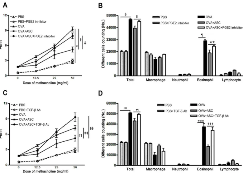
Penh in asthmatic mice increased with the methacholine concentration, and ASC treatment significantly decreased AHR in asthmatic mice. However, treatment with the PGE2 inhibitor or TGF-β-specific neutralizing Ab significantly increased AHR in the OVA+ASC group (p= 0.032 andp= 0.005, respectively) (Fig 2A and 2C).
The numbers of total inflammatory cells and eosinophils were significantly increased in the BALF of the OVA group compared with the PBS group. However, ASC treatment significantly decreased the numbers of total inflammatory cells and eosinophils in asthmatic mice. However, inhibition of PGE2 production or neutralization of TGF-βfailed to reduce BALF total cell numbers (p= 0.045 andp= 0.005, respectively) and eosinophil counts (p= 0.027 andp<0.001,
respectively) (Fig 2B and 2D).
Lung inflammation and goblet cell hyperplasia
asthmatic mice treated with ASCs. Furthermore, ASC administration caused a significant reduction in the inflammation score and PAS-positive cells in asthmatic mice.
When mice were treated with a PGE2 inhibitor or TGF-β-specific neutralizing Ab, lung inflammation and goblet cell hyperplasia increased in the OVA+ASC+PGE2 inhibitor and OVA+ASC+TGF-βAb groups compared with the OVA+ASC group. In addition, blocking PGE2 or TGF-βsignificantly prevented the inflammation score (p= 0.024 andp= 0.035, respectively) and PAS-positive cell reductions (p= 0.003 andp =0.005, respectively) (Fig 3A
and 3B) seen in non-treated animals.
Serum total and OVA-specific IgE, IgG1, and IgG2a
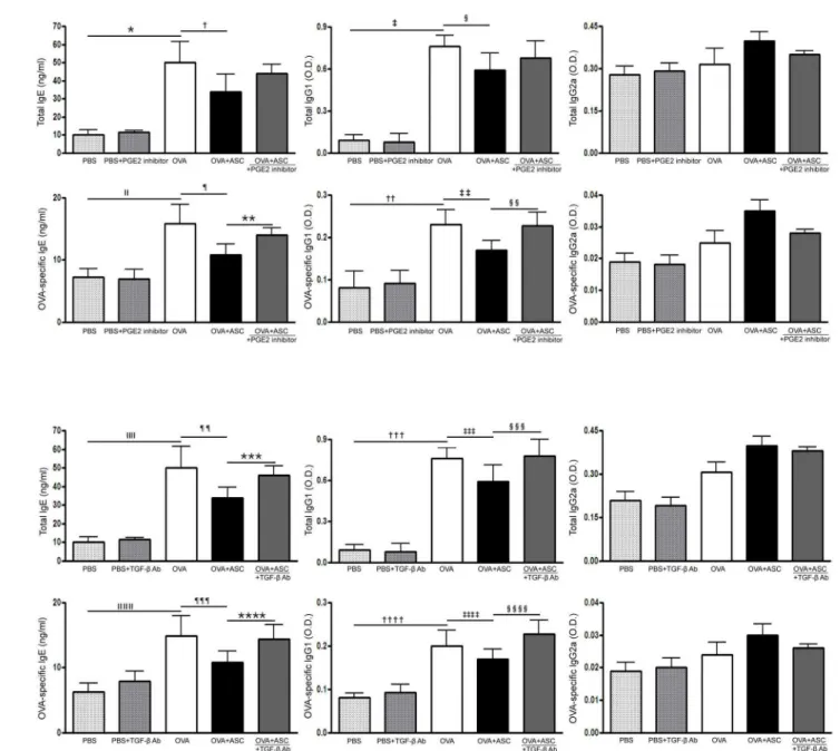
Total and OVA-specific IgE and IgG1 levels were significantly higher in the OVA group than in the PBS group of asthmatic mice. However, systemic administration of ASCs significantly decreased total IgE and OVA-specific IgE in asthmatic mice.
Fig 2. Effect of adipose-derived stem cells (ASCs) on airway hyperresponsiveness (AHR) and inflammatory cells in the bronchoalveolar lavage fluid (BALF).ASCs significantly decreased AHR and the number of total inflammatory cells and eosinophils in asthmatic mice. Treatment with the PGE2 inhibitor or TGF-βneutralizing Ab eliminated the reduction in AHR (A, C) and total cell and eosinophil counts (B, D) induced by ASC treatment. Data are expressed as the mean±SEM of four independent experiments each performed in triplicate.*,†,§,¶,††,ǁǁ,***,†††p<0.001,‡p =0.032,ǁp= 0.045,**
p= 0.027,‡‡p= 0.032, §§p= 0.005, ¶¶p= 0.005.
The PGE2 inhibitor significantly increased OVA-specific IgE and IgG1 levels in the OVA +ASC group (allp= 0.009), but not total IgE and IgG1 levels (Fig 4A). Moreover, neutraliza-tion of TGF-βresulted in a significant increase in total IgE and IgG1 (p= 0.028 andp= 0.037, respectively) and OVA-specific IgE and IgG1 levels (p= 0.035 andp= 0.032, respectively) in the OVA+ASC group (Fig 4B).
Cytokine profiles in the BALF and LLN
OVA-challenged mice showed significantly increased levels of IL-4, IL-5, and IL-13 in BALF. However, ASC treatment significantly decreased IL-4, IL-5, and IL-13 in the BALF and LLN of
Fig 3. Effects of adipose-derived stem cells (ASCs) on lung inflammation and goblet cell hyperplasia.ASCs treatment decreased the infiltration of eosinophils and PAS-positive cells around the airway and blood vessel in asthmatic mice (H&E, PAS ×200). Blocking PGE2 (A) or TGF-β(B) eliminated the beneficial effect of ASCs on lung inflammation and goblet cell hyperplasia. Data are expressed as the mean±SEM of four independent experiments each performed in triplicate.*,§,ǁ,**,§§,ǁǁp<0.001,†p =0.020,‡p= 0.024, ¶p= 0.003,††p =0.030,‡‡p= 0.035, ¶¶p= 0.005.
the asthmatic mice. In contrast, ASC treatment significantly increased IFN-γ, IL-10, and
TGF-βlevels in the BALF and LLN of the asthmatic mice.
The PGE2 inhibitor significantly increased IL-4 (p= 0.027 andp= 0.003, respectively), IL-5 (p<0.001 andp= 0.048, respectively), and IL-13 (p= 0.030 andp= 0.035, respectively) levels
in the BALF and LLN of the OVA+ASC group. However, IFN-γ(p= 0.022 andp= 0.009, respectively), IL-10 (p= 0.003 andp= 0.038, respectively), TGF-β(allp<0.0010) levels were
significantly decreased after inhibition of PGE2 production in the BALF and LLN of the OVA
Fig 4. Effect of adipose-derived stem cells (ASCs) on serum levels of immunoglobulin.Systemic administration of ASCs resulted in a significant decrease in total and OVA-specific IgE in asthmatic mice. (A) A PGE2 inhibitor significantly increased OVA-specific IgE and IgG1 in the OVA+ASC group. (B) TGF-βneutralizing Ab resulted in significant increases in total IgE and IgG1 and OVA-specific IgE and IgG1 in the OVA+ASC group. Data are expressed as the mean±SEM of four independent experiments each performed in triplicate.*,‡,,ǁ,††,,ǁ,ǁ,†††,,ǁǁǁ,††††p<0.001,†,***p =0.028, §p =0.038, ¶,‡‡,¶¶
+ASC group (Figs5Aand6A). In addition, TGF-βneutralizing Ab significantly increased IL-4 (p= 0.028 andp= 0.007, respectively), IL-5 (p =0.004 andp= 0.009, respectively), and IL-13 (allp= 0.029) levels in the BALF and LLN of the OVA+ASC group. However, IFN-γ(p= 0.012 andp= 0.003, respectively), IL-10 (p= 0.046 andp= 0.038, respectively), TGF-β(p =0.007 andp= 0.008, respectively) were significantly decreased after neutralization of TGF-βin the BALF and LLN of the OVA+ASC group (Figs5Band6B).
Fig 5. Effect of adipose-derived stem cells (ASCs) on cytokine levels in the bronchoalveolar lavage fluid.IL-4, IL-5, and IL-13 were significantly higher in the OVA group than PBS group. ASC treatment significantly decreased IL-4, IL-5, and IL-13 but increased IL-10 and TGF—βin asthmatic mice. However, the PGE2 inhibitor (A) or TGF-βneutralizing Ab (B) eliminated these immunomodulatory effects of ASCs. Data are expressed as the mean±SEM of four
independent experiments each performed in triplicate.*,§,ǁ,¶,**,§§§,§§§§, ¶¶¶¶¶p<0.001,†,******p= 0.007,‡p= 0.027,††,¶¶¶p= 0.028,‡‡p =0.030,
§§p =0.010,ǁǁp= 0.022, ¶¶p= 0.032,***,‡‡‡p= 0.038,†††p =0.049,ǁǁǁ,††††p= 0.003,****,‡‡‡‡p =0.004,ǁǁǁǁǁp =0.026, ¶¶¶¶p= 0.029,*****
p= 0.006,†††††p= 0.012,‡‡‡‡‡p= 0.036, §§§§§p =0.042,ǁǁǁǁǁp =0.046.
T cell populations in the LLN
The populations of CD4+CD25+Foxp3+T cells and CD4+IL-10+T cells were markedly increased by administration of ASCs in asthmatic mice. In the OVA+ASC group, CD4+IL-4+T cells were significantly decreased and CD4+IFN-γ+T cells were significantly increased compared with the OVA group. Treatment with the PGE2 inhibitor or TGF-βneutralizing Ab prevented the
Fig 6. Effect of adipose-derived stem cells (ASCs) on cytokine levels in the lung draining lymph nodes.ASCs treatment significantly decreased IL-4, IL-5, and IL-13 levels but increased IFN-γ, IL-10 and TGF—βlevels in asthmatic mice. However, PGE2 inhibitor (A) or TGF-βneutralizing Ab (B) eliminated these immunomodulatory effects of ASCs. Data are expressed as the mean±SEM of four independent experiments each performed in triplicate.*,***
p<0.001,†,††††p =0.003,‡,§,ǁǁǁǁp =0.048,ǁp =0.037, ¶p =0.035,**,****p =0.004,††,§§§p =0.009,‡‡p =0.032, §§,¶¶p= 0.038,ǁǁp =0.006,‡‡‡ p =0.007,‡‡‡p =0.002,ǁǁǁp =0.040, ¶¶¶p =0.029,‡‡‡p =0.049, §§§p =0.020, ¶¶¶¶p =0.008.
increases in CD4+CD25+Foxp3+, CD4+IL-10+, and CD4+IFN-γ+T cell populations and the decrease in the CD4+IL-4+T cell population in the OVA+ASC group (Fig 7A and 7B).
Discussion
MSCs possess remarkable immunosuppressive properties and can inhibit the proliferation and function of major immune cell populations, including T cells, B cells, and natural killer cells
Fig 7. Effects of adipose-derived stem cells (ASCs) on T cells in the lung draining lymph nodes.The CD4+T cells were initially gated and the
percentage of IFN-γ+, IL-4+, IL-10+, and CD25+Foxp3+T cells subsequently analyzed. When treating asthmatic mice with PGE2 inhibitor (A) or TGF-β neutralizing Abs (B), blocking of PGE2 and TGF-βprevented the increases in Foxp3+CD25+, IL-10+, and IFN-γ+T cell populations and the decrease in the
IL-4+T cell population in the OVA+ASC group.
[25,26]. MSCs can also modulate the activities of dendritic cells and induce Tregsin vivoand
in vitro[27,28]. The immunomodulatory function of MSCs has led to increasing interest in using MSCs as promising candidates for the treatment of allergic airway diseases. Several stud-ies have shown that ASC treatment provides a significant reduction in allergic airway inflam-mation and improved lung function [9–15]. Although the immunomodulatory mechanism of MSCs in allergic airway diseases remains to be elucidated, it has been suggested that the induc-tion and expansion of alveolar macrophages and Tregs play a role in alleviating allergic airway inflammation, while several soluble factors play a role in Tregs differentiation [16–19,29].
Alveolar macrophages are the predominant immune effector cells at the air-tissue interface in the lungs [30]. Human MSCs cause a pronounced increase in alveolar macrophages, and selective depletion of this macrophage compartment reversed the therapeutic benefit of human MSC treatment on allergic asthma, indicating a critical role for alveolar macrophages in sup-pressing allergic asthmain vivo[29]. Tregs are a unique T cell population with strong immuno-suppressive properties, and CD4+CD25+Tregs are impaired quantitatively and functionally in allergic airway diseases [31]. CD4+CD25+T cells also play an important role in suppressing air-way eosinophilic inflammation and in the development of airair-way hyperreactivity in asthma [17,31]. The induction of Tregs by MSCs involves not only direct contact between MSCs and CD4+T cells, but also the secretion of soluble factors such as IDO, PGE2, and TGF-β[32]. To our knowledge, this is the first study to investigate the potential role of PGE2 and TGF-βin the modulation of the allergic response through the use of antagonist and neutralizing Abs.
The present study demonstrated that intravenous treatment of ASCs in asthmatic mice pro-vides a significant reduction in allergic airway inflammation and an improvement in lung func-tion. The ratio of Tregs in LLNs was higher in the ASC treatment group than in the asthmatic group, which was similar to previous studies that indicated ASCs preferentially activate CD4+ CD25+T cells subsets, which are the primary mechanisms for the immunosuppressive activity of ASCs [16,17]. Moreover, ASC treatment increased anti-inflammatory cytokines, IL-10 and TGF-β, in the BALF and LLNs, which ultimately led to decreased lung eosinophil infiltration, goblet cell hyperplasia, allergy-specific Th2 cytokines, and Ig production.
Recent studies have suggested a broad role for PGE2 and TGF-β1 in the generation and expansion of Tregs from CD4+CD25-precursors [19,32,33]. PGE2 is the cytokine responsible for lymphocyte and Treg expansion [33], while TGF-βis a key regulator of the signaling path-ways that initiate and maintain Foxp3 expression and suppressive function in CD4+CD25- pre-cursors [34]. Interestingly, human MSCs constitutively express PGE2 and TGF-β1, which are both significantly upregulated by inflammatory mediators [32,35]. Co-culturing T cells with MSCs resulted in elevated levels of PGE2, and treatment with inhibitors of PGE2 production mitigated MSC-mediated immune modulation [36,37]. In this study, a PGE2 synthesis inhibi-tor abolished the suppressive effect induced by ASCs in asthmatic mice. Together with the secretion of PGE2 by ASCs [37], these findings strongly indicate that PGE2 is involved in the suppression of allergic airway inflammation mediated by ASCs. Nemethet al. noted that neu-tralizing Abs against TGF-βand BM-MSCs derived from TGF-β1-KO mice eliminated the beneficial effect of BM-MSCs in a mouse model of ragweed-induced asthma, suggesting that BM-MSCs-derived TGF-β1 was responsible for the immunosuppressive effect in asthmatic mice [19]. The present study supported those findings and clarified an important role of
decreased induction of Treg expansion. These results indicate that although the cell contact mechanism might be involved in ASC induction of CD4+CD25+Foxp3+T cells, soluble factors such as PGE2 and TGF-βplay a major role in this process. Furthermore, the present study cla-rifies the mechanism of ASC-mediated immunosuppression in allergic airway diseases, sup-porting ASC-derived PGE2 and TGF-βas important molecules involved in the induction of Tregs.
We acknowledge the limitations of our study. For example, although the PGE2 inhibitor or TGF-β-neutralizing Ab may enhance allergic inflammation, we did not include a treatment group sensitized with OVA, treated with PGE2 inhibitor or anti-TGF-βAb, and then chal-lenged with OVA. To clarify that ASC-derived PGE2 and TGF-βare important mediators in suppression of allergic airway inflammation by ASCs, it will be of value to inject ASCs derived from microsomal PGE2 synthase-1 or TGF-β-deficient mice.
Conclusions
ASCs are capable of secreting PGE2 and TGF-β, which play a role in inducing Treg expansion. Furthermore, inhibition of PGE2 or TGF-β-specific neutralizing Abs eliminated the beneficial effect of ASC treatment in asthmatic mice, suggesting that PGE2 and TGF-βare the major sol-uble factors involved in suppressing allergic airway inflammation.
Author Contributions
Conceived and designed the experiments: HY HR. Performed the experiments: KC JL MP. Analyzed the data: KC JL HP HY HR. Contributed reagents/materials/analysis tools: KC JL MP. Wrote the paper: KC JL.
References
1. Bousquet J, Khaltaev N, Cruz AA, Denburg J, Fokkens WJ, Togias A, et al. Allergic Rhinitis and its Impact on Asthma (ARIA) 2008 update (in collaboration with the World Health Organization, GA(2)Len and Allergen) Allergy. 2008; 63: 8–160. doi:10.1111/j.1398-9995.2007.01620.xPMID:18331513
2. Cohn L, Elias JA, Chupp GL. Asthma: mechanisms of disease persistence and progression. Annu Rev Immunol. 2004; 22: 789–815. PMID:15032597
3. Wilson MS, Taylor MD, Balic A, Finney CA, Lamb JR, Maizels RM. Suppression of allergic airway inflammation by helminth-induced regulatory T cells. J Exp Med. 2005; 202: 1199–1212. PMID:
16275759
4. Shi HZ, Qin XJ. CD4CD25 regulatory T lymphocytes in allergy and asthma. Allergy. 2005; 60: 986–
995. PMID:15969678
5. Jaffar Z, Sivakuru T, Roberts K. CD4+CD25+ T cells regulate airway eosinophilic inflammation by mod-ulating the Th2 cell phenotype. J Immunol. 2004; 172: 3842–3849. PMID:15004191
6. Bonfield TL, Nolan Koloze MT, Lennon DP, Caplan AI. Defining human mesenchymal stem cell efficacy in vivo. J Inflam (Lond). 2010; 7: 51.
7. Uccelli A, Pistoia V, Moretta L. Mesenchymal stem cells: a new strategy for immunosuprression? Trends Immunol. 2007; 28: 219–226. PMID:17400510
8. Uccelli A, Moretta L, Pistoia V. Immunoregulatory function of mesenchymal stem cells. Eur J Immunol. 2006; 36: 2566–2573. PMID:17013987
9. Park HK, Cho KS, Park HY, Shin DH, Kim YK, Jung JS, et al. Adipose-derived stromal cells inhibit aller-gic airway inflammation in mice. Stem Cells Dev. 2010; 19: 1811–1818. doi:10.1089/scd.2009.0513
PMID:20225940
10. Goodwin M, Sueblinvong V, Eisenhauer P, Ziats NP, LeClair L, Poynter ME, et al. Bone marrow-derived mesenchymal stromal cells inhibit Th2-mediated allergic airways inflammation in mice. Stem Cells. 2011; 29: 1137–1148. doi:10.1002/stem.656PMID:21544902
12. Fu QL, Chow YY, Sun SJ, Zeng QX, Li HB, Shi JB, et al. Mesenchymal stem cells derived from human induced pluripotent stem cells modulate T-cell phenotypes in allergic rhinitis. Allergy. 2012; 67: 1215–
1222. doi:10.1111/j.1398-9995.2012.02875.xPMID:22882409
13. Cho KS, Park HK, Park HY, Jung JS, Jeon SG, Kim YK, et al. IFATS collection: Immunomodulatory effects of adipose tissue-derived stem cells in an allergic rhinitis mouse model. Stem Cells. 2009; 27: 259–265. doi:10.1634/stemcells.2008-0283PMID:18832595
14. Sun YQ, Deng MX, He J, Zeng QX, Wen W, Wong DS, et al. Human pluripotent stem cell-derived mes-enchymal stem cells prevent allergic airway inflammation in mice. Stem Cells. 2012; 30: 2692–2699. doi:10.1002/stem.1241PMID:22987325
15. Cho KS, Roh HJ. Immunomodulatory effects of adipose-derived stem cells in airway allergic diseases. Curr Stem Cell Res Ther. 2010; 5: 111–115. PMID:19941459
16. Cho KS, Park MK, Kang SA, Park HY, Hong SL, Park HK, et al. Adipose-derived stem cells ameliorate allergic airway inflammation by inducing regulatory T cells in a mouse model of asthma. Mediators Inflamm. 2014; 2014: 436476. doi:10.1155/2014/436476PMID:25246732
17. Ge X, Bai C, Yang J, Lou G, Li Q, Chen R. Intratracheal transplantation of bone marrow-derived mesen-chymal stem cells reduced airway inflammation and up-regulated CD4+CD25+regulatory T cells in asthmatic mouse. Cell Biol Int. 2013; 37: 675–686. doi:10.1002/cbin.10084PMID:23483727
18. Fu QL, Chow YY, Sun SJ, Zeng QX, Li HB, Shi JB, et al. Mesenchymal stem cells derived from human induced pluripotent stem cells modulate T-cell phenotypes in allergic rhinitis. Allergy. 2012; 67: 1215–
1222. doi:10.1111/j.1398-9995.2012.02875.xPMID:22882409
19. Nemeth K, Keane-Myers A, Brown JM, Metcalfe DD, Gorham JD, Bundoc VG, et al. Bone marrow stro-mal cells use TGF-βto suppress allergic responses in a mouse model of ragweed-induced asthma. Proc Natl Acad Sci U S A. 2010; 107: 5652–5657. doi:10.1073/pnas.0910720107PMID:20231466
20. Caterson EJ, Nesti LJ, Danielson KG, Tuan RS. Human marrow-derived mesenchymal progenitor cells: isolation, culture expansion, and analysis of differentiation. Mol Biotechnol 2002; 20: 245–256. PMID:11936255
21. Pellaton-Longaretti C, Boudousquie C, Barbier N, Barbey C, Argiroffo CB, Donati Y, et al. CD4+CD25-mTGFbeta+ T cells induced by nasal application of ovalbumin transfer tolerance in a therapeutic model of asthma. Int Immunol. 2011; 23: 17–27. doi:10.1093/intimm/dxq453PMID:21123830
22. Lee CG, Link H, Baluk P, Homer RJ, Chapoval S, Bhandari V, et al. Vascular endothelial growth factor (VEGF) induces remodeling and enhances TH2-mediated sensitization and inflammation in the lung. Nat Med. 2004; 10: 1095–1103. PMID:15378055
23. Tournoy KG, Kips JC, Schou C, Pauwels RA. Airway eosinophilia is not a requirement for allergen-induced airway hyperresponsiveness. Clin Exp Allergy. 2000; 30: 79–85. PMID:10606934
24. Kang JH, Kim BS, Uhm TG, Lee SH, Lee GR, Park CS, et al. Gamma-secretase inhibitor reduces aller-gic pulmonary inflammation by modulating Th1 and Th2 responses. Am J Respir Crit Care Med. 2009; 179: 875–882. doi:10.1164/rccm.200806-893OCPMID:19234107
25. Selmani Z, Naji A, Zidi I, Favier B, Gaiffe E, Obert L, et al. Human leukocyte antigen-G5 secretion by human mesenchymal stem cells is required to suppress T lymphocyte and natural killer fuction and to induce CD4+CD25highFOXP3+ regulatory T cells. Stem Cells. 2008; 26: 212–222. PMID:17932417
26. Spaggiari GM, Capobianco A, Abdelrazik H, Becchetti F, Mingari MC, Moretta L. Mesenchymal stem cells inhibit natural killer-cell proliferation, cytotoxicity, and cytokine production: role of indoleamine 2,3-dioxygenase and prostaglandin E2. Blood. 2008; 111: 1327–1333. PMID:17951526
27. English K, Barry FP, Mahon BP. Murine mesenchymal stem cells suppress dendritic cell migration, maturation and antigen presentation. Immunol Lett. 2008; 115: 50–58. PMID:18022251
28. Shi M, Liu ZW, Wang FS. Immunomodulatory properties and therapeutic application of mesenchymal stem cells. Clin Exp Immunol. 2011; 164: 1–8.
29. Mathias LJ, Khong SM, Spyroglou L, Payne NL, Siatskas C, Thorburn AN, et al. Alveolar macrophage are critical for the inhibition of allergic asthma by mesenchymal cells. J Immunol. 2013; 191: 5914–
5924. doi:10.4049/jimmunol.1300667PMID:24249728
30. Peters-Golden M. The alveolar macrophage: the forgotten cell in asthma. Am J Respir Cell Mol Biol 2004; 31: 3–7. PMID:15208096
31. Hartl D, Koller B, Mehlhorn AT, Reinhardt D, Nicolai T, Schendel DJ, et al. Quantitative and functional impairment of pulmonary CD4+CD25hi regulatory T cells in pediatric asthma. J Allergy Clin Immunol. 2007; 119: 1258–1266. PMID:17412402
32. English K, Ryan JM, Tobin L, Murphy MJ, Barry FP, Mahon BP. Cell contact, prostaglandin E(2) and transforming growth factor beta 1 play non-redundant roles in human mesenchymal stem cell induction of CD4+CD25(High) forkhead box P3+ regulatory T cells. Clin Exp Immunol. 2009; 156: 149–160. doi:
33. Baratelli F, Lin Y, Zhu L, Yang SC, Heuze-Vourc’h N, Zeng G, et al. Prostaglandin E2 induces FOXP3 gene expression and T regulatory cell function in human CD4+ T cells. J Immunol. 2005; 175: 1483–
1490. PMID:16034085
34. Fu S, Zhang N, Yopp AC, Chen D, Mao M, Chen D, et al. TGF-beta induces Foxp3+ T-regulatory cells from CD4+CD25- precursors. Am J Transplant. 2004; 4: 1614–1627. PMID:15367216
35. Ryan JM, Barry F, Murphy JM, Mahon BP. Interferon-gamma does not break, but promotes the immu-nosuppressive capacity of adult human mesenchymal stem cells. Clin Exp Immunol. 2007; 149: 353–
363. PMID:17521318
36. Aggarwal S, Pittenger MF. Human mesenchymal stem cells modulate allogeneic immune cell responses. Blood. 2005; 105: 1815–1822. PMID:15494428