online | memorias.ioc.fiocruz.br
Increased thiol levels in antimony-resistant
Leishmania infantum
isolated from treatment-refractory visceral leishmaniasis in Brazil
Lucas S Magalhães1, Lays GS Bomfim1, Sthefanne G Mota1, Geydson S Cruz1, Cristiane B Corrêa1, Diego M Tanajura1, Michael W Lipscomb2, Valéria M Borges3, Amélia R de Jesus1, Roque P de Almeida1, Tatiana R de Moura1/+
1Universidade Federal de Sergipe, Hospital Universitário, Laboratório de Biologia Molecular, Aracaju, SE, Brasil 2Howard University, Department of Biology, Washington DC, United States of America
3Fundação Oswaldo Cruz-Fiocruz, Instituto Gonçalo Moniz, Salvador, BA, Brasil
BACKGROUND Treatment-refractory visceral leishmaniasis (VL) has become an important problem in many countries.
OBJECTIVES We evaluated the antimony-resistance mechanisms of Leishmania infantum isolated from VL patients refractory or responsive to treatment with pentavalent antimony.
METHODS Strains isolated from antimony-refractory patients (in vitro antimony-resistant isolates) and antimony-responsive patients (in vitro antimony-sensitive isolates) were examined. Morphological changes were evaluated by transmission electron microscopy after trivalent antimony exposure. P-glycoprotein (P-gp) efflux pump activity was evaluated using the pump-specific inhibitor verapamil hydrochloride, and the role of thiol in trivalent antimony resistance was investigated using the enzymatic inhibitor L-buthionine sulfoximine.
FINDINGS Antimony treatment induced fewer alterations in the cellular structure of L. infantum resistant isolates than in that of sensitive isolates. P-gp efflux activity was not involved in antimony resistance in these isolates. Importantly, the resistant isolates contained higher levels of thiol compared to the sensitive isolates, and inhibition of thiol synthesis in the resistant isolates recovered their sensitivity to trivalent antimony treatment, and enhanced the production of reactive oxygen species in promastigotes exposed to the drug.
MAIN CONCLUSIONS Our results demonstrate that isolates from patients with antimony-refractory VL exhibited higher thiol levels than antimony-sensitive isolates. This indicates that redox metabolism plays an important role in the antimony-resistance of New World VL isolates.
Key words: Leishmania - visceral leishmaniasis - antimony - drug resistance
doi: 10.1590/0074-02760170289
Financial support: CNPq, MCTI/CNPQ/Universal 14/2014 (460743/2014-7 TRM) and PROCAD/CASADINHO (552(460743/2014-721/2011-5 RPA); FAPITEC/ SE/FUNTEC/CNPq No. 12/2009 (019.203.02712/2009-8 ARJ); CAPES; Programa Nacional de Incentivo à Pesquisa em Parasitologia Básica, Edital No. 032/2010 (ARJ); NIH (grant #SC2GM103741 - MWL); the Department of Defense (DOD) (grant #W911NF-14-1-0123 - MWL); and the National Science Foundation (NSF) (grant #1428768 - MWL).
+ Corresponding author: tmoura.ufs@gmail.com Received 19 July 2017
Accepted 6 October 2017
Visceral leishmaniasis (VL) is a serious, oft-neglect-ed tropical disease resulting from Leishmania parasite infection, which can be fatal if left untreated (WHO 2010). Specifically, New World VL is caused by Leish-mania infantum. The disease is largely caused by para-sitic adaptive mechanisms in the presence of immune in-sufficiency that fails to control infection. Chemotherapy is the main form of treatment. Since the 1940s, antimony compounds have been the principal therapy for leish-maniasis; however, since the 1980s, cases refractory to pentavalent antimony have continued to increase in inci-dence and prevalence (Mishra et al. 2007).
Therapeutic failure can be due to several factors, in-cluding drug-resistant parasites and/or subtle deficiencies
in host immune mechanisms. Although several VL drug resistance mechanisms are well described, variations in the genetic and transcriptomic profiles of individuals and clinical isolates result in differences in susceptibility and drug resistance (Ponte-Sucre et al. 2013). Unfortunately, few studies have examined the drug sensitivity of clinical isolates from New World VL, focusing instead on labora-tory mutants, the prototypical species L. donovani, and isolates from the Old World (Croft & Olliaro 2011). Un-derstanding the resistance mechanisms and characteris-tics of New World clinical isolates will facilitate the devel-opment of more effective therapies against them.
Previous data from our group demonstrated that iso-lates from cases with VL relapse were resistant to antimo-nial compounds and nitric oxide. These isolates stimulated inflammatory cytokines and were resistant to macrophage-killing mechanisms, factors that may contribute to disease severity, but the mechanism underlying this observation was not clearly delineated (Santos et al. 2012, de Moura et al. 2015). Given the varied results of previous studies and the scarcity of publications on resistant parasites in the Americas, it is important to characterize the physiology of clinical isolates with intrinsic drug resistance. Thus, the aim of this study was to evaluate the mechanisms associ-ated with antimony resistance of L. infantum isolates. This is the first study to evaluate clinical isolates obtained from antimony-refractory patients in the Americas.
MATERIALS AND METHODS
Parasites and culture conditions - Clinical isolates from patients with VL were obtained by bone marrow puncture, before the start of the therapeutic regimen, and inoculated into Novy-MacNeal-Nicolle (NNN) and Schneider’s Insect medium (Gibco, NY, USA) supple-mented with 10% fetal bovine serum (Sigma-Aldrich Co., MO, USA) and 1% penicillin/streptomycin. The Eth-ics Committee of Brazil approved the project (CAAE-0151.0.107.000-07). Two isolates were obtained from antimony-refractory patients (in vitro antimony-resistant isolates, SbR), and two isolates were obtained from re-sponsive patients (in vitro antimony-sensitive isolates, SbS). The clinical follow up of patients who were refrac-tory to leishmanicidal drugs is described in table. Stud-ied parasites were stored in frozen stocks after only one passage in culture, to avoid in vitro mutation. The pa-tients were treated with meglumine, and those classified as partially improved received liposomal amphotericin B. Leishmania isolates were expanded in supplemented Schneider’s medium at 24 ± 1ºC. Promastigotes were ex-amined daily using light microscopy to determine growth curves. The four isolates were tested to determine the half maximal inhibitory concentration (IC50; Table). Briefly, exponentially growing parasites (1 × 10⁵/mL) were treated
with potassium antimonyl tartrate trihydrate (SbIII,
250-2000 μM; Sigma Chemical Co.) in 96-well plates for 48 h. Motile parasites were counted in a Neubauer chamber to determine the viable promastigotes at each concentration, as previously described (Santos et al. 2014).
Transmission electron microscopy (TEM) - Promasti-gotes in late exponential growth phase were washed with phosphate-buffered saline (PBS; Gibco, NY, USA) at 1620 × g for 10 min at 4ºC, resuspended to 1 × 10⁸/mL
in Schneider’s medium (Sigma-Aldrich Co., MO, USA), and incubated for 48 h at 24ºC in the presence or absence of 615 µM SbIII (the average of the IC
50 concentrations of
all tested parasites). After exposure, the cells were washed and then fixed in a solution of 0.1 M sodium cacodylate buffer. The samples were subsequently post-fixed in 0.1 M cacodylate buffer with 1% osmium tetroxide and 0.8% potassium ferricyanide, dehydrated in an acetone gradi-ent, and gradually embedded in Poly/Bed® (Polysciences Inc., PA, USA) prior to sectioning and staining with ura-nyl acetate and lead citrate. Morphological changes in the parasites and their organelles were observed and processed using a JEOL 1230 Transmission Electron Microscope (JEOL Ltd., Tokyo, Japan). The images were analysed in a qualitative and blinded fashion by two observers.
P-glycoprotein-like transport pump activity analysis - Rhodamine 123 (Thermo Fisher Scientific, MA, USA) is a fluorescent probe that can freely enter cells by passive dif-fusion, and its efflux is dependent on P-gp-type transport pumps (Forster et al. 2012). Verapamil hydrochloride (Sig-ma-Aldrich Co., MO, USA) is a classic inhibitor of P-gp-type pumps (Essodaı̈gui et al. 1999). Exponentially grow-ing parasites (1 × 10⁶/mL) were washed with cold PBS, centrifuged at 1620 × g for 10 min at 4ºC, and resuspended in 500 µL Schneider’s medium containing 2.5 µM Rhoda -mine 123 with or without 100 µM verapamil hydrochloride, as previously described (Rai et al. 2013). The cells were in-cubated for 1 h in a darkroom, and then washed and resus-pended in 500 mL PBS for analysis with a BD FACSCanto II flow cytometry system (Becton Dickson, NJ, USA).
P-gp activity in SbIII-resistant isolates - Efflux pump
activity was examined as previously described, with some modifications (Neal et al. 1989, Valiathan et al. 2006). Briefly, promastigotes in exponential growth were washed, centrifuged at 1620 × g for 10 min at 4ºC, then resuspended in Schneider’s medium at 2 × 10⁵/well concentration. The
isolates were then treated with SbIII at their respective IC 50
in the presence or absence of 8 µM verapamil hydrochlo -ride for 48 h at 24ºC. Motile parasites were counted in a Neubauer chamber to determine the concentration of viable promastigotes, as previously described (Santos et al. 2014).
TABLE
Clinical follow up with visceral leishmaniasis patients refractory and responsive to antimony treatment
Isolate Year Circumstances of sampling Treatment Clinical follow-up SbIII IC
50 ± SD µM
SbS Isolate 1 2009 First episode Meglumine Improvement 253.3 ± 19.1
SbS Isolate 2 2009 First episode Meglumine Improvement 146.4 ± 24.9
SbR Isolate 1 2009 6th relapse Meglumine Partial improvement 804.2 ± 193,7*
SbR Isolate 2 2010 3th relapse Meglumine Partial improvement 752.3 ± 126.4*
Determination of promastigote thiol levels - Thiol levels in the promastigotes were determined according to a previously published protocol (Sarkar et al. 2009). Promastigotes in exponential growth were washed (1620 × g, 4ºC, 10 min) and incubated with 1 µM CellTrack -er™ Violet BMQC fluorescent probe (Thermo Fisher Scientific, MA, USA), which binds to thiol components in the parasites with sufficient sensitivity to determine their concentration, for 20 min at 24ºC. The probed cells were washed with PBS and then analysed by flow cy-tometry using a BD FACSCanto II.
Viability analysis following treatment with SbIII and a
thiol synthesis inhibitor - The ability of L-buthionine sulf-oximine (BSO), an inhibitor of thiol synthesis, to reverse SbIII resistance was determined in vitro using an adapted
protocol (Kapoor et al. 2000). Promastigotes in the log phase of growth were washed (1620 × g, 4ºC, 10 min), resuspended in Schneider’s medium to 2 × 10⁵/well in
96-well plates, and then exposed to their respective SbIII IC 50
in the presence or absence of 5 mM BSO (Sigma-Aldrich Co., MO, USA) for 48 h at 24ºC. Viability was determined by counting motile promastigotes in a Neubauer chamber.
Reactive oxygen species (ROS) production after in vitro exposure to SbIII and BSO - ROS were quantified
after exposure to SbIII as previously described by
Fon-seca-Silva et al. (2011). Promastigotes in the log phase of growth were washed (1000 × g, 4ºC, 10 min), resus-pended in Schneider’s medium at 1 × 10⁶/mL in 96-well plates, and incubated with their respective SbIII IC
50 in
the presence or absence of BSO for 48 h at 24ºC. The cells were washed and then resuspended in PBS contain-ing 25 µM 2’,7’-dichlorodihydrofluorescein diacetate (Sigma-Aldrich Co., MO, USA), a chemical component that is converted by oxidative reactions into a fluores-cent component, and incubated for 30 min at 24ºC. The fluorescent parasites were washed in PBS and analysed with a BD FACSCanto II flow cytometer.
Statistical analysis - Data represent the mean ± standard error of the mean (SEM). The normality of the data was analysed by Kolmogorov-Smirnov testing. Statistical anal-ysis was performed using analanal-ysis of variance (ANOVA) with Tukey’s post-test for parametric data, or the Kruskal-Wallis test with Dunn’s post-test for nonparametric data. The analyses were conducted using GraphPad Prism 5.0 software (GraphPad Software Inc., CA, USA). Differences were considered statistically significant when p< 0.05.
RESULTS
Ultrastructural analysis of SbR and SbS L. infantum promastigotes - SbR and SbS L. infantum isolates were treated with 615 µM SbIII to evaluate ultrastructural
changes by TEM. The treatment concentration was based on IC50 values determined by a dose-response curve (Ta-ble). In this curve, we tested a range of 250 to 2000 μM. At 500 μM, only 33% and 16% of the SbS isolates survived, while 70% and 67% of the SbR isolates survived. At 750 μM, only 30% and 13% of the SbS survived, while 57% and 42% of the SbR isolates survived. We used the same concentration of SbIII for all parasite isolates to visualize
the qualitative morphological modifications of suscepti-bility and resistance phenotypes after SbIII exposure.
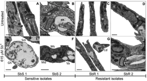
Untreated SbR and SbS isolates did not show any ultrastructural differences by TEM; however, 48 h ex-posure to SbIII caused observable changes in all isolates,
which were more evident in the SbS isolates. SbR isolates displayed changes in cell morphology, increased cyto-plasmic electro density in the cell structure and cytoplas-mic disorganization, and modifications associated with cellular adaptation to stress and sustained cellular viabil-ity. SbS isolates showed more intense cytoplasmic disor-ganization and vacuolization, an absence of subpellicular microtubules, mitochondrial cristae disorganization and swelling, and cells with a near absence of cytoplasmic content (“ghost cells”; Fig. 1A-H). These modifications are all indicative of decreased cell viability.
Fig. 1: ultrastructural images obtained from transmission electron microscopy (TEM) in different conditions. Late log phase promastigotes
were left untreated (A-D) or treated with 615 µM of SbIII (E-H) for 48 h. Images are representative of the major alterations observed in each isolate and condition. SbR: antimony-resistant isolate; SbS: antimony-sensitive isolate. Ed: electrodensity; Gh: “Ghost” cells; Va: cytoplasmic vacuoles; Sm: absence of subpellicular microtubules; Ms: mitochondria swelling; Md: mitochondria disorganization. In the untreated parasites:
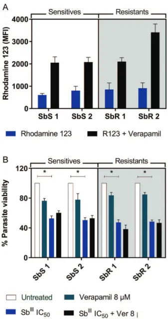
significant in any of the isolates. Additionally, no differ-ences were observed between the resistant and sensitive isolates (p> 0.05; Fig. 2A). Moreover, when evaluating the efflux activity of P-gp channels in the antimony-resistant strains, no significant changes in promastigote viability were observed following in vitro exposure to SbIII in the
presence of verapamil (p> 0.05; Fig. 2B), suggesting that other resistance mechanisms are likely present.
Increased thiol levels contribute to antimony resis-tance - Because increased thiol synthesis promotes anti-mony resistance, we quantified thiol levels in the resis-tant and sensitive isolates. Notably, compared to sensitive isolates, both resistant isolates exhibited approximately 1.24-fold higher thiol levels (p< 0.05; Fig. 3A). To assess whether this increase directly affected SbIII resistance
in vitro, promastigotes were exposed to the drug in the presence of the thiol synthesis inhibitor BSO. Exposure to their IC50 of SbIII significantly reduced the viability of
all isolates (p< 0.05; Fig. 3B); however, in the presence of BSO, resistant isolates were significantly less viable (p< 0.05), confirming that increased thiol levels are re-lated to SbIII resistance. Specifically, the parasite loads
in resistant isolates 1 and 2 were 2.12 and 1.73-fold lower after exposure to SbIII plus BSO compared to SbIII alone.
As the increased thiol metabolism in resistant isolates is known to potentiate the buffering capacity against ROS induced by antimony exposure (Mandal et al. 2007), we tested ROS production in these parasites after antimony treatment. We observed that resistant isolates produced similar amounts of ROS even when exposed to SbIII
con-centrations 4-fold higher than those of sensitive isolates, further supporting enhanced thiol-mediated buffering capacity (Fig. 3C). Moreover, following a thiol synthesis blockade by BSO treatment, only SbR isolates exhibited a significant increase in ROS production (p< 0.05), con-firming the role of increased thiol as a resistance mecha-nism to antimony and ROS-induced death.
Taken together, these data suggest a model for the an-timony resistance mechanism in promastigotes of L. in-fantum isolated from refractory patients from Brazil. In drug-resistant isolates, higher thiol metabolism results in the formation of thiol-metal complexes and drug in-activation. Additionally, the thiol component can buffer against SbIII-induced ROS, allowing parasitic survival at
high concentrations of SbIII (Fig. 4).
DISCUSSION
Chemotherapy is the primary treatment regimen for leishmaniasis, because vaccines are not yet available. However, resistance and toxicity to treatment are major concerns. This study aimed to characterize antimony resistance mechanisms in isolates from patients with treatment-refractory VL in an endemic area of Brazil. Significantly, we found that high thiol metabolism is the mechanism explaining the antimony resistance of these L. infantum isolates.
We recently described antimony and nitric oxide cross-resistance in L. infantum isolates, which induces robust pro-inflammatory cytokine expression in macrophages (Santos et al. 2012, de Moura et al. 2015). However, the
spe-Fig. 2: (A) rhodamine 123 uptake and accumulation mean fluores-cence intensity (MFI) with or without verapamil blockade in Leish-mania infantum promastigotes. Statistical analysis was performed by Kruskal-Wallis test. Results are the mean ± standard error of the mean (SEM) of three independent experiments. (B) Promastigote viability after exposure to SbIII for 48 h with or without verapamil blockade. Each isolate was exposed to its own IC50. Statistical analysis was per-formed by ANOVA with Tukey’s post-test. Statistical significance was defined in p< 0.05. Data shown are from two independent exper-iments performed in quintuplicate. SbS: antimony sensitive isolate; SbR: antimony resistant isolate.
cific mechanisms underlying SbIII resistance have not been
clearly defined. The data in the present study strongly sug-gest that increased thiol levels can explain both resistance to antimony and nitric oxide in these parasites.
Resistance to antimonial compounds utilizes various metabolic pathways. These metabolic changes can lead to altered cellular morphology and structure (Borges et al. 2005). TEM analysis in the present study found that resistant promastigotes treated with SbIII displayed
mod-ifications associated with cellular adaptations to stress, but maintained cellular viability, whereas sensitive iso-lates showed changes that clearly demonstrated the lethal effects of SbIII on these isolates. Generalized changes
were observed in the isolates, reinforcing the notion that antimony compounds act on metabolic pathways essen-tial for parasite survival, as opposed to a single cellular structure or an organelle (Frézard et al. 2009). In addi-tion, these findings reinforce the importance of cellular analysis at the ultrastructural level to fully characterize drug effects (Vannier-Santos & de Castro 2009).
Although the increased expression or activity of ABC superfamily transport pumps such as P-gp has been de-scribed as an antimony resistance mechanism, the pres-ent study showed no differences in transporter activity between resistant and sensitive isolates. Furthermore, no differences in parasite viability were observed with IC50 SbIII treatment in the presence of the channel blocker
vera-pamil hydrochloride, suggesting that another mechanism was likely responsible for antimony resistance. However, it is possible that a more sensitive method could detect effects of these pumps, albeit not as a major mechanism of resistance. More studies should be conducted to evalu-ate the relationship between efflux and antimony resis-tance. Parasites isolated from different geographic re-gions show diverse mechanisms of antimony resistance, and this heterogeneous pattern may be explained by the involvement of several different genes that have evolved to protect these parasites (Jeddi et al. 2014).
The present study demonstrates that the antimony resistance profile of the clinical isolates was primarily based on thiol availability. Notably, the resistant isolates displayed increased thiol expression compared to their sensitive counterparts. Furthermore, the inhibition of thiol synthesis by BSO treatment increased parasite sen-sitivity to SbIII. Previous studies have demonstrated the
essential role of thiols in antimony inactivation (Rai et al. 2013). We observed that antimony-resistant promas-tigotes were also resistant to SbIII-induced ROS, which
was enhanced by thiol synthesis inhibition, specifically in resistant isolates. Interestingly, our group previously demonstrated that L. infantum promastigotes and amas-tigotes show resistance to NO (Santos et al. 2012, de Moura et al. 2015). Additionally, previous studies indi-cate that trypanothione, a thiol molecule with antioxi-dant activity in Leishmania parasites, is able to sequester NO and maintain oxidative homeostasis (Bocedi et al. 2010). Moreover, antimony-resistant L. donovani para-sites display more potent immune-modulating effects such as enhanced interleukin 10 expression in infected macrophages, which is associated with
multidrug-re-Fig. 3: (A) thiol levels were measured (MFI) in promastigotes using
CellTracker™ fluorescent probe (1 µM). Data are the mean ± stan -dard error of the mean (SEM) of five independent experiments. (B) Parasite viability of promastigotes untreated and treated with SbIII (at the IC50 of each isolate) in the presence or absence of L-buthionine sulfoximine (BSO). Data are the mean ± SEM of two independent experiments performed in quintuplicate. (C) ROS production after exposition to IC50 Sb
III with or without BSO measured by
sistant protein 1 overexpression and may contribute to disease severity (Mukherjee et al. 2013). Taken together, these data suggest that there is cross-resistance between SbIII and ROS buffering capacity (Fig. 4). This reinforces
previous evidence of an association between drug and microbicidal resistance mechanisms, and their associa-tion with increased parasite virulence is concerning be-cause of the high prevalence of VL in Brazil (Martins-Melo et al. 2014), including the endemic area containing the patients in the present study, as our group recently showed (Campos et al. 2017). Moreover, previous studies indicate an increased likelihood of in vitro resistance in L. infantum clinical isolates from Brazilian HIV-positive patients (da Luz et al. 2011). In addition, continued inef-fective treatment in humans and reservoirs may lead to the selection of resistant strains persistent in the envi-ronment (Seblova et al. 2014). These results demonstrate the need for a more complete characterization of these clinical isolates to further elucidate the mechanistic re-lationship between drug resistance and evasion from the microbicidal mechanisms of the immune system.
In conclusion, our results demonstrate that isolates from antimony-refractory patients with VL exhibited higher thiol levels than antimony-sensitive isolates. This is the first study with clinical isolates of L. infantum from patients refractory to treatment, and indicates that redox metabolism plays an important role in antimony resistance in New World VL isolates.
ACKNOWLEDGEMENTS
To the Plataforma de Microscopia, teams from Labo-ratório Integrado de Microbiologia e Imunoregulação (LIMI), and Laboratório de Imunoparasitologia (LIP) from Instituto Gonçalo Moniz /Fiocruz-Bahia; and all colleagues from Labo-ratório de Biologia Molecular da UFS/Brazil.
AUTHORS’ CONTRIBUTION
LSM, VMB, ARJ, RPA and TRM - Conceived and de-signed the experiments; LSM, LGSB, SGM and CBM - per-formed the experiments; LSM, CBM, DMT, MWL, VMB, ARJ, RPA and TRM - analysed the data; MWL, ARJ, RPA
and TRM - contributed reagents/materials/analysis tools; LSM, ARJ and TRM - wrote the manuscript
REFERENCES
Bocedi A, Dawood KF, Fabrini R, Federici G, Gradoni L, Pedersen JZ, et al. Trypanothione efficiently intercepts nitric oxide as a harmless iron complex in trypanosomatid parasites. FASEB J. 2010; 24(4): 1035-42.
Borges VM, Lopes UG, de Souza W, Vannier-Santos MA. Cell struc-ture and cytokinesis alterations in multidrug-resistant Leishma-nia (LeishmaLeishma-nia) amazonensis. Parasitol Res. 2005; 95(2): 90-6.
Campos R, Santos M, Tunon G, Cunha L, Magalhães L, Moraes J, et al. Epidemiological aspects and spatial distribution of human and canine visceral leishmaniasis in an endemic area in northeastern Brazil. Geospat Health. 2017; 12: 503.
Croft SL, Olliaro P. Leishmaniasis chemotherapy - challenges and op-portunities. Clin Microbiol Infect. 2011; 17(10): 1478-83.
da Luz RI, Romero GAS, Dorval ME, Cruz I, Cañavate C, Dujardin JC, et al. Drug susceptibility of Leishmania infantum (syn. Leish-mania chagasi) isolates from Brazilian positive and
HIV-negative patients. J Antimicrob Chemother. 2011; 66(3): 677-9.
de Moura TR, Santos MLB, Braz JM, Santos LFVC, Aragão MT, de Oliveira FA, et al. Cross-resistance of Leishmania infantum iso-lates to nitric oxide from patients refractory to antimony treat-ment, and greater tolerance to antileishmanial responses by mac-rophages. Parasitol Res. 2015; 115(2): 713-21.
Essodaı̈gui M, Frézard F, Moreira ESA, Dagger F, Garnier-Suillerot A. Energy-dependent efflux from Leishmania promastigotes of substrates of the mammalian multidrug resistance pumps. Mol Biochem Parasitol. 1999; 100(1): 73-84.
Fonseca-Silva F, Inacio JDF, Canto-Cavalheiro MM, Almeida-Ama-ral EE. Reactive oxygen species production and mitochondrial dysfunction contribute to quercetin induced death in Leishmania amazonensis. PLoS ONE. 2011; 6(2): e14666.
Forster S, Thumser AE, Hood SR, Plant N. Characterization of rhoda-mine-123 as a tracer dye for use in in vitro drug transport assays. PLoS ONE. 2012; 7(3): e33253.
Frézard F, Demicheli C, Ribeiro RR. Pentavalent antimonials: new
perspectives for old drugs. Molecules. 2009; 14(7): 2317-36.
field isolates of Leishmania species from the Western
Mediterra-nean area. Antimicrob Agents Chemother. 2014; 58(8): 4866-74.
Kapoor P, Sachdev M, Madhubala R. Inhibition of glutathione syn-thesis as a chemotherapeutic strategy for leishmaniasis. Trop
Med Int Health. 2000; 5(6): 438-42.
Mandal G, Wyllie S, Singh N, Sundar S, Fairlamb AH, Chatterjee M. Increased levels of thiols protect antimony unresponsive Leishma-nia donovani field isolates against reactive oxygen species
gener-ated by trivalent antimony. Parasitology. 2007; 134(Pt12): 1679-87.
Manzano JI, García-Hernández R, Castanys S, Gamarro F. A new ABC half-transporter in Leishmania major is involved in resistance to antimony. Antimicrob Agents Chemother. 2013; 57(8): 3719-30.
Martins-Melo FR, Lima MS, Ramos AN, Alencar CH, Heukelbach J. Mortality and case fatality due to visceral leishmaniasis in Bra-zil: a nationwide analysis of epidemiology, trends and spatial pat-terns. PLoS ONE. 2014; 9(4): e93770.
Messaritakis I, Christodoulou V, Mazeris A, Koutala E, Vlahou A, Papadogiorgaki S, et al. Drug resistance in natural isolates of Leishmania donovani s.l. promastigotes is dependent of Pgp170
expression. PLoS ONE. 2013; 8(6): e65467.
Mishra J, Saxena A, Singh S. Chemotherapy of leishmaniasis: past, present and future. Curr Med Chem. 2007; 14(10): 1153-69.
Mukherjee A, Padmanabhan PK, Singh S, Roy G, Girard I, Chatterjee M, et al. Role of ABC transporter MRPA, gamma-glutamylcyste-ine synthetase and ornithgamma-glutamylcyste-ine decarboxylase in natural antimony-resistant isolates of Leishmania donovani. J Antimicrob Che-mother. 2007; 59(2): 204-11.
Mukherjee B, Mukhopadhyay R, Bannerjee B, Chowdhury S, Mukherjee S, Naskar K, et al. Antimony-resistant but not anti-mony-sensitive Leishmania donovani up-regulates host IL-10 to overexpress multidrug-resistant protein 1. Proc Natl Acad Sci USA. 2013; 110(7): E575-82.
Neal RA, van Bueren J, McCoy NG, Iwobi M. Reversal of drug resis-tance in Trypanosoma cruzi and Leishmania donovani by vera-pamil. Trans R Soc Trop Med Hyg. 1989; 83(2): 197-8.
Ouellette M, Légaré D, Haimeur A, Grondin K, Roy G, Brochu C, et al. ABC transporters in Leishmania and their role in drug resis-tance. Drug Resist Updat. 1998; 1(1): 43-8.
Pérez-Victoria JM, Torres APTC, Gamarro F, Castanys S. ABC transporters in the protozoan parasite Leishmania. Int Microbiol.
2001; 4(3): 159-66.
Ponte-Sucre A, Diaz E, Padrón-Nieves M. Drug resistance in Leishmania parasites. Vienna: Springer; 2013. 459 pp.
Rai S, Goel S, Dwivedi U, Sundar S, Goyal N. Role of efflux pumps and intracellular thiols in natural antimony resistant isolates of Leishmania donovani. PLoS ONE. 2013; 8(9): e74862.
Santos DM, Petersen ALOA, Celes FS, Borges VM, Veras PST, de Oliveira CI. Chemotherapeutic potential of 17-AAG against cu-taneous leishmaniasis caused by Leishmania (Viannia) brazilien-sis. PLoS Negl Trop Dis. 2014; 8(10): e3275.
Santos PL, Costa RV, Braz JM, Santos LFVC, Batista AC, Vascon-celos CRO, et al. Leishmania chagasi naturally resistant to nitric oxide isolated from humans and dogs with visceral leishmaniasis in Brazil. Nitric Oxide. 2012; 27(1): 67-71.
Sarkar A, Mandal G, Singh N, Sundar S, Chatterjee M. Flow cyto-metric determination of intracellular non-protein thiols in Leish-mania promastigotes using 5-chloromethyl fluorescein diacetate. Exp Parasitol. 2009; 122(4): 299-305.
Seblova V, Oury B, Eddaikra N, Aït-Oudhia K, Pratlong F, Gazanion E, et al. Transmission potential of antimony-resistant Leishmania
field isolates. Antimicrob Agents Chemother. 2014; 58(10): 6273-6.
Singh N, Chatterjee M, Sundar S. The over expression of genes of thiol metabolism contribute to drug resistance in clinical isolates of
vis-ceral leishmaniasis (kala azar) in India. Parasit Vectors. 2014; 7: 596.
Valiathan R, Dubey ML, Mahajan RC, Malla N. Leishmania
don-ovani: effect of verapamil on in vitro susceptibility of promasti-gote and amastipromasti-gote stages of Indian clinical isolates to sodium
stibogluconate. Exp Parasitol. 2006; 114(2): 103-8.
Vannier-Santos MA, de Castro SL. Electron microscopy in antipara-sitic chemotherapy: a (close) view to a kill. Curr Drug Targets.
2009; 10(3): 246-60.