Corresponding author: Dra. Tiane Martin de Moura. Depto. Ciências Básicas da Saúde/UFCSPA. Rua Sarmento Leite 245, sala 204, 90050-170 Porto Alegre, Rio Grande do Sul, Brasil.
Phone: 55 51 3303-8825 e-mail: [email protected] Received 19 January 2015 Accepted 18 May 2015
Infl uence of a subinhibitory concentration of vancomycin
on the
in vitro
expression of virulence-related genes
in the vancomycin-resistant
Enterococcus faecalis
Tiane Martin de Moura
[1],
Fabrício Souza Campos
[2],
Juliana Caierão
[1],
Ana Claudia Franco
[2],
Paulo Michel Roehe
[2],
Pedro Alves d’Azevedo
[1],
Jeverson Frazzon
[3]and
Ana Paula Guedes Frazzon
[2][1]. Departamento Ciências Básicas da Saúde, Universidade Federal de Ciências da Saúde de Porto Alegre, Porto Alegre, Rio Grande do Sul, Brazil. [2]. Departamento de Microbiologia, Parasitologia e Imunologia, Universidade Federal do Rio Grande do Sul, Porto Alegre, Rio Grande do Sul, Brazil. [3]. Instituto de Ciência e Tecnologia de Alimentos, Universidade Federal do Rio Grande do Sul, Porto Alegre, Rio Grande do Sul, Brazil.
ABSTRACT
Introduction: Exposure to subinhibitory concentrations (SICs) of antimicrobials may alter the bacterial transcriptome. Methods: Here, we evaluated the expression of nine virulence-related genes in vancomycin-resistant enterococci (VRE) urinary tract infection isolates grown at SICs of vancomycin. Results: A Subinhibitory concentrations of vancomycin interferes with gene modulation, but does not affect the phenotype of a VRE strain in vitro. Conclusions: Subinhibitory concentrations of vancomycin may regulate the expression of virulence factors in vivo or contribute to the selection of vancomycin-resistant strains.
Keywords: VRE. Biofi lm. Expression.
Enterococcus faecalis, an important microorganism that
causes healthcare-associated infections, is part of the normal human microbiota. Enterococcus faecalis is the fi fth most
common pathogen that causes catheter-associated urinary tract infections (UTIs)(1). Several different virulence factors have
been reported in clinical strains of E. faecalis,including biofi lm formation and the expression of surface adhesion components. The ability of E. faecalis to adhere to medical devices such as ureteral stents and catheters and to develop biofi lms is likely associated with its pathogenicity(2).
The biofi lm on plastic (BOP) operon is essential for biofi lm formation(3). Adhesion of collagen of enterococci (ACE) and
aggregation substance (ASC10) are proteins that are also crucial for biofi lm production(4) (5). The sulfur assimilation system
(SUF) is responsible for iron-sulfur cluster biogenesis and is essential for cellular survival, particularly during aerobic growth or oxidative stress. The SUF operon, which is associated with virulence, is controlled by the regulatory protein sensor for oxidative stress (OxyR) and the ferric-uptake regulator (FUR)(6).
Vancomycin-resistant enterococci (VRE) have emerged as important nosocomial pathogens worldwide. The VanA
phenotype (associated with vanA) is responsible for high levels of resistance to vancomycin(7). The clinical use of vancomycin
is a subject of major concern, because continuous monitoring is required to maintain a concentration of approximately 15mg/L of the drug in patients’ sera. Occasionally, because of the complex pharmacokinetics of vancomycin, this value is not achieved and patients are subjected to subinhibitory concentrations (SICs) of the drug. Consequently, frequent incidences of vancomycin failure and poor outcomes have been observed recently. Under these conditions, there is a high probability of selecting resistant or heteroresistant isolates(8).
Exposure to SICs of antimicrobials may alter the transcriptomic and phenotypic responses of pathogenic bacteria(9) (10); however, the effect of SICs of antimicrobials on
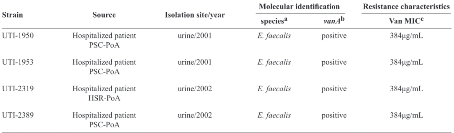
virulence remains unclear. The aim of this study was to evaluate the expression of virulence-related genes in an E. faecalis VRE strain isolated from a urinary tract infection and grown under SICs of vancomycin. The bacterial strains used in this study are listed in Table 1. The growth capacities of four E. faecalis strains were examined, and the strain with the highest optical density (OD) value of biofi lm formation was selected for transcriptional profi ling. All the E. faecalis strains were streaked onto Tryptic
Moura TM et al. - VRE expression is aff ected by vancomycin
TABLE 1 - Bacterial strains used in this study.
Molecular identifi cation Resistance characteristics
Strain Source Isolation site/year
speciesa vanAb Van MICc
UTI-1950 Hospitalized patient urine/2001 E. faecalis positive 384μg/mL
PSC-PoA
UTI-1953 Hospitalized patient urine/2001 E. faecalis positive 384μg/mL
PSC-PoA
UTI-2319 Hospitalized patient urine/2002 E. faecalis positive 384μg/mL
HSR-PoA
UTI-2389 Hospitalized patient urine/2002 E. faecalis positive 384μg/mL
PSC-PoA
Van: vancomycin; MIC: minimal inhibitory concentration; UTI: urinary tract infections; PSC:Pronto Socorro Central; PoA: Porto Alegre;
HSR: Hospital Santa Rita; E.:Enterococcus.addlE. faecalis gene; bvancomycin resistance gene A-type; cMinimal inhibitory concentration by broth microdilution method.
nine different sample concentrations: 50%, 25%, 12%, 10%, 8%, 6%, 4%, and 2% (2×YT-U50-2×YT-U2) and pure urine (100%). The positive control contained only 2× YT broth. The concentration of vancomycin chosen for the assays (64µg/mL) was based on the range 64-1,000µg/mL, which has been shown to produce high levels of resistance(7).
All the strains were used to inoculate plates containing 2× YT agar and incubated at 35°C ON. All the isolates were then evaluated for their ability to form biofi lms in ten different media: 2× YT broth (control), eight different concentrations of urine (2-50%) and 100% urine under two conditions – drug-free condition (DFC) and vancomycin condition (VC) – by using the crystal violet assay(11). Four to eight colonies were diluted
in 0.9% sterile saline (w/v) until the turbidity matched that of a 0.5 McFarland standard [approximately 1 × 10⁸
colony-forming units per milliliter (CFU/ml)]. Eight wells of a 96-well fl at-bottomed microplate were fi lled with 180μL of each media type and 20µL of bacterial suspension. The optical density (OD) was measured at 492nm in a spectrophotometer, and the OD of each strain was determined by comparing the arithmetic mean of the absorbance of the wells with the mean absorbance of the negative controls. The strains were categorized on the basis of their ODs into the following groups: non-adherent, weak biofi lm producers, moderate biofi lm producers, and strong biofi lm producers(11). Non-inoculated 2× YT wells were used as negative
controls, and Staphylococcus epidermidis ATCC 35984 was used as a positive control. The OD492 of each well was measured; all tests were carried out in triplicate. The effects of the different media on bacterial growth and biofi lm formation were evaluated using the paired t-test (level of signifi cance= 0.05).
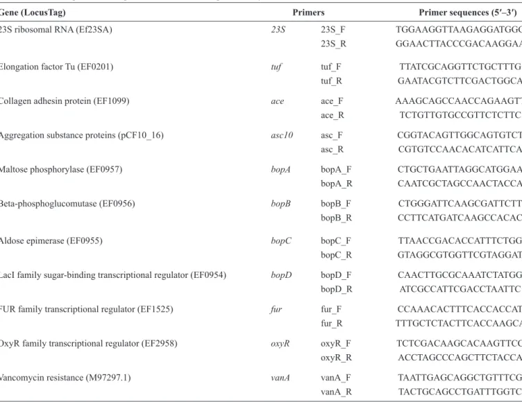
Eleven genes (vanA, bopA, bopB, bopC, bopD, ace,
asc10, fur, oxyR, tuf, and 23S) were analyzed by quantitative polymerase chain reaction (qPCR). The oligonucleotides used in this study are listed in Table 2. Only one strain, exhibiting the
maximum OD value, was selected for transcriptional profi ling. The qPCR analyses were performed at least three times. Five-hundred microliters of ON culture were inoculated in 49.5mL
of 2× YT-U10 in the absence (DFC) and presence (VC) of SICs of vancomycin. Aliquots were collected and extracted at the early-exponential phase (OD650 0.3), total ribonucleic acid (RNA) was purifi ed and quantifi ed, and cDNAs were synthesized(12). Quantitative PCRs were performed as uniplex
reactions in fi nal volumes of 20µL with 100pg of cDNA using the EcoTM Real Time PCR System (Illumina® San Diego, USA)
and universal cycling protocols.
We analyzed the effect of different concentrations of urine on biofi lm formation by phenotypic characterization and on the basis of the OD values. Biofi lm formation in the VRE strains was negatively infl uenced by the presence of pure urine at 35 °C (no growth was observed). With urine concentrations of 2-50%, all the strains showed growth (OD, 0.016 ± 0.004); however, they were unable to form biofi lms under both conditions. The UTI-2389 isolate produced the highest OD value (0.021 ± 0.004). No signifi cant association was observed between biofi lm formation and urine concentration. These fi ndings might be a consequence of the in vitro environment, which lacked essential components
of the in vivo urinary system such as urothelial cells, glucose, mineral salts, and albumin. Moreover, it has been suggested that biological signals in human urine play an important role in modulating virulence at enterococci infection sites(13). On
the basis of our biofi lm assay, all the strains could be classifi ed
as non-adherent according to both DFC and VC. Although
maximal biofi lm induction has been observed at a concentration of 3/4th the minimal inhibitory concentration (MIC)(10), in
other studies, vancomycin at concentrations ≤ 1/2 the MIC(9)
had little or no effect on S. epidermidis biofi lm formation.
Thus, we concluded that the low concentration of vancomycin used in our study (1/6th the MIC) produced no effect on biofi lm
formation, which is consistent with the OD values observed in our study.
TABLE 2 - List of genes and oligonucleotides used for qPCR analyses.
Gene (LocusTag) Primers Primer sequences (5′–3′) 23S ribosomal RNA (Ef23SA) 23S 23S_F TGGAAGGTTAAGAGGATGGG
23S_R GGAACTTACCCGACAAGGAA
Elongation factor Tu (EF0201) tuf tuf_F TTATCGCAGGTTCTGCTTTG
tuf_R GAATACGTCTTCGACTGGCA
Collagen adhesin protein (EF1099) ace ace_F AAAGCAGCCAACCAGAAGTT
ace_R TCTGTTGTGCCGTTCTCTTC
Aggregation substance proteins (pCF10_16) asc10 asc_F CGGTACAGTTGGCAGTGTCT
asc_R CGTGTCCAACACATCATTCA
Maltose phosphorylase (EF0957) bopA bopA_F CTGCTGAATTAGGCATGGAA
bopA_R CAATCGCTAGCCAACTACCA
Beta-phosphoglucomutase (EF0956) bopB bopB_F CTGGGATTCAAGCGATTCTT
bopB_R CCTTCATGATCAAGCCACAC
Aldose epimerase (EF0955) bopC bopC_F TTAACCGACACCATTTCTGG
bopC_R GTAGGCGTGGTTCGTAGGAT
LacI family sugar-binding transcriptional regulator (EF0954) bopD bopD_F CAACTTGCGCAAATCTATGG bopD_R ATCGCCATTCGACCTAATTC
FUR family transcriptional regulator (EF1525) fur fur_F CCAAACACTTTCACCACCAT
fur_R TTTGCTCTACTTCACCAAGCA
OxyR family transcriptional regulator (EF2958) oxyR oxyR_F TCTCGACAAGCACAAGTTCC oxyR_R ACCTAGCCCAGCTTCTACCA
Vancomycin resistance (M97297.1) vanA vanA_F TAATTGAGCAGGCTGTTTCG
vanA_R TACTGCAGCCTGATTTGGTC
qPCR: quantitative polymerase chain reaction; RNA: ribonucleic acid; LacI: lactose; FUR: ferric-uptake regulator; OxyR: oxidative stress regulator; tuf: elongation factor Tu; ace: adhesion of collagen of enterococci; asc10: aggregation substance; bop: biofi lm on plastic; vanA: vancomycin resistance gene A-type; F: primer sense, R:primer antisense. The oligonucleotides were designed using bioinformatics tools (https://www.genscript.com/ssl-bin/app/primer)
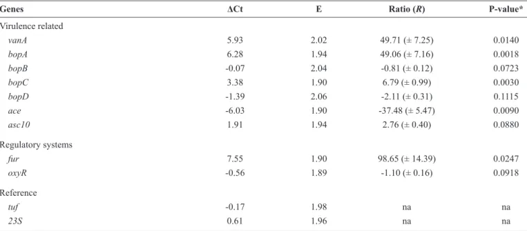
shown in Table 3. Signifi cant upregulation of vanA,which is vancomycin-inducible(7),was detected under the VC condition.
We used this result to investigate the up/downregulation of the other selected genes. Genes of the bop operon were partially regulated in the presence of SICs of vancomycin. Treatment with SICs of vancomycin signifi cantly upregulated bopA and bopC
and slightly downregulated bopB and bopD. Similar partial regulation of the bop operon in a VRE strain has been reported previously(14) under growth conditions lacking antimicrobial
agents, which suggests that this operon behaves erratically, even under normal growth conditions. Under the conditions tested, the expression of ace was signifi cantly downregulated. However,
in a previous study, ace expression was scarcely detected(15)
in cells at the mid- and late exponential phases and was not detectable in cells during the stationary phase, suggesting that
ace transcription is very low under standard in vitro growth conditions. The expression of acs10 was induced slightly by SICs of vancomycin. ASC10 is expressed in the presence of
an intercellular signal cCF10, which is a peptide pheromone(4).
Moreover, antibiotics can act as signaling molecules; therefore, vancomycin could have induced asc10 expression. We also detected minor downregulation of oxyR. During intracellular oxidative stress, the E. faecalis SUF system is induced by OxyR
and requires the expression of the integration host factor(6).
In this in vitro study, no hydrogen peroxide production was observed, which may explain the lack of stimulation of oxyR. In contrast to oxyR, the expression of the ferric uptake regulator fur was signifi cantly upregulated in response to the stress caused
by vancomycin, indicating that this gene is not regulated in the same manner as oxyR. For example, fur may operate in response to a cofactor in cell homeostasis, because the SUF system is the housekeeping machinery for E. faecalis(6).
TABLE 3 - Delta cycle threshold (ΔCt) and ratio values of the expression of virulence related genes in a vancomycin-resistant enterococcus strain grown in 2× YT + 10% urine in absence (DFC) and presence (VC) of subinhibitory concentrations of vancomycin.
Genes ΔCt E Ratio (R) P-value*
Virulence related
vanA 5.93 2.02 49.71 (± 7.25) 0.0140 bopA 6.28 1.94 49.06 (± 7.16) 0.0018 bopB -0.07 2.04 -0.81 (± 0.12) 0.0723 bopC 3.38 1.90 6.79 (± 0.99) 0.0030 bopD -1.39 2.06 -2.11 (± 0.31) 0.1115
ace -6.03 1.90 -37.48 (± 5.47) 0.0090
asc10 1.91 1.94 2.76 (± 0.40) 0.0880
Regulatory systems
fur 7.55 1.90 98.65 (± 14.39) 0.0247
oxyR -0.56 1.89 -1.10 (± 0.16) 0.0918
Reference
tuf -0.17 1.98 na na
23S 0.61 1.96 na na
ΔCt: cycle threshold variation; YT: yeast extract, tryptone and NaCl broth; DFC: drug-free condition; VC: vancomycin condition; E: effi ciency of the reaction; R: the relative expression ratio is the average value of the normalized based on the reference genes; vanA: vancomycin resistance gene A-type; bop: biofi lm on plastic; ace: adhesion of collagen of enterococci; asc10: aggregation substance; fur: ferric-uptake regulator; oxyR: oxidative stress regulator; tuf: elongation
factor Tu; 23S ribosomal RNA; na: not applicable; *Level of signifi cance = 0.05.
in Enterococcus faecalis. Although there was an increase in
the expression of several VRE genes in the presence of SICs of vancomycin, no differences in their phenotypes were observed in vitro. Although the data presented here are from in vitro assays, SICs of vancomycin may contribute to a similar increase in the expression of virulence factors in vivo, leading to adverse clinical outcomes and to the selection of vancomycin-resistant strains. In-vitro experiments may not effectively represent the complex in vivo conditions during enterococcal urinary tract infection. Therefore, additional in vitro and in vivo studies should be carried out to corroborate our fi ndings. The current study is limited because the strains tested were isolated at local hospitals before the study commenced, meaning that we have no patient records detailing their treatments or clinical outcomes. Our main interest was to investigate the behavioral response of a clinical VRE strain against SICs of vancomycin. Therefore, we did not perform tests with other antimicrobials or compare our results to a reference strain.
REFERENCES
The authors declare that there is no confl ict of interest.
CONFLICT OF INTEREST
1. Sievert DM, Ricks P, Edwards JR, Schneider A, Patel J, Srinivasan
A, et al. National Healthcare Safety Network (NHSN) Team and Participating NHSN Facilities 2013. Antimicrobial-resistant
pathogens associated with healthcare-associated infections:
summary of data reported to the National Healthcare Safety Network at the Centers for Disease Control and Prevention,
2009-2010. Infect Control Hosp Epidemiol 2013; 34:1-14.
2. Mohamed JA, Huang DB. Biofi lm formation by enterococci. J Med
Microbiol 2007; 56 (Pt 12):1581-1588.
3. Hufnagel M, Koch S, Creti R, Baldassarri L, Huebner J. A putative sugar-binding transcriptional regulator in a novel gene locus in
Enterococcus faecalis contributes to production of biofi lm and
prolonged bacteremia in mice. J Infect Dis 2004; 189:420-430.
4. Waters CM, Dunny GM. Analysis of functional domains of
the Enterococcus faecalis pheromone-induced surface protein
aggregation substance. J Bacteriol 2001; 183:5659-5667.
5. Chuang-Smith ON, Wells CL, Henry-Stanley MJ, Dunny GM.
Acceleration of Enterococcus faecalis biofi lm formation by
aggregation substance expression in an ex vivo model of cardiac
valve colonization. PLoS One 2010; 5:e15798.
6. Riboldi GP, de Mattos EP, Frazzon J. Biogenesis of [Fe-S] cluster
in Firmicutes: an unexploited fi eld of investigation. Antonie Van Leeuwenhoek 2013; 104:283-300.
7. Courvalin P. Vancomycin resistance in gram-positive cocci. Clin Infect Dis 2006; 42:S25-S34.
8. van Hal SJ, Fowler Jr VG. Is it time to replace vancomycin in the treatment of methicillin-resistant Staphylococcus aureus
infections? Clin Infect Dis 2013; 56:1779-1788.
9. Cargill JS, Upton M. Low concentrations of vancomycin stimulate
biofi lm formation in some clinical isolates of Staphylococcus
epidermidis. J Clin Pathol 2009; 62:1112-1116.
10. Kaplan JB, Jabbouri S, Sadovskaya I. Extracellular
DNA-dependent biofi lm formation by Staphylococcus epidermidis
RP62A in response to subminimal inhibitory concentrations of antibiotics. Res Microbiol 2011; 162:535-541.
11. Stepanovic S, Vukovic D, Dakic I, Savic B, Svabic-Vlahovic M. A
modifi ed microtiter-plate test for quantifi cation of staphylococcal biofi lm formation. J Microbiol Methods 2000; 40:175-179.
12. Moura TM, Cassenego AP, Campos FS, Ribeiro AM, Franco AC, d'Azevedo PA, et al. Detection of vanC1 gene transcription in vancomycin-susceptible Enterococcus faecalis. Mem Inst
Oswaldo Cruz 2013; 108:453-456.
13. Shepard BD, Gilmore MS. Differential expression of virulence-related genes in Enterococcus faecalis in response to biological cues in serum and urine. Infect Immun 2002; 70:4344-4352.
14. Vebø HC, Solheim M, Snipen L, Nes IF, Brede DA. Comparative
genomic analysis of pathogenic and probiotic Enterococcus
faecalis isolates, and their transcriptional responses to growth in
human urine. PLoS One 2010; 5: e12489.
15. Nallapareddy SR, Murray BE. Ligand-signaled upregulation
of Enterococcus faecalis ace transcription, a mechanism for