Original Article
Hypocholesterolemic mechanism of phenolics-enriched extract from
Moringa oleifera
leaves in HepG2 cell lines
Peera Tabboon
1, Bungorn Sripanidkulchai
1, and Kittisak Sripanidkulchai
2*
1 Center for Research and Development of Herbal Health Products, Faculty of Pharmaceutical Sciences,
2 Department of Anatomy, Faculty of Medicine,
Khon Kaen University, Mueang, Khon Kaen, 40002 Thailand.
Received: 13 August 2015; Accepted: 28 September 2015
Abstract
Previous studies have demonstrated the hypolipidemic activity of Moringa oleifera (MO) leaves via lowering serum levels of cholesterol, but the mechanism of action is unknown. In this study, we demonstrated the hypocholesterolemic mechanism of a phenolics-enriched extract of Moringa oleifera leaf (PMO) in HepG2 cells. When compared to the control treatment, PMO significantly decreased total intracellular cholesterol, inhibited the activity of HMG CoA reductase in a dose-dependent manner and enhanced LDL receptor binding activity. Moreover, PMO also significantly increased the genetic expressions of HMG CoA reductase and LDL receptor.
Keywords: Moringa oleifera, LDL receptor, hypolipidemic, HepG2 cell, HMG CoA reductase, phenolics
1. Introduction
Several epidemiological studies have shown that hypercholesterolemia is one of the risk factors for athero-sclerosis and cardiovascular disease (CVD) (Yang, 2005). The liver is a major organ involved in cholesterol metabolism. Inhibition of the rate limiting enzyme 3-hydroxy-3-methyl-glutaryl coenzyme A reductase (HMG CoA reductase) in endogenous cholesterol biosynthesis and up-regulation of the low density lipoprotein (LDL) receptor are the most effec-tive ways for lowering blood cholesterol and reducing cardio-vascular event rates (Brown and Goldstein, 1997).
HMG CoA reductase inhibitors (Statins) are the drugs of choice for lowering blood cholesterol in people with or at high risk of cardiovascular disease. Although the drugs can decrease rates of mortality and morbidity, they also cause adverse side effects such as myopathy or liver dysfunction (Thompson et al., 2003). Therefore, studies of naturally
occurring compounds as the regulators of cholesterol meta-bolism are of interest. Some natural compounds found in the human diet such as the plant flavonoid, quercetin, have positive effects on cholesterol metabolism both in vitro and
in vivo (Moon et al., 2012; Jung et al., 2013), and catechins in green tea have been reported to lower CVD risk (Zheng et al., 2011). Several phenolic compounds in green tea,includ-ing catechin, epicatechin gallate (ECG), epigallocatechin (EGC), and epigallocatechin gallate (EGCG) have shown remarkable effects on the clearance rate of cholesterol due to an increase in the expression of the LDL receptor (Brusill and Roach, 2006).
Moringa oleifera (MO), locally known as drumstick tree or horseradish tree, belongs to the Moringaceae family. It is a quick growing tree and widely distributed in tropical areas. The whole parts of MO have been used as traditional medicines because of their wide spectrum of biological activi-ties, including antibacterial, fungal, antiviral, anti-inflammatory, and antioxidant effects (Guevara et al., 1996; Fahey, 2005; Signh et al., 2009). Due to its hypolipidemic effects both in animal models and clinical trials (Ghasi et al., 2000; Chumark et al., 2008; Nambiar et al., 2010), the products
* Corresponding author.
Email address: [email protected]
from the dried leaves of MO was widely marketed as a functional food. Although MO has been reported to decrease lipid absorption (Jain et al., 2010) and inhibit cholesterol biosynthesis (Chumark et al., 2008), the exact mechanism of action has not yet been established. The present study aimed to investigate the mechanism underlying the hypocholes-terolemic effect of Moringa oliefera leaf extract in human hepatoblastoma (HepG2) cells by determination of the activity, and mRNA expression of HMG CoA reductase enzyme and the LDL receptor.
2. Materials and Methods
2.1 Chemicals
Methanol (HPLC grade), isopropanol, 2,2-diphenyl-1-picrylhydrazyl (DPPH), and formalin, (Fisher Scientific Co Ltd., United Kingdom); acetonitrile (HPLC grade), (Labscan, Bkk, Thailand); molecular biology agarose (Bio-Rad, Spain); 1kb DNA ladder, blue/orange 6X loading dye (Promega, U.S.A.); primer -actin, 3-hydroxy-3-methylglutraryl co-enzyme A (HMG CoA) reductase, and low density lipoprotein (LDL) receptor (Eurofins MWG Operon, Germany); omiscript reverse transcription Kit, TopTaq MasterMix kit (QIAGEN, Germany); novel juice (GeneDirex); RNA extraction kit (GE Healthcare, UK); Dulbecco’s modified eagle medium (DMEM), fetal bovine serum (FBS), and 1,1’-dioctadecyl-3,3,3’,3’-tetramethylidocarbocyanin percholorate (Dil-LDL), (Invitrogen, UK); tryphan blue, neutral red, epigallocatechin-gallate (EGCG), and an HMG CoA reductase assay kit (Sigma, USA); cholesterol enzymatic kit (Randox, UK) and other analytical grade chemicals were used.
2.2 Preparation of phenolics-enriched extract from Moringa oleifera leaf
Fresh leaves of Moringa oleifera were collected in July, 2013 from Khon Kaen Province, Thailand and the voucher specimen (HB186/56) of plant was kept at the Center for Research and Development of Herbal Health Products, Faculty of Pharmaceutical Sciences, Khon Kaen University. The samples were dried and pulverized with an electric grinder to obtain a free flowing powder. One kilogram of dried leaf powder was extracted with seven liters of 70% ethanol in a stainless steel pot for three days, then filtered through cotton and filter paper Whatman® No. 1. The extract was
concen-trated using a vacuum evaporator at 50°C and then freeze-dried in a lyophilizer. The freeze-dried phenolics-enriched extract of
Moringa oleifera leaves (PMO) was kept at -20°C until further used.
2.3 High performance liquid chromatography (HPLC) analysis of PMO
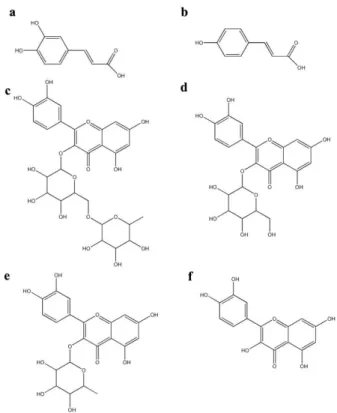
Six commercial available phenolic compounds (caffeic acid, p-coumaric acid, rutin, isoquercetin, quercitrin, and
quercetin) (Figure 1) were used as reference standards for PMO standardization. The HPLC analysis was performed on an Agilent Technologies 1260 machine using an Agilent hypersil ODS column (5 µm, 4.6 x 250 mm) at 30°C. The gradi-ent mobile phase consisted of solution A (acetonitrile:water: phosphoric acid = 80:19.8:0.2) and solution B (0.2% phos-phoric acid) at a flow rate of 1.0 ml/min. The gradient solvent elution was 100% solution B initially, and the gradient program was as follows: 0-5 min: isocratic at 0% solution A; 5-10 min: gradient to 5% solution A; 10-15 min: isocratic at 5% solution A; 15-20 min: gradient to 10% solution A; 20-30 min: isocratic at 10% solution A; 30-35 min: gradient to 15% solution A; 35-45 min, isocratic at 15% solution A; 45-50 min: gradient to 20% solution A; 50-55 min: isocratic at 20% solution A; 55-80 min: gradient to 50% solution A. The UV detection wavelength was set at 210 nm and the injection sample volume was 20 µl.
2.4 HMG CoA reductase activity assay
Based on the oxidation of NADPH, the HMG CoA reductase activity was evaluated by a kit assay following manufacturer’s instruction. The reaction was started by the addition of HMG CoA reductase to the assay mixture contain-ing buffer, NADPH, HMG CoA, and test samples; then the absorbance at 340 nm was continuously determined for 20 min. The initial velocity of each sample was measured as the decreasing rate of absorbance, and the rate of reaction in the
units of Abs340/min was then calculated for the enzyme
specific activity.
2.5 Cell culture
HepG 2, human hepatoma cells were grown in 25 cm2
polystyrene flasks containing DMEM medium supplemented with 10% (v/v) heat-inactivated fetal bovine serum, 1% peni-cillin-streptomycin at 37°C in a humidified atmosphere at 5% CO2. The culture medium was changed twice a week and the
cells were subcultured once a week.
2.6 Measurement of intracellular cholesterol
HepG2 cells were seeded in a 100 mm cell culture disc. After reaching 80% confluence, the media was discarded and cells were pre-incubated in serum-free DMEM for 12 hrs, then treated with various concentrations of test sample in DMEM free serum and incubated at 37°C in a humidified atmosphere at 5% CO2 for 24 hrs. A modification of the method was used
to extract cholesterol (Mizoguchi et al., 2004). After washing with 10 mM phosphate buffer saline, pH 7.4 (PBS), the cells were extracted with a mixture of hexane-isopropanol (3:2) and dried using a vacuum evaporator at 50°C. The dried residue was suspended in 0.2 ml of isopropanol and assayed for cholesterol content using an enzymatic test kit according to the manufacturer’s instructions. The remaining cellular protein was dissolved in 0.5 N NaOH for protein determina-tion (Lowry et al., 1951).
2.7 LDL uptake assay
LDL-receptor activity of HepG2 cells was determined by measuring the uptake of fluorescent labeled Dil-LDL (Teupser et al., 1996). The cells were seeded overnight at a density of 106 cells/well in a 12-well tissue culture plate and
pre-incubated in serum-free DMEM for 12 hrs, then treated with various concentrations of test sample for 24 hrs. After washing with PBS, the cells were incubated with a fluores-cent dye (Dil-LDL) at a confluores-centration of 6 g/mL for 4 hrs, twice washed with PBS, then 0.5 ml of isopropanol was added to each well and the cultures were gently shaken on a TITRAMAX 1000 shaker (Heidolp, Germany) for 15 min. The isopropanol containing fluorescent dye was centrifuged at 500 g for 15 min and the fluorescent intensity in the super-natant was determined at 520 nm (excitation) and 578 nm (emission) wavelengths by a microplate reader (Beckman coulter DTX 990 multimode detector, USA). The remaining cellular protein was dissolved in 0.5 N NaOH for protein determination (Lowry et al., 1951).
2.8 Expression of HMG CoA reductase and LDL receptor genes
Semi-quantitative-reverse transcription-polymerase chain reaction (RT-PCR) was used to study the effect of PMO
on the gene expression of HMG CoA reductase and LDL receptor. The overnight incubated cells (at a density of 106
cells/well), were pre-incubated with serum-free DMEM for 12 hrs; then treated with various concentration of PMO at 37°C in 5% CO2 for 24 hrs. The cells were harvested and total RNA
was extracted by RNA isolation kit according to the manu-facturer’s instructions. The purity of RNA was confirmed with the spectrophotometric absorbance ratio at 260/280 nm and RNA quantity was determined at the absorbance of 260 nm. Total RNA (40 ng) were subjected to RT-PCR using a Two Step RT-PCR kit and performing by thermo cycler (Biometra, Germany). Specific oligonucleotides were based on published sequences (Suarez et al., 2004). The product sizes were 471, 408, and 661 bp, and 26 , 29, and 30 cycles for HMG CoA reductase, LDL receptor and -actin, respectively. Each cycle consisted of 90°C denaturation for 1 min, 60°C primer anneal-ing for 1 min, and 72°C primer extension for 2 min. cDNA was strained (NOVEL JUICE) and analyzed by electrophoresis on 1.5% agarose gel. RT-PCR product density was determined by Gel Documentation and a system analysis machine (Gel Documentation InGenius L, Bio-rad Lab, Hercules, CA, USA). The data were expressed as the relative mRNA expression level with -actin.
2.9 Statistical analysis
All experiments were performed in triplicate and the results were expressed as mean+S.D. One-Way ANOVA and multiple comparisons were used to test for significant differ-ences (P<0.05) using SPSS version 19.0.
3. Results
3.1 Phenolic content of PMO
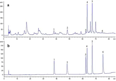
By comparing the retention times of HPLC chromato-grams and spiking with each standard compound, at least six phenolic compounds were determined to be present in PMO, which were 17.63±0.13 mg/g of caffeic acid (RT=38.09 min), 28.6±1.13 mg/g of p-coumaric acid (RT=47.95 min), 11.47± 1.07 mg/g of rutin or quercetin 3-rutinoside (RT=62.14 min), 80.23±0.78 mg/g of isoquercetin or quercetin 3-glucoside (RT=63.17 min), 62.48±0.65 mg/g of quercitrin or quercetin 3-rhamnoside (RT=67.98 min), and 1.88±0.15 mg/g of quercetin (RT=75.30 min), and several unidentified peaks (Figure 2). The content summation of these six phenolics was 202.3 mg/g (20.23%), indicating the phenolics-enriched extracted of MO (PMO). Among these phenolic compounds, isoquercetin and quercitrin were the major constituents.
3.2 PMO decreased intracellular cholesterol
3.3 PMO inhibited HMG CoA reductase activity
PMO significantly inhibited HMG CoA reductase activity in a dose-dependent manner. The inhibitions were 23.08±10.47, 48.10±14.02, 49.43±7.70, and 51.18±9.80 % at 50, 100, 200, and 400 µg/ml of PMO, respectively (Figure 3b).
3.4 PMO increased LDL receptor binding activity
PMO increased the LDL receptor binding activity. The amount of LDL uptake by HepG2 cells was initially sig-nificantly greater than that of control with the addition of 100 µg/ml of PMO, and attained a plateau onward from 200-400
g/ml of PMO (Figure 3c). It is interesting to observe that PMO at doses of 100-400 g/ml increased the activity of LDLR greater than the positive control EGCG at 100 ìM.
3.5 Effect of PMO on the expression of cholesterol meta-bolism-related genes
PMO significantly increased the genetic expression of both LDL receptor and HMG CoA reductase (Figure 4). PMO (25-400 g/ml) had a significantly higher effect on the expres-sion of LDL receptor gene than EGCG (100 M). Similarly, PMO at 100-400 g/ml concentrations significantly enhanced the expression of HMG CoA reductase gene at a level compa-rable to that of EGCG (100 M).
4. Discussion
The aim of the present study was to elucidate the underlying mechanism of the hypocholesterolemic effect of PMO. Since the liver plays a central role ensuring the
Figure 2. HPLC chromatograms of PMO (a) and six standard phenolic compounds (b), 1 = caffeic acid, 2 = p-coumaric acid, 3 = rutin, 4 = isoquercetin, 5 = quercitrin, 6 = quercetin.
systemic homeostasis of glucose and lipid metabolism via the ability to metabolise cholesterol and triglycerides, as well as the synthesis of lipoproteins (Wu et al., 1984; Javitt, 1990), HepG2 cells were used as a liver model.
The findings that PMO decreased intracellular choles-terol and HMG CoA reductase activity in HepG2 cells were in agreement with a previous report that PMO decreased the cholesterol accumulation in rat liver (Gashi et al., 2000). The direct inhibitory effect of PMO on HMG CoA reductase activity can deplete intracellular cholesterol, similar to the previous report that statins can inhibit HMG CoA reductase activity and decrase intracellular cholesterol in HepG2 cells (Scharnagle et al., 2001). EGCG was used as a positive control in this study, because it was earlier reported to block HMG CoA reductase activity in liver microsomes (Cuccioloni et al., 2011), and decrease intracellular cholesterol in HepG2 cells (Bursill et al., 2001).
The significant increase in genetic expression of HMG CoA reductase and LDL receptor genes as well as the signifi-cant stimulation of the LDL receptor binding activity, suggest a vital role of PMO in the regulation of cholesterol metabo-lism. In the cholesterol feedback regulation to control the level of intracellular cholesterol and lipid metabolism, a group of key lipogenic transcription factors, called sterol regulatory element binding proteins (SREBPs), affect several genes related to lipid metabolism such as HMG CoA reductase and LDL receptor genes (Wang et al., 1993; Vallett et al., 1996). In general, the SREBPs are synthesized as inactive proteins. When the intracellular cholesterol is depleted, the SREBPs are translocated from ER to Golgi bodies, which are activated by two proteinase enzymes, S1P and S2P. Then active SREBPs are translocated to the nucleus and promote the transcription of SREBPs target genes such as HMG CoA reductase and LDL receptor gene (Bengoechea-Alonso and Ericsson, 2007). Interestingly, the findings that PMO enhanced the expression of LDL receptor and HMG-CoA reductase mRNA, and increased LDL receptor activity are similar to those of a previous report on the effect of HMG CoA reductasae inhibitor statin (Scharnagl et al., 2001).
Surprisingly, PMO increased the expression of the HMG CoA reductase mRNA, but the intracellular cholesterol was still depleted. This phenomenon may suggest competitive inhibi-tion against the substrate HMG CoA of PMO, as previously observed for mevastatin (Endo et al., 1976). Moreover, PMO may modulate the phosphorylation of HMG CoA reductase to the inactive form, as observed for garlic extract (Liu and Yeh, 2002). Therefore, PMO may regulate cholesterol meta-bolism via the cholesterol feedback regulation. However, the mechanism of PMO on the HMG CoA reductase activity, SREBP expression and activity should be further investigated. Taken together, our results provide the underlying mecha-nism to support the previous finding that MO extracts decreased blood cholesterol in animals (Ghasi et al., 2000; Chumark et al., 2008). Furthermore, our results suggest that PMO affected cholesterol levels by blocking cholesterol synthesis, thereby reducing the liver cholesterol and increas-ing the expression and production of LDL receptors.
A number of phenolic compounds were reported to have hypocholesterolemic effect. For example, quercetin at 75 M concentration significantly induced LDL gene expres-sion, accompanied by an increase in nuclear SREBPs (Moon
et al., 2012). Rutin was shown to inhibit oleic acid induced lipid accumulation via inhibition of transcription of HMG CoA reductase in HepG2 cells (Wu et al., 2011). Moreover, rutin and quercitin in buckwheat protein were reported to have cholesterol-lowering activity (Fabjan et al., 2003; Tomotake et al., 2007). A diet containing 0.2% of caffeic acid significantly reduced both HMG CoA reductase activity and total cholesterol in high-cholesterol diet induced rats (Yeh
et al., 2009), isoquercetin significantly decreased total cholesterol and triglyceride in diabetic mice and high-choles-terol diet induced rabbits (Kamada et al., 2005; Zhang et al., 2011) and p-coumaric acid also significantly decreased total cholesterol and triglyceride in the isoproterenol induced myocardial infarcted rats (Roy and Stanely Mainzen Prince, 2013). With the findings that PMO contained considerable amount of caffeic acd, p-coumaric acid, rutin, isoquercetin, quercitrin and quercetin, therefore, these six phenolic compounds may confer the hypolipidemic effect.
5. Conclusions
Our data demonstrate a vital role of PMO in LDL-cholesterol metabolism as an inducer of LDL receptor expres-sion and binding activity by blocking HMG CoA reductase activity. These findings suggest the underlying mechanism of hypocholesterolemic action of Moringa oleifera, which occurred via the inhibition of HMG CoA reductase activity, enhancement of LDL receptor binding activity and up-regula-tion of LDL receptor gene expression.
Acknowledgements
This work was financially supported by the Higher Education Research Promotion and National Research
versity Project of Thailand, Office of the Higher Education Commission, through the Food and Functional Food Research Cluster, Center for Research and Development of Herbal Health Products, Faculty of Pharmaceuticals Sciences, Khon Kaen University, Thailand.
References
Bengoechea-Alonso, M.T. and Ericsson, J. 2007. SREBP in signal transduction: cholesterol metabolism and beyond. Current Opinion in Cell Biology. 19, 215-222. Brown, M.S. and Goldstein, J.L. 1997. The SREBP pathway:
regulation of cholesterol metabolism by proteolysis of a membrane-bound transcription factor. Cell. 89, 331-340.
Brusill, C.A. and Roach, P.D. 2006. Modulation of cholesterol metabolism by the green tea polyphenol (-)-epigallo-catechin gallate in cultured human liver (HepG2) cells. Journal of Agricultural and Food Chemistry. 54, 1621-1626.
Brusill, C.A., Roach, P.D., Bottema, C.D. and Pal, S. 2011. Green tea upregulates the low-density lipoprotein receptor through the sterol-regulated element binding protein in Hepg2 liver cells. Journal of Agricultural and Food Chemistry. 49, 5639-5645.
Chumark, P., Khunawat, P., Sanvarinda, Y., Phornchirasilp, S., Morales, N.P., Phivthong-Ngam, L., Ratanachamnong, P., Srisawat, S. and Pongrapeeporn, K.U. 2008. The in vitro and ex vivo antioxidant properties, hypo-lipidemic and antiatherosclerotic activities of water extract of Moringa oleifera Lam. leaves. Journal of Ethnopharmacology. 116, 439-446.
Cuccioloni, M., Mozzicafreddo, M., Spina, M., Tran, C.N., Falconi, M., Eleuteri, A.M. and Angeletti, M. 2011. Epigallocatechin-3-gallate potently inhibits the in vitro activity of hydroxyl-3-methyl-glutaryl-CoA reductase. Journal of Lipid Research. 52, 897-907.
Endo, A., Kuroda, M. and Tanzawa, K. 1976. Competitive inhibition of 3-hydroxy-3-methylglutaryl coenzyme A reductase by ML-236A and ML-236B fungal meta-bolites, having hypocholesterolemic activity. FEBS Letters. 72, 323-326.
Fabjan, N., Rode, J., Košir, I.J., Wang, Z., Zhang, Z. and Kreft, l. 2003. Tartary buckwheat (Fagopyrum tataricum Gaertn.) as a source of dietary rutin and quercitrin. Journal of Agricultural and Food Chemistry. 51, 6452-6455.
Fahey, J.W. 2005. Moringa oleifera:a review of the medical evidence for its nutritional, therapeutic and prophy-lactic properties. Part1. Trees for Life Journal. 1, 5. Ghasi, S., Nwobodo, E. and Ofili, J.O. 2000.
Hypocholestero-lemic effects of crude extract of leaf of Moringa oleifera Lam in high-fat diet fed wistar rats. Journal of Ethnopharmacology. 69, 21-25.
Guevara, A.P., Vargas, C. and Uy, M. 1996. Anti-inflammatory and antitumor activities of seed extracts of malunggay,
Moringa oleifera L. (Moringaceae). Plant Physiology and Biochemistry. 125, 175-184.
Jain, P.G., Patil, S.D., Haswani, N.G., Girase, M.V. and Surana, S.J. 2010. Hypolipidemic activity of Moringa oleifera
Lam., Moringaceae, on high fat diet induced hyper-lipidemia in albino rats. Revista Brasileira de Farmacognosia. 20, 969-973.
Javitt, N.B. 1990. HepG2 cells as a resource for metabolic studies: lipoprotein, cholesterol, and bile acids. FASEB Journal. 4, 161-168.
Jung, C.H., Cho, I., Ahn, J., Jeon, T.I. and HA, T.Y. 2013. Quercetin reduces high-fat diet-induced fat accumula-tion in the liver by regulating lipid metabolism genes. Phytotherapy Research. 27, 139-143.
Kamada, C., Da Silva, E.L., Ohnishi-Kameyama, M. and Terao, J. 2005. Attenuation of lipid peroxidation and hyper-lipidemia by quercetin glucoside in the aorta of high cholesterol-fed rabbit. Free Radical Research. 39, 185-194.
Liu, L. and Yeh, Y.Y. 2002. S-alk(en)yl cysteines of garlic inhibit cholesterol synthesis by deactivating HMG-CoA reductase in cultured rat hepatocytes. Journal of Nutrition. 132, 1129-1134.
Lowry, O.H., Rosebrough, N.J., Farr, A.L. and Randall, R.J. 1951. Protein measurement with the folin phenol reagent. Journal of Biological Chemistry. 193, 265-275. Mizoguchi, T., Edano, T. and Koshi, T. 2004. A method of direct measurement for the enzymatic determination of cholesteryl esters. Journal of Lipid Research. 45, 396-401.
Moon, J., Lee, S.M., Do, H.J., Cho, Y., Chung, J.H. and Shin, M.J. 2012. Quercetin up-regulates LDL receptor ex-pression in HepG2 cells. Phytotherapy Research. 26, 1688-1694.
Nambiar, V.S., Guin, P., Paranami, S. and Daniel, M. 2010. Impact of antioxidant from drumstick leaves on lipid profile of hyperlipidemics. Journal of Herbal Medicine and Toxicology. 4, 165-172.
Roy, A.J. and Stanely Mainzen Prince, P. 2013. Preventive effects of p-coumaric acid on cardiac hyperthrophy and alterations in electrocardiogram, lipids, and lipo-proteins in experimentally induced myocardial infracted rats. Food and Chemical Toxicology. 60, 348-354.
Scharnagl, H., Schinker, R., Gierens, H., Nauck, M., Wieland, H. and Marz, W. 2001. Effect of atorvastatin, simvastatin, and lovastatin on the metabolism of cholesterol and triglycerides in HepG2 cells. Bio-chemical Pharmacology. 62, 1545-1555.
Singh, B.N., Singh, B.R., Singh, R.L., Prakash, D., Dhakarey, R., Upadkyay, G. and Singh, H.B. 2009. Oxidative DNA damage protective activity, antioxidant and anti-quorum sensing potentials of Moringa oleifera. Food and Chemical Toxicology. 47, 1109-1116.
2004. Synergistic upregulation of low-density lipo-protein receptor activity by tamoxifen and lovastatin. Cardiovascular Research. 64, 346-355.
Teupser, D., Thiery, J., Walli, A.K. and Seidel, D. 1996. De-termination of LDL-and scavenger-receptor activity in adherent and non-adherent cultured cells with a new single-step fluorometric assay. Biochimica et Biophysica Acta. 1303, 193-198.
Thompson, P.D., Clarkson, P. and Karas, R.H. 2003. Statin-associated myopathy. Journal of the American Medi-cal Association. 289, 1681-1690.
Tomotake, H., Yamamoto, N., Kitabayashi, H., Kawakami, A., Kayashita, J., Ohinata, H., Karasawa, H. and Kato, N. 2007. Preparation of tartary buckwheat protein product and its improving effect on cholesterol metabolism in rats and mice fed cholesterol-enriched diet. Journal of Food Science. 72, S528-S533.
Vallett, S.M., Sanchez, H.B., Rosenfeld, J.M. and Osborne, T.F. 1996. A direct role for sterol regulatory element binding protein in activation of 3-hydroxy-3-methyl-glutaryl coenzyme A reductase gene. Journal of Bio-logical Chemistry. 271, 12247-12253.
Wang, X., Briggs, M.R., Hua, X., Yokoyama, C., Goldstein, J.L. and Brown, M.S. 1993. Nuclear protein that binds sterol regulatory element of low density lipoprotein receptor promoter. I. Identification of the protein and delineation of its target nucleotide sequence. Journal of Biological Chemistry. 268, 14490-14496.
Wu, C.H., Lin, M.C., Wang, H.C., Yang, M.Y., Jou, M.J. and Wang, C.J. 2011. Rutin inhibits oleic acid induced lipid accumulation via reducing lipogenesis and oxidative stress in hepatocarcinoma cells. Journal of Food Science. 76, T65-72.
Wu, G.Y., Wu, C.H., Rifici, V.A. and Stockert, R.J. 1984. Activity and regulation of low density lipoprotein receptors in a human hepatoblastoma cell line. Hepatology. 4, 1190-1194.
Yang, B. 2005. A change of heart: how the people of framing-ham, massachusetts, helped unravel the mysteries of cardiovascular disease. Discovery Medicine. 5, 104-111.
Yeh, Y.H., Lee, Y.T. and Hsieh, H.S. 2009. Dietary caffeic acid, ferulic acid and courmaric acid supplements on cholesterol metabolism and antioxidant activity in rats. Journal of Food and Drug Analysis. 17, 123-132. Zhang, R., Yao, Y., Wang, Y. and Ren, G. 2011. Antidiabetic
activity of isoquercetin in diabetic KK-Ay mice. Nutrition and Metabolism. 8, 85-91.