Effect of estradiol and bisphenol A on human
hepatoblastoma cell viability and telomerase activity
B.L. Xu
1, Q.Z. Zhao
2, X.Y. Gao
1and G.J. Hou
11Children
’s Hospital of Zhengzhou, Zhengzhou, China
2Basic Medical College, Zhengzhou University, Zhengzhou, China
Abstract
Sex hormones from environmental and physiological sources might play a major role in the pathogenesis of hepatoblastoma in children. This study investigated the effects of estradiol and bisphenol A on the proliferation and telomerase activity of human hepatoblastoma HepG2 cells. The cells were divided into 6 treatment groups: control, bisphenol A, estradiol, anti-estrogen ICI 182,780 (hereinafter ICI), bisphenol A+ICI, and estradiol+ICI. Cell proliferation was measured based on average absorbance using the Cell Counting-8 assay. The cell cycle distribution and apoptotic index were determined byflow cytometry. Telomerase activity was detected by polymerase chain reaction and a telomeric repeat amplification protocol assay. A higher cell density was observed in bisphenol A (Po0.01) and estradiol (Po0.05) groups compared with the control group. Cell numbers in S and G2/M
phases after treatment for 48 h were higher (Po0.05), while the apoptotic index was lower (Po0.05) and telomerase activities at
48 and 72 h (Po0.05) were higher in these groups than in the control group. The cell density was also higher in bisphenol A+ICI
(Po0.01) and estradiol+ICI (Po0.05) groups compared with the ICI group. Furthermore, cell numbers were increased in S and
G2/M phases (Po0.05), while the apoptotic index was lower (Po0.05) and telomerase activities at 48 and 72 h were higher
(Po0.05) in these groups than in the ICI group. Therefore, bisphenol A and estradiol promote HepG2 cell proliferationin vitroby
inhibition of apoptosis and stimulation of telomerase activity via an estrogen receptor-dependent pathway.
Key words: Estradiol; Bisphenol A; HepG2; Telomerase activity
Introduction
Hepatoblastoma (HB) is the most common malignant, solid liver tumor in children, arising from multipotent stem cells that differentiate into liver and bile duct epithelial cells in undifferentiated embryonic tissue (1). It accounts for about 25% of pediatric liver tumors and 50–69% of malignant
hepatic tumors among primary embryonal tumors (2). HB is presumed to be caused by abnormal hyperplasia and differentiation of healthy liver cells, although the details of its pathogenesis remain unknown (3).
It has been suggested that environmental factors and sex hormones play major roles in the etiology of HB (4). Estrogen maintains the function of sex organs and reg-ulates metabolism in humans (5). Environmental sources of estrogen include pesticides, plastics, detergents, combus-tion products, as well as industrial and agricultural waste products (6). Once estrogen enters the body, it can affect the endocrine system and promote the growth of hormone-sensitive tumors. Correlations between estrogen and tumor-igenesis have been investigated in various studies (7–10). It
has been demonstrated that estrogen regulates the expres-sion of specific biomarkers in breast cancer (11). Another study has suggested that estrogen promotes angiogenesis
and, consequently, the proliferation of hemangiomas in children (12). However, there is little known about the role of estrogen in hepatoblastoma.
Telomerase is a type of reverse transcriptase consisting of a ribonucleoprotein complex with a RNA template and various catalytic and regulatory subunits. It is expressed in 98% of immortalized cell lines and 490% of malignant tumors (13). Telomerase activity is principally responsible for the infinite proliferative capacity of tumors (14).
This study investigated the effects of physiological and environmental estrogen on HB by treating human hepato-blastoma HepG2 cells with 17b-estradiol (E2) and bisphenol A (BPA), and then evaluating cell proliferation, apoptosis, and telomerase activity. The mechanism of action of these hormones was examined using anti-estrogen ICI 182,780 (hereinafter ICI).
Material and Methods
Cells and reagents
The HepG2 cell line was provided by the Medical School of Zhengzhou University. Phenol red-free Roswell
Correspondence: G.J. Hou:<[email protected]>.
Park Memorial Institute (RPMI) 1640 medium and fetal bovine serum (FBS) were obtained from Gibco (USA). ICI was purchased from Santa Cruz Biotechnology Co., Ltd. (China). E2 was from IBL International (Germany). The cell counting kit (CCK)-8 was from Dojindo (Japan). Dimethyl sulfoxide (DMSO) was obtained from Zhengzhou Chengxiang Chemical Technology Ltd. (China). The reverse transcription kit was purchased from Invitrogen (USA). SYBR Green supermix was from Toyobo (Japan), and the Telo TAGGG Telomerase PCR enzyme-linked immunosorbent assay (ELISA) kit was from Nanjing KeyGEN Biotech Co., Ltd. (China). Penicillin and strepto-mycin were purchased from Beijing BioDee Biotechnology Co. Ltd (China).
Primer and probe design
Primers and probes were synthesized by Invitrogen. The sequence of forward primer TS was 50-AATCCG TCGAGCAGAGTT-30, which was labeled with biotin at the 50-end. The sequence of reverse primer CX was 50-CCCTTACCCTTACCCTTACCCTTA-30. The probe sequence was 50-CCCTAACCCTAACCCTAA-30labeled with digoxin at the 50-end.
Cell culture
HepG2 cells were cultured in RPMI 1640 medium containing 10% FBS, 100 U/mL penicillin, and 100 U/mL streptomycin at 37°C with 5% CO2and saturated humidity.
After the cells had attached to the culture dish, the medium was replaced with phenol red-free RPMI 1640 medium, and the cells were cultured for 24 h. The cells were examined daily by phase contrast microscopy.
Reagent preparation and determination of effective doses
BPA, E2, and ICI were dissolved in DMSO and stored at 20°C. Working solutions were prepared by diluting the stock solutions in phenol red-free RPMI 1640 medium. HepG2 cells were resuspended at 1106cells/mL and
seeded in a 96-well plate with 200mL each well. After adherence, the culture medium was removed, and cells were washed twice with phosphate-buffered saline (PBS) before BPA or E2 was added at various concentrations (0, 210–5, 2
10–4, 2
10–3, 2
10–2, 2
10–1, 2
100,
2101, and 2102mg/mL BPA; 0, 110–5, 1
10–4, 110–3, 1
10–2, 1
10–1, 1
100, 1101, and
1102ng/mL E2). Normal liver cells were similarly treated with the various concentrations of BPA or E2. ICI was used at 1106
M according to a previous report (15).
Treatment groups
Cells were divided into 6 treatment groups as follows: control (DMSO only), BPA, E2, ICI, BPA+ICI, and E2+ICI. The volume of DMSO in each group was o0.1% of the total volume.
Analysis of cell proliferation
Cells were seeded at 1105 cells/well in a 96-well
plate. After adherence, the culture medium was removed, and cells were washed twice with PBS. CCK-8 solution (10mL) was added to each well at 0, 24, 48, 72, 96, and 120 h, and the cells were cultured for an additional 3 h before the absorbance at 450 nm (A450nm) was
deter-mined using a microplate reader (Bio-Rad). A growth curve was generated from the measured values.
Examination of the cell cycle distribution and apoptosis
Cells were collected at the logarithmic growth phase and seeded at 3105 cells/25 mL culture flask. After 24 h, the cells were washed twice with PBS and subjected to the various treatments. After 48 h, 1–5106cells were
collected by trypsinization and centrifuged at 12,000gfor 5 min at 4°C. The cells were then repeatedly washed with PBS and fixed in pre-cooled 70% alcohol at 20°C overnight. After washing with PBS, the cells were treated with RNase A (10mL of a 20mg/mL stock solution in 500mL PBS) for 30 min at 37°C, followed by centrifugation at 8,000gfor 5min at 4°C. The cells were then incubated with 10mL of a propidium iodide solution (50mg/mL in 500mL PBS) for 30 min at room temperature in the dark. Cell cycle and apoptosis analyses were carried out byflow cytometry (BD Biosciences, USA) using CellQuest soft-ware (BD Biosciences, USA). A total of 10,000 cells was used to analyze and the cell cycle distribution with FlowJo software (USA).
Analysis of telomerase activity
A PCR-telomeric repeat amplification protocol (TRAP)–ELISA kit (16,17) was used to determine the
telomerase activity of HepG2 cells according to the manufacturer’s instructions. Briefly, the cells were col-lected at each time point and washed twice with normal saline. A lysis solution (200mL) was then added to dissolve the cells. After 30 min of incubation, the cells were centrifuged at 12,000g for 20 min at 4°C, and the supernatant was stored at 80°C until use. Two
washed four times with cleaning solution (10 mM Hepes-KOH, 1.5 mM MgCl2, 10 mM KCl, and 1 mM DTT). PCR
product (5mL) was mixed with 20mL denaturation solution (0.5% NaOH), followed by incubation for 10 min at room temperature and then addition of 225mL hybridization solution. A total of 100mL of the mixture was transferred to the panel, and hybridization was carried out at 37°C for 3 h. After three washes with washing buffer, 100mL peroxidase-conjugated anti-digoxin antibody was added to the panel, followed by incubation for 30 min at 37°C. Afterfive washes with washing buffer, 100mL substrate buffer (containing 3,30,5,50-tetramethylbenzidine) was added to the panel. Color development was allowed to proceed for 10–15 min.
The reaction was terminated by addition of 2M sulfuric acid. Telomerase activity was determined by measuring the A450nm (reference wavelength: 630 nm) on a microplate
reader. Negative and positive control measurements corre-sponded to A450nmo0.2 and A450nm41.0, respectively.
Statistical analysis
Data are reported as means±SD. Comparisons between groups were performed by one-way analysis of variance. MATLAB software (MathWorks, USA) was used for all statistical analyses. Po0.05 was considered to be statistically significant.
Results
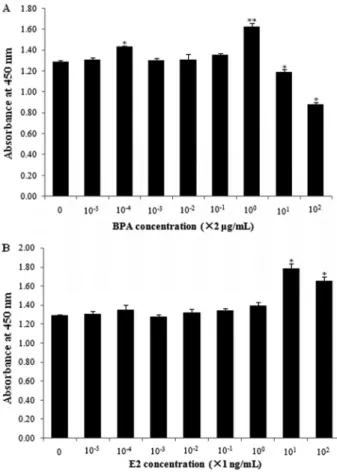
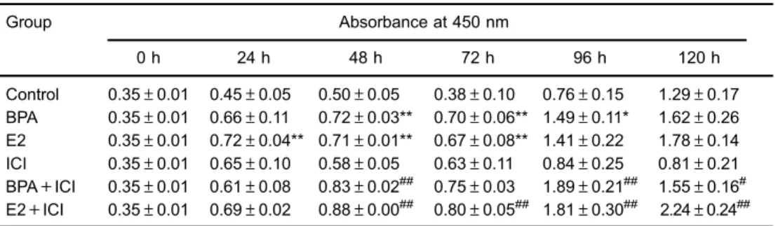
E2 and BPA stimulate HepG2 cell proliferation After 120 h of treatment, the effective concentrations of E2 and BPA to stimulate HepG2 cell proliferation were 2mg/mL and 10 ng/mL, respectively (Figure 1). For normal liver cells, both BPA and E2 had inhibitory effects on their growth (Figure 2). At various time points, BPA, E2, BPA+ICI, and E2+ICI groups had the highest prolifera-tion rates, whereas ICI alone had little effect on cell growth (Figure 3 and Table 1). Cell numbers in the BPA group at 48, 72, and 96 h were significantly higher (Po0.01 or Po0.05) and those in the E2 group were higher at 24, 48, and 72 h (Po0.01) compared with control cells. Com-pared with the ICI group, cell numbers were higher in the BPA+ICI group at 48, 96, and 120 h (Po0.01 or Po0.05) and in the E2+ICI group at 48, 72, 96, and 120 h (Po0.01). These results indicate that E2 and BPA induce the proliferation of HepG2 cells.
Cell cycle regulation is affected by E2 and BPA The proportions of cells in the various cell cycle phases were obviously different in E2- and BPA-treated cells and control cells (Po0.05; Figure 4). Similarly, there were significant differences in the cell cycle distributions of BPA +ICI and E2+ICI groups compared with the ICI group (Po0.05). No statistical difference was found between BPA and BPA+ICI groups or between E2 and E2+ICI groups (P40.05). These results indicate that E2 and BPA stimulate HepG2 cell proliferation by altering the cell cycle.
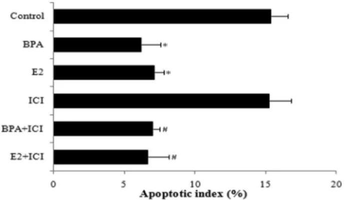
E2 and BPA inhibit apoptosis of HepG2 cells
Flow cytometry showed that the apoptotic index was markedly reduced in cells treated with E2 and BPA compared with the control group (Po0.05; Figure 5). Similarly, compared with cells treated with ICI alone, apoptosis rates were reduced in BPA+ICI and E2+ICI groups (Po0.05). Differences between control and ICI groups, BPA and BPA+ICI groups, and E2 and E2 +ICI groups were not statistically significant (P40.05). These data demonstrate that E2 and BPA inhibit apoptosis of HepG2 cells.
Telomerase activity is induced by E2 and BPA Telomerase activity in HepG2 cells was evaluated by a PCR-TRAP-ELISA. Compared with the control group, telomerase activity was enhanced in BPA- and E2-treated cells at 48 and 72 h (Po0.05; Figure 6). Similarly, compared with the ICI group, telomerase activity was higher in BPA+ICI and E2+ICI groups at 48 and 72 h (Po0.05). There were no differences between ICI and control groups at any time point (P40.05).
Discussion
HB is the most common type of pediatric liver tumor. It is highly malignant, associated with poor outcomes, even after treatment, and characterized by occult occur-rence, vascular enrichment, rapid growth, and early metastasis (18,19). The pathogenesis of HB remains
unclear, but it is thought to arise from mutations and changes in hormone levels caused by environmental factors (4,20). For example, some childhood cases of HB have been linked to the use of oral contraceptives by the mother (4), suggesting the influence of estrogen. The estrogen antagonist tamoxifen has been shown to stimulate HepG2 cell activity and inhibit apoptosis (21). Furthermore, estrogen antagonists inhibit expression of
Table 1.Effect of bisphenol A (BPA) and estradiol (E2) on HepG2 cell viability.
Group Absorbance at 450 nm
0 h 24 h 48 h 72 h 96 h 120 h
Control 0.35±0.01 0.45±0.05 0.50±0.05 0.38±0.10 0.76±0.15 1.29±0.17 BPA 0.35±0.01 0.66±0.11 0.72±0.03** 0.70±0.06** 1.49±0.11* 1.62±0.26 E2 0.35±0.01 0.72±0.04** 0.71±0.01** 0.67±0.08** 1.41±0.22 1.78±0.14 ICI 0.35±0.01 0.65±0.10 0.58±0.05 0.63±0.11 0.84±0.25 0.81±0.21 BPA+ICI 0.35±0.01 0.61±0.08 0.83±0.02## 0.75±0.03 1.89±0.21## 1.55±0.16#
E2+ICI 0.35±0.01 0.69±0.02 0.88±0.00## 0.80±0.05## 1.81±0.30## 2.24±0.24##
Data are reported as means±SD. * Po0.05,**Po0.01 compared to control group;#Po0.05,##Po0.01 compared to ICI (ICI 182,780) group (one-way ANOVA).
Figure 2.Inhibitory effect of bisphenol A (BPA) and estradiol (E2) on normal liver cell survival. The inhibitory action was stronger with increased dosage of BPA and E2.
Figure 3.Growth curve of HepG2 cells treated with bisphenol A (BPA) or estradiol (E2) alone or in combination with the anti-estrogen ICI 182,780 (ICI).
the estrogen receptor and HepG2 cell proliferationin vitro in time- and concentration-dependent manners (22). Consistent with these previous findings, the present results showed that E2 and the environmental estrogen BPA promote HepG2 cell proliferation, which may be due to inhibition of apoptosis because treatment with these agents had no effect on the cell cycle distribution. Co-treatment with ICI did not alter the effects of E2 or BPA, indicating that these agents act via a non-estrogen receptor-dependent pathway in accordance with the known mechanism of estrogen receptor signaling (21).
The role of telomerase in tumor malignancy has been highlighted by many studies. According to a previous report, the hyperproliferation of tumor cells in 90% of human malignancies is linked to inappropriate telomerase activity (23). A study of 100 immortalized cell lines derived from 18 types of tumor tissues demonstrated that 98 cell lines had abnormally high telomerase activity by TRAP in contrast to cells from non-cancerous tissue that were negative for telomerase activity (24). Here, we showed that E2 and BPA stimulate telomerase activity in HepG2 cells. Therefore, inhibition of apoptosis by these two agents may be achieved by stimulation of telomerase activity, which suppresses telomere shortening, chromo-somal damage, and ultimately apoptosis (24). Thisfinding is substantiated by the observation that tamoxifen induces apoptosis of HepG2 cells by suppression of telomerase function (21). Taken together, these results indicate that therapeutic agents targeting telomerase may be effective for the treatment of HB. Moreover, ourfindings provide an insight into the mechanisms underlying the tumorigenic effects of physiological and environmental estrogens.
Acknowledgments
We thank the Medical School of Zhengzhou University for providing the HepG2 cells.
References
1. Zhang SC, Wang WL, Cai WS, Jiang KL, Yuan ZW. Engineered measles virus Edmonston strain used as a novel oncolytic viral system against human hepatoblastoma.BMC Cancer2012; 12: 427, doi: 10.1186/1471-2407-12-427. 2. Miller RW, Young JLJr, Novakovic B. Childhood cancer.
Cancer1995; 75 (1 Suppl): 395–405.
3. Zhang Q, Ming J, Zhang S, Guo D, Qiu X. A rare case of adult hepatoblastoma with neuroendocrine differentiation misdiagnosed as neuroendocrine tumor. Int J Clin Exp Pathol2013; 6: 308–313.
4. Pilotti G, Bosco M, Leo D, Ricci C, Suria G. [Hepatoblastoma in an infant after estroprogestational intake by the mother during pregnancy].Pediatr Med Chir1983; 6: 323–325. 5. Faulds MH, Zhao C, Dahlman-Wright K, Gustafsson JA. The
diversity of sex steroid action: regulation of metabolism by estrogen signaling.J Endocrinol2012; 212: 3–12, doi: 10.1530/JOE-11-0044.
6. Nilsson EE, Skinner MK. Environmentally induced epige-netic transgenerational inheritance of disease suscepti-bility.Transl Res2015; 165: 12–17, doi: 10.1016/j.trsl.2014. 02.003.
7. Mathieu D, Zafrani ES, Anglade MC, Dhumeaux D. Association of focal nodular hyperplasia and hepatic hemangioma.Gastroenterology1989; 97: 154–157. 8. Saegusa T, Ito K, Oba N, Matsuda M, Kojima K, Tohyama K,
et al. Enlargement of multiple cavernous hemangioma of the liver in association with pregnancy.Intern Med1995; 34: 207–211, doi: 10.2169/internalmedicine.34.207.
9. Glinkova V, Shevah O, Boaz M, Levine A, Shirin H. Hepatic haemangiomas: possible association with female sex hormones.Gut 2004; 53: 1352–1355, doi: 10.1136/gut.2003. 038646.
10. Aktas B, Muller V, Tewes M, Zeitz J, Kasimir-Bauer S, Loehberg CR, et al. Comparison of estrogen and
Figure 6. Effect of bisphenol A (BPA) and estradiol (E2) on telomerase activity in HepG2 cells. Cells were treated with BPA or E2 alone or in combination with the anti-estrogen ICI 182,780 (ICI). Telomerase activity was assessed by the PCR-telomeric repeat amplification protocol (TRAP). Data are reported as means ±SD. **Po0.01 compared to control group;##Po0.01 compared to ICI group (one-way ANOVA test).
progesterone receptor status of circulating tumor cells and the primary tumor in metastatic breast cancer patients. Gynecol Oncol2011; 122: 356–360, doi: 10.1016/j.ygyno.2011.04.039. 11. Bartlett JM, Brookes CL, Robson T, van de Velde CJ,
Billingham LJ, Campbell FM, et al. Estrogen receptor and progesterone receptor as predictive biomarkers of response to endocrine therapy: a prospectively powered pathology study in the Tamoxifen and Exemestane Adjuvant Multi-national trial.J Clin Oncol2011; 29: 1531–1538, doi: 10.1200/ JCO.2010.30.3677.
12. Verma K, Tran D, Bryan B, Mitchell D. Meta-analysis of infantile hemangioma endothelial cell microarray expression data reveals significant aberrations of gene networks involved in cell adhesion and extracellular matrix composi-tion.Angiology2013; 1: 2.
13. Kim NW, Piatyszek MA, Prowse KR, Harley CB, West MD, Ho PL, et al. Specific association of human telomerase activity with immortal cells and cancer.Science1994; 266: 2011–2015, doi: 10.1126/science.7605428.
14. Holt SE, Wright WE, Shay JW. Regulation of telomerase activity in immortal cell lines. Mol Cell Biol 1996; 16: 2932–2939.
15. Zhu H, Xiao X, Zheng J, Zheng S, Dong K, Yu Y. Growth-promoting effect of bisphenol A on neuroblastomain vitro andin vivo.J Pediatr Surg2009; 44: 672–680, doi: 10.1016/ j.jpedsurg.2008.10.067.
16. Hoos A, Hepp HH, Kaul S, Ahlert T, Bastert G, Wallwiener D. Telomerase activity correlates with tumor aggressiveness and reflects therapy effect in breast cancer.Int J Cancer 1998; 79: 8–12.
17. Wu YY, Hruszkewycz AM, Delgado RM, Yang A, Vortmeyer AO, Moon YW, et al. Limitations on the quantitative
determination of telomerase activity by the electrophoretic and ELISA based TRAP assays.Clin Chim Acta2000; 293: 199–212, doi: 10.1016/S0009-8981(99)00238-7.
18. Haas JE, Muczynski KA, Krailo M, Ablin A, Land V, Vietti TJ, et al. Histopathology and prognosis in childhood hepato-blastoma and hepatocarcinoma.Cancer1989; 64: 1082–1095. 19. Karski EE, Dvorak CC, Leung W, Miller W, Shaw PJ, Qayed M, et al. Treatment of hepatoblastoma with high-dose chemotherapy and stem cell rescue: the pediatric blood and marrow transplant consortium experience and review of the literature.J Pediatr Hematol Oncol2014; 36: 362–368, doi: 10.1097/MPH.0000000000000130.
20. Nielsen J, Jensen RB, Juul A. Increased sex hormone-binding globulin levels in children and adolescents with thyrotoxicosis.Horm Res Paediatr2013; 79: 157–161, doi: 10.1159/000348837.
21. Brandt S, Heller H, Schuster KD, Grote J. Tamoxifen induces suppression of cell viability and apoptosis in the human hepatoblastoma cell line HepG2 via down-regulation of telomerase activity. Liver Int 2004; 24: 46–54, doi: 10.1111/j.1478-3231.2004.00887.x.
22. Jaber BM, Mukopadhyay R, Smith CL. Estrogen receptor-alpha interaction with the CREB binding protein coactivator is regulated by the cellular environment.J Mol Endocrinol 2004; 32: 307–323.
23. Skordalakes E. Telomerase structure paves the way for new cancer therapies.Future Oncol2009; 5: 163–167, doi: 10.2217/ 14796694.5.2.163.