Surface Cysteine
Proteases Induce Cleavage of the Intestinal
Epithelial Cytoskeletal Protein Villin via
Myosin Light Chain Kinase
Amol Bhargava1,2,3, James A. Cotton1,2,3, Brent R. Dixon4, Lashitew Gedamu1,3, Robin M. Yates5,6, Andre G. Buret1,2,3*
1Department of Biological Sciences, University of Calgary, Calgary, Alberta, Canada,2Inflammation Research Network, University of Calgary, Calgary, Alberta, Canada,3Host-Parasite Interactions, University of Calgary, Calgary, Alberta, Canada,4Bureau of Microbial Hazards, Food Directorate, Health Products and Food Branch, Health Canada, Ottawa, Ontario, Canada,5Department of Comparative Biology and Experimental Medicine, University of Calgary, Calgary, Alberta, Canada,6Department of Biochemistry and Molecular Biology, University of Calgary, Calgary, Alberta, Canada
Abstract
Giardia duodenalisinfections are among the most common causes of waterborne diarrhoeal disease worldwide. At the height of infection,G.duodenalistrophozoites induce multiple pathophysiological processes within intestinal epithelial cells that contribute to the develop-ment of diarrhoeal disease. To date, our understanding of pathophysiological processes in giardiasis remains incompletely understood. The present study reveals a previously unap-preciated role forG.duodenaliscathepsin cysteine proteases in intestinal epithelial patho-physiological processes that occur during giardiasis. Experiments first established that
Giardiatrophozoites indeed produce cathepsin B and L in strain-dependent fashion. Co-incubation ofG.duodenaliswith human enterocytes enhanced cathepsin production by Assemblage A (NF and S2 isolates) trophozoites, but not when epithelial cells were
exposed to Assemblage B (GSM isolate) trophozoites. Direct contact betweenG. duodena-lisparasites and human intestinal epithelial monolayers resulted in the degradation and redistribution of the intestinal epithelial cytoskeletal protein villin; these effects were abol-ished when parasite cathepsin cysteine proteases were inhibited. Interestingly, inhibition of parasite proteases did not prevent degradation of the intestinal tight junction-associated protein zonula occludens 1 (ZO-1), suggesting thatG.duodenalisinduces multiple patho-physiological processes within intestinal epithelial cells. Finally, this study demonstrates thatG.duodenalis-mediated disruption of villin is, at least, in part dependent on activation of myosin light chain kinase (MLCK). Taken together, this study indicates a novel role for para-site cathepsin cysteine proteases in the pathophysiology ofG.duodenalisinfections.
OPEN ACCESS
Citation:Bhargava A, Cotton JA, Dixon BR, Gedamu L, Yates RM, Buret AG (2015)Giardia duodenalis
Surface Cysteine Proteases Induce Cleavage of the Intestinal Epithelial Cytoskeletal Protein Villin via Myosin Light Chain Kinase. PLoS ONE 10(9): e0136102. doi:10.1371/journal.pone.0136102
Editor:Matthew Bogyo, Stanford University, UNITED STATES
Received:May 6, 2015
Accepted:July 29, 2015
Published:September 3, 2015
Copyright:© 2015 Bhargava et al. This is an open access article distributed under the terms of the
Creative Commons Attribution License, which permits unrestricted use, distribution, and reproduction in any medium, provided the original author and source are credited.
Data Availability Statement:All relevant data are within the paper.
Introduction
Giardia duodenalis(syn.G.intestinalis,G.lamblia) is a non-invasive protozoan parasite of the upper small intestines of mammals, including humans. This parasite is a common cause of waterborne diarrhoeal disease worldwide, and is estimated to infect over 280 million
individu-als annually with 20,000 cases reported per year in the US alone [1,2]. Importantly, infection
has been shown to cause stunting and failure to thrive in young children and in food-producing
animals [3–6]. Furthermore,G.duodenalisinfections can also result in the development of
extraintestinal or post-infectious complications [7,8]. Due to the high burden ofG.
duodena-lis-related illness in the developing world, its impairment on development and socioeconomic improvements, and its close association with poverty, this parasite has been included on the
World Health Organization’s (WHO) Neglected Diseases Initiative since 2006 [9]. Therefore,
the impact thatG.duodenalishas on society warrants a better understanding of the disease
pathophysiology. To date, it has been established thatG.duodenaliscauses increased rates of
enterocyte apoptosis, intestinal epithelial barrier dysfunction, shortening of small intestinal brush border microvilli in a CD8+ lymphocyte dependent manner, anion hypersecretion, and
increased rates of intestinal transit (reviewed in [10]). Furthermore,G.duodenalisis divided
into eight distinct genetic Assemblages, designated A through H, whereby only Assemblages A
and B isolates are infective to humans [11,12]. The idea thatG.duodenalisAssemblages A and
B are distinct species is frequently debated [13,14], and it remains to be determined whether
pathogenicity, or the parasite’s immuno-modulating capabilities [15], are assemblage- or
iso-late-dependent.
Only a small number ofG.duodenalisvirulence factors have been identified. Of these, the
best characterized are ventral adhesive disc proteins and surface lectins that ensure attachment, the four pairs of flagella that confer re-colonization and movement, and variant surface
pro-teins (VSPs) that evade host IgA-directed clearance [1,15,16].G.duodenalisalso produces an
arginine deiminase that prevents intestinal epithelial cells (IECs) from utilizing arginine,
thereby impairing intestinal epithelial proliferation and nitric oxide production [17–20].
Although the above-mentioned factors may indirectly contribute to host disease, parasite
prod-ucts directly involved inG.duodenalis-mediated pathogenesis remain largely unknown.
Cathepsin cysteine proteases contain an active site cysteine and histidine residue, and are cate-gorized as clan CA cysteine proteases; in addition, these proteases are further subdivided into cathepsin B (catB) or cathepsin L (catL) superfamilies (reviewed in [21]). Cathepsin-like cyste-ine proteases are critical to the pathogenesis and pathophysiology of several protozoan
para-sites (reviewed in ([22,23]), includingLeishmania donovaniandEntamoeba histolytica[24,
25]. TheG.duodenalisgenome contains genes for numerous catB- and catL-like cysteine
prote-ases [26,27] and cysteine protease activity has been reported inG.duodenaliscultures [28–31].
However, these proteases remain incompletely characterized and their role in giardiasis remains obscure. Thus far, it has been established that cathepsin-like cysteine proteases are
required for trophozoite encystation and excystation [32,33]. Moreover, recent reports
dem-onstrated thatG.duodenaliscatB-like cysteine proteases degrade interleukin-8 (CXCL8),
lead-ing to attenuated CXCL8-induced neutrophil chemotaxis [34]. However, the effect of these cathepsin-like cysteine proteases on live enterocytes has yet to be evaluated. A better under-standing of such proteases in the initiation of disease at the epithelial enterocyte level will
fur-ther clarifyG.duodenalispathogenesis, and help identify specific parasitic proteases that may
become therapeutic targets [35].
AsG.duodenalistrophozoites are non-invasive, the intestinal epithelium represents a pri-mary point of a contact between host and parasite. This layer is comprised of a single layer of IECs that separate the external environment of the intestinal lumen from underlying host
(AIHS) and the Crohn's and colitis foundation of Canada (CCFC). The funders had no role in study design, data collection and analysis, decision to publish, or preparation of the manuscript.
tissues (reviewed in [36]). As such, IECs and their constituent proteins represent an ideal target forG.duodenalisparasite factors. Indeed, previous work has demonstrated thatG.duodenalis
trophozoites induce various pathophysiological processes within IECs [37–40]; however,
spe-cific parasite factors have yet to be identified.
Villin is a unique cytoskeletal protein expressed in gastrointestinal, renal, and urogenital epithelial cells. Within the gut, villin is primarily expressed in the microvilli of IEC where it provides an essential function in regulating the organization of epithelial brush border microfilaments during periods of physiological stress [41]. This protein is capable of poly-merizing and depolypoly-merizing actin, via its ability to cap, sever, nucleate and bundle actin
fila-ments [42].Giardiais known to shorten epithelial brush border microvilli, thereby
contributing to malabsorptive diarrhea [38,43,44]. Research has also demonstrated that
vil-lin is a pro-survival factor, as its overexpression results in delayed epithelial apoptosisin
vitro, and its deletionin vivoenhances susceptibility to dextran sodium sulfate
(DSS)-induced colitis due to increased rates of IEC apoptosis [45]. In its pathogenic cascade,
Giar-diahas been found to cause epithelial apoptosis [38,46]. In addition, villin has been shown
to participate in IEC migration and wound healing [42,47–50]. Collectively, these results
demonstrate that villin is a critical homeostatic protein with multiple functions that extend beyond the maintenance of IEC microvilli. The multifunctional purpose of this protein, in addition to its close relationship with F-actin, warrants further research on its interaction withG.duodenalis. Indeed, previous research has demonstrated thatG.duodenalis trophozo-ites disrupt intestinal epithelial F-actin [39]. Moreover, remodelling of intestinal epithelial
villin has been observed during late stages ofG.duodenalisinfectionin vivovia CD4+ and
CD8+ immune responses [51]. However, it remains to be determined whetherG.duodenalis
parasite products directly target villin. The objectives of this study were to characterize
cyste-ine protease activity inG.duodenalistrophozoites during co-incubations with IECs, and to
determine whether there is a role for these proteases in intestinal epithelial cytoskeletal villin breakdown and disruption.
Materials and Methods
Reagents
Cell Culture
Previous research has validated the human adenocarcinoma Caco-2 cell line (ATCC HTB-37)
as a reliable model for studying giardiasisin vitro[34,39,56,57]. Caco-2 cells were grown in
Minimum Essential Medium Eagle (MEME) supplemented with 100μg/ml streptomycin, 100
Units/ml penicillin, 200 mM L-glutamine, 5mM sodium pyruvate (all from Sigma-Aldrich), and 20% heat-inactivated fetal bovine serum (FBS) (VWR, Radnor, PA). The cells were subcul-tured using 2X Trypsin-EDTA into 6-well plates (Becton Dickinson, Sparks, MD) or Lab-Tek chamber slides (Nalgene Nunc International, Naperville, IL) when flasks were at approximately
80% confluence. The media in the subcultures and flasks was replaced every 2–3 days. The cells
were incubated at 37°C, 5% CO2, and 96% humidity. Cells were used between passages 22 and 34.
Parasites
G.duodenalisNF trophozoites (Assemblage A) were originally obtained following an epidemic
of human giardiasis in Newfoundland, Canada [37,39],G.duodenalisS2 isolate parasites
(Assemblage A) were obtained from a sheep [37,58], andG.duodenalisGS/M isolate
(Assem-blage B) was obtained from the American Type Culture Collection (ATCC 50581) [59].
Tro-phozoites were cultured axenically in Keister’s modified TY1-S-33 medium [60,61]
supplemented with piperacillin (Sigma-Aldrich). Trophozoites were grown and passaged in 15 ml polystyrene conical tubes (Benton Dickinson Falcon) at 37°C under anaerobic conditions. Experiments were performed when cultures were at peak density.
Giardia duodenalis
trophozoite isolation
Confluent tubes ofG.duodenalistrophozoites were collected by cold shock on ice for 30 min.
Following this, 15ml tubes were pooled into 50 ml polypropylene tubes, centrifuged at 500gfor
10 min at 4°C, and resulting supernatants were aspirated. The pellets were resuspended in 10ml of chilled sterile 1X phosphate buffered saline (PBS) (Sigma-Aldrich) and centrifuged at
500gfor 10 min at 4°C. The resulting pellet was resuspended in Caco-2 growth media, the
tro-phozoites enumerated using a hemocytometer, and their concentration was adjusted to a mul-tiplicity of infection (MOI) of 10 parasites per 1 host cell (MOI of 10:1).
Giardia duodenalis
trophozoite DNA extraction
In preparation for DNA extraction, trophozoites were harvested by placing culture tubes on ice for 10 min. Tubes were then gently inverted to mix the contents, and 2 ml of each suspension were pipetted into 15 ml centrifuge tubes (Falcon), and PBS pH 7.4 was added to a final volume
of 10 ml. Tubes were centrifuged at 1,000gfor 5 min. The supernatants were decanted and the
pellets were washed twice more by repeating the centrifugation. Trophozoites were suspended in a final volume of 1 ml PBS. Total DNA was extracted from the trophozoites using the DNeasy Tissue Kit (Qiagen Inc., Mississauga, ON), using a modified protocol. Two hundred microliters of the suspended trophozoites were transferred to 1.5 ml microcentrifuge tubes,
and lysed overnight at 56°C using 180μl of lysis buffer and 20μl of proteinase K (20 mg/ml)
supplied with the DNeasy Tissue Kit. The manufacturer’s instructions were then followed to
purify the DNA. Nucleic acid was eluted with 100μl of elution buffer. Positive control,G.
PCR Protocols
A nested-PCR was performed to amplify fragments of theGiardia16S rRNA gene as described
[62]. PCR amplification of fragments of the glutamate dehydrogenase (gdh) gene forGiardia,
and a restriction fragment length polymorphism (RFLP) assay for genotyping, was performed as described [63].
DNA Sequencing
DNA sequencing of the products of both 16S rRNA and gdh PCR was performed at the McGill University and Genome Quebec Innovation Centre in Montreal, QC, using a 3730xl DNA Analyser (Applied Biosystems, Foster City, CA). PCR products were purified and bi-directional sequencing was performed using the same primers as the original amplifications. DNA sequences were assembled, edited and aligned using SeqScape v2.5 (ABI). Resulting consensus
sequences were then aligned with representative sequence data fromG.duodenalis
Assem-blages and trimmed to identical lengths of 189 bp or 367 bp for 16S rRNA and gdh genes, respectively, using Bioedit v7.1.3.0 [64]. Consensus sequences were compared to reference sequences in GenBank using NCBI standard nucleotide BLAST (blastn). Phylogenetic trees were generated using Molecular Evolutionary Genetics Analysis (MEGA v5.2.1) (http://www. megasoftware.net/) with a Kimura two parameter model neighbour-joining analysis, with 100 bootstraps and pair-wise deletion.
Giardia duodenalis
trophozoite viability
Motility ofG.duodenalistrophozoites was used to assess parasite viability after 2- or 24-hour
co-incubation. Subsequently, Caco-2 cell supernatants were collected, vortexed, and 10μL of
cell supernatant was analyzed on a hemocytometer. The ratio of motile, or swimming, tropho-zoites to total trophozoite counts was assessed as a marker of viability [65].
Giardia duodenalis
modulation of intestinal epithelial villin
G.duodenalistrophozoites (NF, S2, or GS/M) were co-incubated with confluent Caco-2 mono-layers at an MOI of 10:1 for 2 or 24 hours. Identical experiments were performed, except
tro-phozoites were initially pre-treated with E-64d (10μM), Ca-074Me (10μM), or vehicle control
(dimethyl sulfoxide; DMSO) 3 hours prior (see below) to co-incubation within vitroCaco-2
monolayers. Similarly, Caco-2 monolayers were pre-treated with one of E-64d (10μM),
Ca-074Me (10μM), ML-9 (40μM), Z-DEVD-FMK (50μM), or vehicle control (DMSO) prior to
co-incubation withG.duodenalistrophozoites for 2 or 24 hours. In complimentary
experi-ments,G.duodenalisNF trophozoites were co-incubated with Caco-2 monolayers grown to
confluence on the bottom of 12-well plates; in this instance,G.duodenalistrophozoites were
seeded into the top compartment of 0.4μm transwells to prevent direct contact betweenin
vitromonolayers and parasites. Incubation conditions were maintained at 37°C, 5% CO2, and 96% humidity for experimental duration. At the end of the incubation period, cell supernatants were collected and processed for cathepsin activity assays (see below), while monolayers were washed once with PBS and subsequently processed for Western blotting analysis (see below).
In parallel experiments, Caco-2 monolayer cellular lysates were co-incubated withG.
duode-nalisNF trophozoite sonicates for 2 hours. Briefly, culture media from confluent Caco-2 monolayers was removed, and cells were washed once with PBS. Following this, the PBS was
aspirated, the Caco-2 monolayers were collected in 200μL protease inhibitor-free
Dismembranator, Fisher Scientific). Caco-2 lysates protein concentrations were adjusted to 3mg/ml using the Bradford assay method (BioRad Laboratories, Hercules, CA). At the same
time,G.duodenalisNF trophozoites were adjusted to a concentration of 1x10⁷trophozoites/ml
and sonicated three times at level 4 for 30 seconds on ice (550 Versonic Dismembranator,
Fisher Scientific). The resulting Caco-2 lysates andG.duodenalistrophozoite sonicates were
co-incubated for 2 hours in the presence of 10 mM DTT and in the presence or absence of
E-64 (200μM) or Ca-074Me (200μM). After 2 hours, samples were processed for Western
blot-ting analysis (see below).
Whole cell protein extraction for Western blotting
At the end of the co-incubation for 2 or 24 hours, trophozoites were removed from the co-cul-ture via three ice-cold PBS washes. Caco-2 monolayers were subsequently collected in RIPA buffer supplemented with a protease inhibitor cocktail tablet (Complete-Mini, Roche Diagnos-tics, Laval, QC). After a 30 min incubation at 4°C, cellular lysates were sonicated at level 3 for 5
seconds and centrifuged at 10,000gfor 10 min at 4°C. The resulting supernatant was collected
and protein concentrations determined using the Bradford assay method [66]. Protein samples were then normalized to 1.0 mg/ml and then combined at a 1:1 ratio with 2X electrophoresis buffer (100 mM Tris-HCl, pH 6.8, 4% sodium dodecyl sulfate (SDS), 0.2% bromophenol blue,
20% glycerol, 200 mMβ-mercaptoethanol) to further dilute the samples to a final
concentra-tion of 0.5 mg/ml. The samples were then denatured at 95°C for 3 min and stored at-20°C until further analysis.
Western blotting
Protein samples were separated via SDS-PAGE (7–12%) and transferred to nitrocellulose
membranes (Whatman, Buckinghamshire, England) over 1 hour at 100V. The membranes were blocked using 5% fat-free milk solution in 1X Tris-buffered saline + 0.1% Tween (TBS-T) for 1 hour. Primary antibodies were diluted in the same 5% milk solution and incubated with the membranes overnight at 4°C. After three 15-min washes with TBS-T, HRP-conjugated sec-ondary antibodies, also diluted in 5% milk solution, were added to the membranes for 1 hour at room temperature. The membranes were washed three times with TBS-T for 15 min each and then visualized using ECL-plus chemifluorescence detection system (GE Healthcare, Pitts-burgh, PA). ECL-plus was added the membranes for 5 min. The membranes were visualized on ECL film (GE Healthcare). The films were scanned for densitometric analysis using the soft-ware ImageJ (http://rsbweb.nih.gov/ij/). The membranes were stripped using 0.5M acetic acid (45 min incubation) and 0.2 M sodium hydroxide (5 min incubation), and re-probed with a GAPDH antibody to ensure equal loading of the gels. In all instances, GAPDH was used as the loading control.
Inhibition of
G
.
duodenalis
cysteine proteases
Previous research has demonstrated that concentrations of 10μM E-64d or Ca-074Me do not
affectG.duodenalistrophozoite viability, but significantly reduce cathepsin-like cysteine
prote-ase activity [34,67]. Therefore, experiments involvingG.duodenalistrophozoite and Caco-2
monolayer co-incubations were repeated with the administration of either 10μM E-64d, 10μM
Ca-074Me, or vehicle control (DMSO) for 2 or 24 hours. Following this, samples were collected and processed for cathepsin activity assays and Western blotting analysis. In separate
experi-ments, confluent tubes ofG.duodenalistrophozoites were treated with E64d (10μM),
Ca-074Me (10μM), or vehicle control (DMSO) for 3 hours. Following this,G.duodenalis
monolayers for 2 or 24 hours. Samples were again collected for cathepsin activity assays and Western blotting analysis.
Sample extraction for cathepsin activity assays
After co-incubation ofG.duodenalistrophozoites with Caco-2 monolayers (as described
above), supernatants, parasites, and IECs were collected and processed for cathepsin activity assays. Cell supernatants were collected, centrifuged at 500g, 4°C for 10 min and stored for
fur-ther use. The supernatant pellet, representingG.duodenalistrophozoites, was resuspended in
ice-cold PBS, centrifuged at 500g, 4°C for 10 min, and the trophozoite pellet re-suspended in RIPA buffer; this fraction was then sonicated at level 4 for 30 seconds on ice and centrifuged at 10,000g, 4°C for 10 min. A Bradford assay was performed and samples normalized to 1.0 mg/
ml. AdherentG.duodenalistrophozoites were removed from Caco-2 monolayers by modifying
a previously described protocol [68]. In short, a sterile 10μM formononetin solution was made
in the Caco-2 growth medium, added to Caco-2 monolayers, and then allowed to incubate at
37°C, 5% CO2for 60 min. Following this, the formononetin solution was aspirated, and
mono-layers were washed with ice-cold PBS three times. Monomono-layers were then collected and lysed in RIPA buffer not containing a protease inhibitor table, sonicated on level 3 for 5 seconds, and
then centrifuged for 10 min at 10,000gat 4°C. A Bradford assay was performed on collected
Caco-2 lysates and samples were then normalized to 3.0 mg/ml. All samples were stored at-70°C until further use.
Cathepsin activity assays
Assessment of cathepsin cysteine protease activity was performed via recording the liberation of 7-aminomehtylcoumarin (AMC) from fluorogenic substrates, whereby cathepsin protease
activity correlates to an increase in detectable relative light units (RFUs) over time [53,54]. To
assess supernatant cathepsin cysteine protease activity, samples were thawed and incubated at a 1:2 ratio with cathepsin assay buffer (100 mM sodium acetate, 10 mM DTT, 0.1% Triton
X-100, 1 mM EDTA, 0.5% DMSO, 200μM ZFR-AMC or ZRR-AMC). Assessment of
intra-tro-phozoite or intracellular Caco-2 cathepsin cysteine protease activity involved incubating sam-ples at a 1:3 ratio with cathepsin assay buffer. All samsam-ples were incubated in 96-well clear, flat-bottom plates and at 37°C for 5 min and subsequently measured kinetically using a microplate
reader (SpectraMax M2e, Molecular Devices, Sunnyvale, CA) at 37°C with excitation and
emis-sion wavelengths of 354nm and 445nm, respectively. Measurements were recorded every 30 seconds for 5 min. For all experiments, cathepsin assay buffer was adjusted to a pH of 7.2 to mimic the luminal pH of the upper small intestine.
Immunofluorescence
Caco-2 monolayers grown to confluence in Lab-Tek chamber slides (Nalgene Nunc
Interna-tional, Naperville, IL) (8–10 days) were co-incubated for 2 or 24 hours with 1.0x10⁷G.
duode-nalisNF trophozoites pretreated for 3 hours with E64d (10uM) or vehicle control (dimethyl sulfoxide; DMSO) at 37°C, 5% CO2. In another set of experiments, Caco-2 monolayers were
pre-treated with ML-9 (40μM) for 30 min prior to co-incubation withG.duodenalisNF
subsequently administered to cells for 1 hour at 37°C. Following two PBS washes, the cells were incubated with fluorescent secondary antibodies (also prepared in 2% FBS-PBS) for 1 hour at 37°C. The cells were subsequently washed twice with PBS. Nuclei were counterstained with
1μM Hoechst fluorescent staining 33258 (Invitrogen Life Technologies) for 30 min at 37°C.
The cells were washed once with PBS and the slides were mounted with Aqua Poly/Mount (Polyscience Inc.; Warrington, PA). Micrographs were obtained using a Leica DMR Micro-scope, with a Retiga 2000R (Q Imaging, BC) at 400x. All images were collected with the same gain and exposure time lengths. Micrographs presented in the Results section are representa-tive images of 2 replicate monolayers from three separate experiments.
Statistical analysis
All data are representative of at least three separate experiments and expressed as
means ± SEM, where applicable. All statistical analyses were performed using the software, Graphpad Prism 6, which ensures normality of data prior to analysis. Comparisons between
groups were made using a Student t-test or one-way ANOVA, followed by Tukey’s test for
multiple comparison analysis. Statistical significance was established at p<0.05.
Results
Giardia duodenalis
NF and S2 trophozoites are Assemblage A isolates
PCR-RFLP of the glutamate dehydrogenase (gdh) gene revealed identical banding patterns for
both NF and S2G.dudoenalisisolates, matching the expected banding pattern forG.
duodena-lisAssemblage A (Fig 1). DNA sequence data on both 16S rRNA and gdh PCR products
Fig 1. Restriction fragment length polymorphism (RFLP) profile of a fragment of the gdh gene amplified by PCR from NF and S2 isolates ofGiardia duodenalis.The ladder is a 100 bp molecular weight marker (Promega Corp., Madison, WI). NF and S2 isolates ofGiardia duodenalistrophozoites were each run in replicate (i.e., NFa and b, S2a and b).
demonstrated that both the NF and S2 isolates were 100% homologous to reference sequences ofG.duodenalisAssemblage A. Phylogenetic trees for both loci are shown inFig 2.
Giardia duodenalis
trophozoites contain and release catB and L cysteine
proteases
Previous research has demonstrated thatG.duodenalistrophozoites contain, as well as release,
cathepsin cysteine proteases into cell supernatants [28–31,34]. However, the kinetics of
cathepsin cysteine protease activity within multipleG.duodenalistrophozoites, IECs, or cell
supernatants during co-incubation of parasites and IECs remain unknown. Therefore, cathep-sin cysteine protease activity levels were assessed within parasites, IECs, and cell supernatants, following co-incubation. Cathepsin cysteine protease activity was determined by calculating
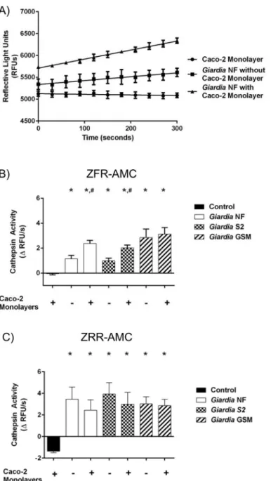
the slope from the RFU versus time, as illustrated inFig 3A. Figs3B, 3C,4B, 4C,5B and 5C
illustrate these calculated slopes and are shown as histograms. As demonstrated by an increase
Fig 2. Phylogenetic trees of the NF and S2 isolates ofGiardia duodenalis.Phylogenetic trees are based on the DNA sequences of amplified fragments of the 16s rRNA (A) and glutamate dehydrogenase (gdh) genes (B).
in the number of detectable RFUs over time, hydrolysis of the catB and L fluorogenic substrate
ZFR-AMC occurred inG.duodenalisNF trophozoite sonicates alone (Fig 3A), or cell
superna-tants from co-incubation (Fig 4A) with Caco-2 monolayers; in contrast, hydrolysis was not
Fig 3.Giardia duodenalistrophozoite sonicates hydrolyze cathepsin cysteine protease substrates in an isolate-independent manner.This figure illustrates cathepsin activity within trophozoite sonicates.G. duodenalistrophozoites (isolates NF, S2, or GS/M) were incubated in the presence or absence of Caco-2 monolayers for 24 hours. Following this,G.duodenalistrophozoites were collected and sonicated to assess for intra-trophozoite cathepsin activity (A to C). The slope value was calculated from the RFU vs time for each experimental group. Line graphs are not shown for all to avoid redundancy, but a representative graph is shown for isolate NF. As a representative figure,G.duodenalisNF trophozoite sonicates were incubated with the catB/L substrate ZFR-AMC (200μM: 5 min: 37°C: pH 7.2). Proteolytic activity is represented as the change in RFUs over time (A).G.duodenalistrophozoite sonicates (isolates NF, S2, or GS/M) or culture media alone (baseline) were incubated with the catB/L fluorogenic substrate ZFR-AMC (B) or the catB-specific fluorogenic substrate (C) (ZRR-AMC) (200μM: 5 min: 37°C: pH 7.2). Proteolytic activity was calculated by determining the change or slope in RFUs over time. Data are mean+/-SEM, n = 3.
observed inside the Caco-2 cells incubated in the presence or absence ofG.duodenalisNF tro-phozoites (Fig 5A).
Fig 4.Giardia duodenalistrophozoites release catB and L cysteine proteases into cell supernatants. This figure illustrates cathepsin activity in parasite supernatants (released activity from trophozoites).G. duodenalistrophozoites (isolates NF, S2, or GS/M) were incubated in the presence or absence of Caco-2 monolayers for 24 hours. Supernatants were collected and analyzed for cathepsin cysteine protease activity (A to C). As a representative figure, supernatants collected fromG.duodenalisNF incubations in the presence or absence of Caco-2 monolayers were incubated with the catB/L fluorgenic substrate ZFR-AMC
(200μM: 5 min: 37°C: pH 7.2). Proteolytic activity is represented as the change in RFUs over time (A).
Supernatants were collected and incubated with the catB/L fluorogenic substrate ZFR-AMC (B) or the catB-specific fluorogenic substrate (C) (ZRR-AMC) (200μM: 5 min: 37°C: pH 7.2). Proteolytic activity was calculated by determining the change or slope in RFUs over time.).*p<0.05 vs Control monolayers #p<0.05 vs corresponding isolate incubated without Caco-2 monolayers. Data are mean+/-SEM, n = 3.
Fig 5.Giardia duodenalistrophozoites do not induce catB/L activity within Caco-2 monolayers.This figure illustrates cathepsin activity within Caco-2 cells post co-incubation with trophozoites.G.duodenalis trophozoites (isolates NF, S2, or GS/M) were co-incubated with Caco-2 monolayers for 24 hours. Caco-2 cell lysates were collected and analyzed for cathepsin cysteine protease activity (A to C). As a representative figure, Caco-2 lysates co-incubated withG.duodenalisNF trophozoites were incubated with the catB/L fluorogenic substrate ZFR-AMC (200μM: 5 min: 37°C: pH 7.2). Proteolytic activity is represented as the change in RFUs over time (A). Caco-2 lysates were collected and incubated with the catB/L fluorogenic substrate ZFR-AMC (B) or the catB-specific fluorogenic substrate (C) (ZRR-AMC) (200μM: 5 min: 37°C: pH 7.2). Proteolytic activity was calculated by determining the change or slope in RFUs over time. Data are mean +/-SEM, n = 3.
Slope values were determined whenG.duodenalistrophozoites (NF, S2, or GS/M) were co-incubated in Caco-2 growth media in the presence or absence of Caco-2 monolayers. Hydroly-sis of the catB/L substrate ZFR-AMC (Fig 3B) and catB-selective substrate ZRR-AMC (Fig 3C)
was observed in sonicates from testedG.duodenalisisolates (NF, S2, or GS/M) and values were
not statistically significant from each other (Fig 3B and 3C). A significant increase in
ZFR-AMC (Fig 4B) and ZRR-AMC (Fig 4C) hydrolysis was detected in cell supernatants
col-lected fromG.duodenalistrophozoites incubated Caco-2 growth media in the presence or
absence of Caco-2 monolayers, when compared against control groups. Interestingly,
co-incu-bation ofG.duodenalisNF or S2 trophozoites with Caco-2 monolayers significantly increased
cell supernatant hydrolysis of ZFR-AMC, compared against the same isolate incubated in the absence of Caco-2 cells (Fig 4B). This trend was not observed when hydrolysis of ZRR-AMC was analyzed (Fig 4C). Hydrolysis of ZFR-AMC (Fig 5B) and ZRR-AMC (Fig 5C) was not
sig-nificantly increased in Caco-2 monolayers incubated in the presence or absence ofG.
duodena-listrophozoites. Therefore,G.duodenalistrophozoites do not appear to increase catB/L activity
inside Caco-2 monolayers. These results demonstrate that catB/L cysteine protease activities
are active at similar levels withinG.duodenalistrophozoites, and that parasites release cysteine
proteases into cell supernatants. However, this activity was further increased in two Assem-blage A isolates (NF and S2) following their exposure to Caco-2 monolayers, while this further increase could not be detected when cells were co-incubated with the Assemblage B GS/M.
G.duodenalisNF trophozoites were used for the rest of the study to assess whether and how parasite catB/L proteases may affect host IECs. Initial experiments sought to determine
whether catB/L activity could be inhibited withinG.duodenalistrophozoites via the
broad-spectrum clan CA cysteine protease inhibitor E64d (10μM) or the catB-specific inhibitor
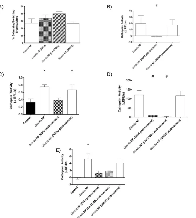
Ca-074Me (10μM). Parasite viability was not affected by 3-hour treatment with E-64d or
Ca-074Me; compared against controlG.duodenalistrophozoites, no significant difference in the
proportion of motile to non-motile trophozoites in groups treated with E64d, Ca-074Me, or vehicle control (DMSO) was observed (Fig 6A). Next, Caco-2 monolayers were co-incubated withG.duodenalisNF trophozoites pretreated with E64d for 2 or 24 hours; this treatment
sig-nificantly reduced the hydrolysis of ZFR-AMC withinG.duodenalissonicates (Fig 6B) and cell
supernatants (Fig 6C), following their 2-hour co-incubation with Caco-2 monolayers.
Simi-larly, hydrolysis of ZFR-AMC was significantly reduced withinG duodenalissonicates (Fig 6D)
and cell supernatants (Fig 6E) when parasites were pre-treated with E-64d or Ca-074Me for 3-hours and subsequently co-incubated with Caco-2 monolayers for 24 hours. Collectively,
these results demonstrate thatG.duodenaliscatB and L proteases are sensitive to inhibition
using commercial protease inhibitors.
Giardia duodenalis
cathepsin cysteine proteases promote villin
breakdown in a contact-dependent manner
Recent research has focused on the ability ofG.duodenalisto disrupt tight junctional proteins
[37,38,40,69]. Less research has focused on this parasite’s ability to affect intestinal epithelial
cytoskeletal proteins.G.duodenalisdisrupts cytoskeletal actin filaments in a myosin light chain
kinase (MLCK)-dependent manner [39]. Moreover, host CD4+ and CD8+ T-lymphocytes are
responsible for villin cleavage during late-stageG.duodenalisinfectionin vivo[51]. It is not
known whetherG.duodenalisparasite products are capable of directly targeting intestinal
epi-thelial cytoskeletal proteins such as villin. As previous findings have shown thatEntamoeba
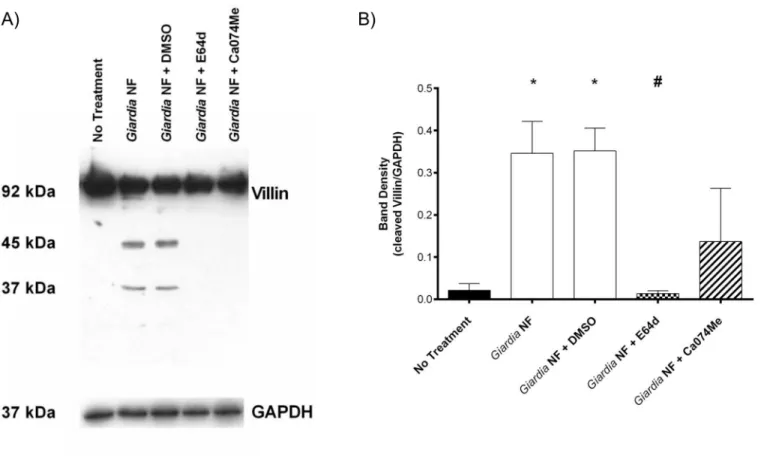
Co-incubation ofG.duodenalisNF trophozoites with Caco-2 monolayers for 2 hours yielded a ~ 45kDa cleavage product as determined by Western blotting (Fig 7A and 7B). Full-length villin (90kDa) was not altered, as determined via densitometry (Fig 7B). Similar results
Fig 6. Pre-treatment ofGiardia duodenalistrophozoites with E-64d or Ca-074Me inhibits cathepsin cysteine protease activity.G.duodenalisNF trophozoites were treated with E-64d (10uM), Ca074Me (10uM), or vehicle control (DMSO) for 3 hours and then co-incubated with Caco-2 monolayers for 2 (B and C) or 24 (D and E) hours.G.duodenalistrophozoites were collected and sonicated to assess for intra-trophozoite cathepsin cysteine protease activity. Supernatants were collected and assessed for the viability ofG.duodenalistrophozoites by examining the ratio of motile: non-motile trophozoites (A).G. duodenalissonicates were incubated with catB/L fluorogenic substrate ZFR-AMC (B and D) or the catB fluorogenic substrate ZRR-AMC (C and E) (200μM: 5 min: 37°C: pH 7.2). Proteolytic activity was calculated by determining the change in RFUs over time.*p<0.05 vs Control cells; #p<0.05 vs G.duodenalisNF trophozoites. Data are mean +/- SEM, n = 3.
were observed when parasites and Caco-2 monolayers were co-incubated for 24 hours as illus-trated from Western blotting and densitometric analyses (Fig 7C and 7D). Densitometry also
demonstrated that E-64d pre-treatment ofG.duodenalisNF trophozoites prior to
co-Fig 7.Giardia duodenalisNF trophozoite proteases that are sensitive to inhibition with E-64d promote villin cleavage in Caco-2 monolayers.G. duodenalisNF isolate trophozoites were pre-treated with E64d (10uM) or vehicle control (DMSO) prior to co-incubation with Caco-2 monolayers for 2 (A and B) or 24 (C and D) hours. Caco-2 lysates were collected and processed for Western blotting to examine for villin protein at 2 (A) and 24 (C) hours. Western blots are representative of three independent experiments performed in triplicate. Densitometry was performed to compare protein levels of cleaved villin vs loading control GAPDH for the 2 (B) or 24 (D) hour co-incubation.*p<0.05 vs No Treatment. Data are mean +/- SEM, n = 3.
incubation with monolayers resulted in decreased detection of the villin cleavage product at both 2 (Fig 7B) and 24 (Fig 7D) hours. Follow-up experiments were performed to determine whether co-incubation of parasite sonicates with Caco-2 cellular lysates for 2 hours also resulted in villin cleavage. As demonstrated via Western blotting and densitometry,
co-incuba-tion ofG.duodenalistrophozoite sonicates with Caco-2 cellular lysates resulted in increased
detection of villin cleavage fragments (Fig 8A and 8B). Importantly, this was significantly reversed when experiments were performed in the presence of E-64d (Fig 8A and 8B). These
results suggest thatG.duodenalisparasite products are capable of cleaving villin within
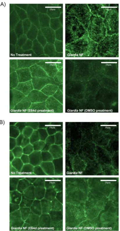
intesti-nal epithelial cellular lysates. Similarly, immunofluorescent staining indicated that
co-incuba-tion ofG.duodenalistrophozoites with Caco-2 monolayers resulted in redistribution of villin
protein at 2 (Fig 9A) and 24 (Fig 9B) hours. Importantly, pre-treatment ofG.duodenalis
tro-phozoites with E-64d, at least partially, prevented redistribution of villin within IECs at 2 and 24 hours (Fig 9A and 9B).
To determine whether secreted products were sufficient to cause a villin break down,
para-sites and Caco-2 monolayers were separated by 0.4μm transwells and co-incubated for 24
hours; interestingly, Western blotting and subsequent densitometry indicated that villin break-down was not seen in these groups (Fig 10A and 10B). Together, these results demonstrate that G.duodenalisclan CA cysteine proteases are involved in the cleavage of cytoskeletal villin in Caco-2 monolayers. Moreover, these data suggest that a parasite surface clan CA cysteine pro-tease contributes to villin cleavage and redistribution within Caco-2 monolayers.
Fig 8. Co-incubation ofGiardia duodenalisNF trophozoite sonicates and Caco-2 lysates results in villin cleavage and is prevented by E64d.G. duodenalisNF isolate trophozoite sonicates and Caco-2 monolayers were co-incubated for 2 hours in the presence of E-64d (10uM), Ca-74Me (10uM), or vehicle control (DMSO). Samples were processed Western blotting to examine for villin protein (A). Densitometry was performed to compare protein levels of cleaved villin vs loading control GAPDH (B).*p<0.05 vs No Treatment. Data are mean +/- SEM, n = 3.
Fig 9.Giardia duodenalistrophozoites produce proteases that are sensitive to inhibition with E-64 that induce disruption of villin within Caco-2 monolayers.G.duodenalisNF isolate trophozoites were pre-treated with E64d (10uM) or vehicle control (DMSO) prior to co-incubation with Caco-2 monolayers for 2 (A) or 24 (B) hours. Caco-2 monolayers were processed for immunofluorescence to examine for villin expression at 2 (A) or 24 (B) hours at 400X magnification. Micrographs are representative of three independent experiments performed in duplicate. n = 3.
Giardia duodenalis clan CA cysteine proteases do not contribute to ZO-1
breakdown
Previous research has demonstrated thatG.duodenalistrophozoites promote the breakdown
and redistribution of the tight junction-associated protein zonula occludens 1 (ZO-1) [37–39,
69]. Therefore, we decided to assess whetherG.duodenalisclan CA cysteine proteases affect ZO-1 in a manner similar to villin. As determined via Western blotting and corresponding
densitometry, co-incubation ofG.duodenalistrophozoites with Caco-2 monolayers resulted in
loss of full-length ZO-1 protein at 2 (Fig 11A and 11B) and 24 (Fig 11C and 11D) hours.
How-ever, pre-treatment ofG.duodenalistrophozoites with E-64d, and subsequent incubation with
Caco-2 monolayers, did not affect degradation of ZO-1 at 2 (Fig 11A and 11B) or 24 hours (Fig 11C and 11D). These observations suggest that degradation and redistribution of villin and ZO-1 may occur via separate mechanisms.
Giardia duodenalis
-induced villin breakdown is partially dependent on
myosin light chain kinase
AsGiardiais known to disrupt tight junctional proteins by activating MLCK and caspase-3 [38,39]), we decided to assess whetherG.duodenalisclan CA cysteine proteases induce villin disruption via activation of MLCK or caspase-3. Therefore, experiments were performed whereby Caco-2 monolayers were pre-treated with the MLCK-selective inhibitor ML-9 or the
caspase-3-specific inhibitor Z-DEVD-FMK prior to co-incubation withG.duodenalis
Fig 10.Giardia duodenalisinduces villin cleavage in Caco-2 monolayers in a contact-dependent manner.G.duodenalisNF isolate trophozoites were co-incubated directly contacting Caco-2 monolayers or separated from Caco-2 monolayers via 0.4um Transwells for 24 hours. Caco-2 lysates were collected and processed for Western blotting to examine for villin protein (A). Western blots are representative of three independent experiments performed in triplicate. Densitometry was performed to compare protein levels of cleaved villin vs loading control GAPDH (B).*p<0.05 vs No Treatment. Data are mean
+/-SEM, n = 3.
trophozoites for 2 or 24 hours. Pre-treatment of Ca2 monolayers with ML-9 prior to
co-incubation withG.duodenalistrophozoites for 2 hours did not affect villin cleavage, as
deter-mined via Western blotting analysis (Fig 12A and 12B). Interestingly, villin degradation was significantly reduced in Caco-2 monolayers pre-treated with ML-9 prior to co-incubation with
Fig 11. Pre-treatment with E-64d does not inhibitGiardia duodenalistrophozoites from-inducing ZO-1 degradation in Caco-2 monolayers.G. duodenalisNF isolate trophozoites were pre-treated with E64d (10uM) or vehicle control (DMSO) prior to co-incubation with Caco-2 monolayers for 2 (A and B) or 24 (C and D) hours. Caco-2 cell lysates were collected and processed for Western blotting to examine for ZO-1 protein at 2 (A) and 24 (C) hours. Western blots are representative of three independent experiments performed in triplicate. Densitometry was performed to compare full length ZO-1 levels vs loading control GAPDH after 2 (B) or 24 (D) hours.*p<0.05 vs Control cells. Data are mean +/- SEM, n = 3.
G.duodenalistrophozoites for 24 hours (Fig 12C and 12D). No significant difference in villin cleavage was observed when Ca2 monolayers were pre-treated with Z-DEVD-FMK and
co-incubated withG.duodenalistrophozoites for 2 (Fig 13A and 13B) or 24 hours (Fig 13C and
13D). Collectively, these results demonstrate thatG.duodenalis-mediated villin degradation and redistribution is, at least partially, dependent on MLCK activation.
Fig 12.Giardia duodenalistrophozoites induce MLCK-dependent cleavage of villin in Caco-2 monolayers in a time-dependent manner.G.
duodenalisNF isolate trophozoites were co-incubated with Caco-2 monolayers for 2 (A and B) or 24 (C and D) hours in the presence or absence of the MLCK inhibitor ML-9. Caco-2 cell lysates were processed for Western blotting to examine for villin protein at 2 (A) and 24 (C) hours. Western blots are
representative of three independent experiments performed in triplicate. Densitometry was performed to compare protein levels of cleaved villin vs loading control GAPDH after a 2 (B) or 24 (D) hour co-incubation.*p<0.05 vs No Treatment. Data are mean +/- SEM, n = 3.
Fig 13.Giardia duodenalistrophozoites promote villin cleavage in Caco-2 monolayers independent of caspase-3 activity.G.duodenalisNF isolate trophozoites were co-incubated with Caco-2 monolayers for 2 (A and B) or 24 (C and D) hours in the presence or absence of Z-DEVD-FMK, a selective caspase-3 inhibitor. Caco-2 cell lysates were collected and processed for Western blotting to examine for villin protein at 2 (A) and 24 (C) hours. Western blots are representative of three independent experiments performed in triplicate. Densitometry was performed to compare protein levels of cleaved villin vs loading control GAPDH at 2 (B) and 24 (D) hours.*p<0.05 vs No Treatment. Data are mean +/- SEM, n = 3.
Discussion
Results from this study reveal a previously unrecognized role for parasite surface clan CA
cyste-ine proteases in the pathophysiology ofG.duodenalisinfections, and implicate them as
poten-tial virulence factors. CatB/L activity was observed withinG.duodenalistrophozoites and in
parasite supernatants following exposure to host IECs. This activity was inhibited when para-sites were pre-treated with the broad-spectrum clan CA, cysteine protease inhibitor E-64d [52]
or the catB-specific inhibitor Ca-074Me [55]. Co-incubation of the parasite with IEC’s
increased the production of catB and L activity for Assemblage A, but not for the Assemblage B isolate. However, no cathepsin activity was observed within the Caco-2 cells following exposure toG.duodenalistrophozoites from any isolate, ruling out any translocation of the cysteine
pro-teases into the cells from the supernatant.G.duodenalistrophozoites degraded and
redistrib-uted the intestinal epithelial cytoskeletal protein villin; this required direct contact between the parasite and IEC, and was prevented when parasites were pre-treated with E-64d prior to incu-bation with intestinal monolayers. In addition, this study elucidated that degradation and redistribution of the tight junction protein ZO-1 was not dependent on factors sensitive to
inhibition with E-64d, thereby suggesting thatG.duodenalismay induce multiple
pathophysio-logical responses within host IECs, some mediated by parasite clan CA cysteine proteases, some not. Finally, this study demonstrates that degradation and disruption of villin is partially dependent upon MLCK activation. At early time points, MLCK inhibition in intestinal
mono-layers exposed toG.duodenalistrophozoites did not prevent villin degradation; however,
MLCK inhibition preventedG.duodenalis-induced villin degradation at later time points.
Col-lectively, these results suggest thatG.duodenalissurface clan CA cysteine proteases induce
vil-lin degradation and redistribution within IECs and that this effect, at least in part, is mediated by MLCK activation within IECs.
To elucidate the role of parasite cathepsin cysteine proteases in the pathophysiology ofG.
duodenalisinfections, we used a well-establishedin vitrocell culture model [18,19,34,57,71, 72]. Furthermore, our experiments used inhibitors, which have previously been shown to suc-cessfully attenuate catB and L cysteine proteases in mammalian and protozoan parasites [24, 52,55,73], includingG.duodenalis[34,67]. Results from this study corroborate previous
observations thatG.duodenalistrophozoites promote the disruption of intestinal epithelial
barrier proteins in vitro [37–40,44,69], andin vivo[46], and disrupt cytoskeletal protein villin
[51]. These studies implicated host lymphocytes in the disruption of the intestinal epithelial cytoskeleton. In contrast, our study demonstrates that parasite clan CA cysteine proteases can directly disrupt intestinal epithelial villin.
Within the gastrointestinal tract, the cytoskeletal protein villin is exclusively expressed by IECs, and its primary function is to maintain microvillous brush border integrity during peri-ods of stress via its ability to promote the assembly and disassembly of the actin cytoskeleton (reviewed in [49]). Pathogen-mediated actin cytoskeleton disruptions can aid in the
establish-ment of infection [74–78]. Interestingly, inhibition of contact between parasites and IECs via
chemical disruption ofG.duodenalislipid rafts prevents actin cytoskeletal remodelling [71].
Consistent with these observations, results from the present study demonstrate thatG.
duode-nalistrophozoites were unable to induce villin degradation and redistribution within IECs when the two were separated by transwells. Collectively, these results suggest that intestinal epithelial actin cytoskeletal remodelling during giardiasis is dependent at least in part on
con-tact betweenG.duodenalistrophozoites and host IECs. It remains to be elucidated whether
actin cytoskeletal remodelling via villin activation is necessary for establishment ofG.
Disruption of villin and the related protein ezrin occurs during the late stages ofG. duode-nalisinfectionin vivo, and is dependent on CD4+ and CD8+ lymphocytes [51]. Data from the present study indicate that early disruption of villin within IECs is dependent on clan CA
cyste-ine proteases. Together, these results suggest thatG.duodenalisuses more than one mechanism
to disrupt villin and the intestinal epithelial brush border. A variety of invasive gastrointestinal pathogens are reliant on the intestinal epithelial expression of villin and its ability to remodel
the actin cytoskeleton to facilitate entry and dissemination through host tissues [79–81].
Fur-thermore, althoughSalmonellasp. and enteropathogenicEscherichia coliinduce villin
redistri-bution, this occurs in the absence of villin proteolysis [82,83]. Therefore,G.
duodenalis-mediated disruption of villin may significantly affect the ability of other gastrointestinal
patho-gens to colonize and cause disease. Moreover,G.duodenalisinfections have been reported to
occur simultaneously with other pro-inflammatory gastrointestinal pathogens [84–87]. A
recent report examining giardiasis in Tanzanian children found that individuals infected with G.duodenalishad reduced incidence of diarrhoeal disease and fever, and lower serum
inflam-matory scores when compared to individuals not infected withG.duodenalis[88]. More
research is warranted to assess whetherG.duodenalisinfections may prevent other pathogens
from establishing and/or inducing disease within their host.
Several parasites contain and produce cathepsin cysteine proteases (reviewed in [22,23]).
TheG.duodenalisgenome contains genes for multiple cathepsin B and L cysteine proteases, but their functions have only begun to be described [26]. Results illustrated herein indicate that G.duodenaliscathepsin cysteine proteases are involved in the degradation and redistribution
of the intestinal epithelial cytoskeletal protein villin, in keeping with the observation that
Ent-amoeba histolyticacysteine proteases promote villin breakdown [70]. The role of villin break-down in the establishment of infection by enteric parasites requires further investigation. Prior
to this study,G.duodenaliscathepsin cysteine proteases had been known to play a role in
tro-phozoite encystation and excystation [33], and degrading the potent neutrophil
chemoattrac-tant interleukin-8 (CXCL8) [34]. Imporchemoattrac-tantly,G.duodenalis-mediated attenuation of CXCL8
did not require direct contact between parasites and host IEC monolayers [34]. These results
suggestG.duodenalistrophozoites possess multiple types of cathepsin cysteine proteases, and
each may have a different role in disease pathogenesis. Indeed, single-celled parasites can pos-sess both surface-associated and secreted cathepsin cysteine proteases which may have unique
roles in pathogenesis [89–92].
In conclusion, our data reveal a novel role forG.duodenaliscathepsin cysteine proteases in
the degradation and redistribution of the intestinal epithelial cytoskeletal protein villin. These results support previous observations that disruption of the intestinal epithelial cytoskeleton requires intimate contact between parasites and intestinal epithelial cells. The present findings
also suggest thatGiardiacysteine proteases disrupt villin at least in part by engaging epithelial
MLCK. Our findings are the first to indicate thatG.duodenaliscathepsin cysteine proteases
represent potential parasite virulence factors capable of inducing pathophysiological responses within host intestinal epithelial cells.
Acknowledgments
Author Contributions
Conceived and designed the experiments: AB JAC LG RMY AGB. Performed the experiments: AB JAC. Analyzed the data: AB JAC BRD. Contributed reagents/materials/analysis tools: LG RMY AGB. Wrote the paper: AB JAC AGB.
References
1. Ankarklev J, Jerlstrom-Hultqvist J, Ringqvist E, Troell K, Svard SG. Behind the smile: cell biology and disease mechanisms of Giardia species. Nature reviews Microbiology. 2010; 8(6):413–22. doi:10. 1038/nrmicro2317PMID:20400969.
2. Yoder JS, Harral C, Beach MJ, Centers for Disease C, Prevention. Giardiasis surveillance—United States, 2006–2008. Morbidity and mortality weekly report Surveillance summaries. 2010; 59(6):15–25. PMID:20535095.
3. Olson ME, Guselle NJ, O'Handley RM, Swift ML, McAllister TA, Jelinski MD, et al. Giardia and Crypto-sporidium in dairy calves in British Columbia. The Canadian veterinary journal La revue veterinaire canadienne. 1997; 38(11):703–6. PMID:9360789; PubMed Central PMCID: PMC1576818. 4. Heimer J, Staudacher O, Steiner F, Kayonga Y, Havugimana JM, Musemakweri A, et al.
Age-depen-dent decline and association with stunting of Giardia duodenalis infection among schoolchildren in rural Huye district, Rwanda. Acta tropica. 2015; 145:17–22. doi:10.1016/j.actatropica.2015.01.011PMID: 25683729.
5. Ignatius R, Gahutu JB, Klotz C, Steininger C, Shyirambere C, Lyng M, et al. High prevalence of Giardia duodenalis Assemblage B infection and association with underweight in Rwandan children. PLoS neglected tropical diseases. 2012; 6(6):e1677. doi:10.1371/journal.pntd.0001677PMID:22720102; PubMed Central PMCID: PMC3373622.
6. Olson ME, McAllister TA, Deselliers L, Morck DW, Cheng KJ, Buret AG, et al. Effects of giardiasis on production in a domestic ruminant (lamb) model. American journal of veterinary research. 1995; 56 (11):1470–4. PMID:8585658.
7. Halliez MC, Buret AG. Extra-intestinal and long term consequences of Giardia duodenalis infections. World journal of gastroenterology: WJG. 2013; 19(47):8974–85. doi:10.3748/wjg.v19.i47.8974PMID: 24379622; PubMed Central PMCID: PMC3870550.
8. Wensaas KA, Langeland N, Hanevik K, Morch K, Eide GE, Rortveit G. Irritable bowel syndrome and chronic fatigue 3 years after acute giardiasis: historic cohort study. Gut. 2012; 61(2):214–9. doi:10. 1136/gutjnl-2011–300220PMID:21911849.
9. Savioli L, Smith H, Thompson A. Giardia and Cryptosporidium join the 'Neglected Diseases Initiative'. Trends in parasitology. 2006; 22(5):203–8. doi:10.1016/j.pt.2006.02.015PMID:16545611.
10. Cotton JA, Beatty JK, Buret AG. Host parasite interactions and pathophysiology in Giardia infections. International journal for parasitology. 2011; 41(9):925–33. doi:10.1016/j.ijpara.2011.05.002PMID: 21683702.
11. Caccio SM, Ryan U. Molecular epidemiology of giardiasis. Molecular and biochemical parasitology. 2008; 160(2):75–80. doi:10.1016/j.molbiopara.2008.04.006PMID:18501440.
12. Lasek-Nesselquist E, Welch DM, Sogin ML. The identification of a new Giardia duodenalis assemblage in marine vertebrates and a preliminary analysis of G. duodenalis population biology in marine systems. International journal for parasitology. 2010; 40(9):1063–74. doi:10.1016/j.ijpara.2010.02.015PMID: 20361967; PubMed Central PMCID: PMC2900473.
13. Xu F, Jerlstrom-Hultqvist J, Andersson JO. Genome-wide analyses of recombination suggest that Giar-dia intestinalis assemblages represent different species. Molecular biology and evolution. 2012; 29 (10):2895–8. doi:10.1093/molbev/mss107PMID:22474166.
14. Franzen O, Jerlstrom-Hultqvist J, Castro E, Sherwood E, Ankarklev J, Reiner DS, et al. Draft genome sequencing of giardia intestinalis assemblage B isolate GS: is human giardiasis caused by two different species? PLoS pathogens. 2009; 5(8):e1000560. doi:10.1371/journal.ppat.1000560PMID:19696920; PubMed Central PMCID: PMC2723961.
15. Palm D, Weiland M, McArthur AG, Winiecka-Krusnell J, Cipriano MJ, Birkeland SR, et al. Developmen-tal changes in the adhesive disk during Giardia differentiation. Molecular and biochemical parasitology. 2005; 141(2):199–207. doi:10.1016/j.molbiopara.2005.03.005PMID:15850703.
16. Adam RD. Biology ofGiardia lamblia. Clinical microbiology reviews. 2001; 14(3):447–75. doi:10.1128/ CMR.14.3.447–475.2001PMID:11432808; PubMed Central PMCID: PMC88984.
parasitology. 2008; 159(2):85–91. doi:10.1016/j.molbiopara.2008.02.005PMID:18359106; PubMed Central PMCID: PMC3658456.
18. Eckmann L, Laurent F, Langford TD, Hetsko ML, Smith JR, Kagnoff MF, et al. Nitric oxide production by human intestinal epithelial cells and competition for arginine as potential determinants of host defense against the lumen-dwelling pathogen Giardia lamblia. Journal of immunology. 2000; 164(3):1478–87. PMID:10640765.
19. Stadelmann B, Hanevik K, Andersson MK, Bruserud O, Svard SG. The role of arginine and arginine-metabolizing enzymes during Giardia—host cell interactions in vitro. BMC microbiology. 2013; 13:256. doi:10.1186/1471–2180–13–256PMID:24228819; PubMed Central PMCID: PMC4225669. 20. Stadelmann B, Merino MC, Persson L, Svard SG. Arginine consumption by the intestinal parasite
Giar-dia intestinalis reduces proliferation of intestinal epithelial cells. PloS one. 2012; 7(9):e45325. doi:10. 1371/journal.pone.0045325PMID:23028934; PubMed Central PMCID: PMC3446895.
21. Turk V, Stoka V, Vasiljeva O, Renko M, Sun T, Turk B, et al. Cysteine cathepsins: from structure, func-tion and regulafunc-tion to new frontiers. Biochimica et biophysica acta. 2012; 1824(1):68–88. doi:10.1016/ j.bbapap.2011.10.002PMID:22024571.
22. McKerrow JH, Caffrey C, Kelly B, Loke P, Sajid M. Proteases in parasitic diseases. Annual review of pathology. 2006; 1:497–536. doi:10.1146/annurev.pathol.1.110304.100151PMID:18039124. 23. Sajid M, McKerrow JH. Cysteine proteases of parasitic organisms. Molecular and biochemical
parasi-tology. 2002; 120(1):1–21. PMID:11849701.
24. Somanna A, Mundodi V, Gedamu L. Functional analysis of cathepsin B-like cysteine proteases from Leishmania donovani complex. Evidence for the activation of latent transforming growth factor beta. The Journal of biological chemistry. 2002; 277(28):25305–12. doi:10.1074/jbc.M203034200PMID: 12000761.
25. Kissoon-Singh V, Mortimer L, Chadee K. Entamoeba histolytica cathepsin-like enzymes: interactions with the host gut. Advances in experimental medicine and biology. 2011; 712:62–83. doi:10.1007/978– 1–4419–8414–2_5PMID:21660659.
26. Aurrecoechea C, Brestelli J, Brunk BP, Carlton JM, Dommer J, Fischer S, et al. GiardiaDB and TrichDB: integrated genomic resources for the eukaryotic protist pathogens Giardia lamblia and Trichomonas vaginalis. Nucleic acids research. 2009; 37(Database issue):D526–30. doi:10.1093/nar/gkn631PMID: 18824479; PubMed Central PMCID: PMC2686445.
27. DuBois KN, Abodeely M, Sajid M, Engel JC, McKerrow JH. Giardia lamblia cysteine proteases. Parasi-tology research. 2006; 99(4):313–6. doi:10.1007/s00436–006–0149–4PMID:16598471.
28. Williams AG, Coombs GH. Multiple protease activities in Giardia intestinalis trophozoites. International journal for parasitology. 1995; 25(7):771–8. PMID:7558562.
29. Rodriguez-Fuentes GB, Cedillo-Rivera R, Fonseca-Linan R, Arguello-Garcia R, Munoz O, Ortega-Pierres G, et al. Giardia duodenalis: analysis of secreted proteases upon trophozoite-epithelial cell interaction in vitro. Memorias do Instituto Oswaldo Cruz. 2006; 101(6):693–6. PMID:17072486. 30. Coradi ST, Guimaraes S. Giardia duodenalis: protein substrates degradation by trophozoite proteases.
Parasitology research. 2006; 99(2):131–6. doi:10.1007/s00436–005–0124–5PMID:16521040. 31. de Carvalho TB, David EB, Coradi ST, Guimaraes S. Protease activity in extracellular products
secreted in vitro by trophozoites of Giardia duodenalis. Parasitology research. 2008; 104(1):185–90. doi:10.1007/s00436–008–1185-zPMID:18797927.
32. Ward W, Alvarado L, Rawlings ND, Engel JC, Franklin C, McKerrow JH. A primitive enzyme for a primi-tive cell: the protease required for excystation of Giardia. Cell. 1997; 89(3):437–44. PMID:9150143. 33. DuBois KN, Abodeely M, Sakanari J, Craik CS, Lee M, McKerrow JH, et al. Identification of the major
cysteine protease of Giardia and its role in encystation. The Journal of biological chemistry. 2008; 283 (26):18024–31. doi:10.1074/jbc.M802133200PMID:18445589; PubMed Central PMCID:
PMC2440617.
34. Cotton JA, Bhargava A, Ferraz JG, Yates RM, Beck PL, Buret AG. Giardia duodenalis cathepsin B pro-teases degrade intestinal epithelial interleukin-8 and attenuate interleukin-8-induced neutrophil chemo-taxis. Infection and immunity. 2014. doi:10.1128/IAI.01771–14PMID:24733096.
35. Busatti HG, Santos JF, Gomes MA. The old and new therapeutic approaches to the treatment of giardi-asis: where are we? Biologics: targets & therapy. 2009; 3:273–87. PMID:19707415; PubMed Central PMCID: PMC2726062.
36. Turner JR. Intestinal mucosal barrier function in health and disease. Nature reviews Immunology. 2009; 9(11):799–809. doi:10.1038/nri2653PMID:19855405.
manner. Infection and immunity. 2002; 70(7):3673–80. PMID:12065509; PubMed Central PMCID: PMC128105.
38. Scott KG, Meddings JB, Kirk DR, Lees-Miller SP, Buret AG. Intestinal infection with Giardia spp. reduces epithelial barrier function in a myosin light chain kinase-dependent fashion. Gastroenterology. 2002; 123(4):1179–90. PMID:12360480.
39. Teoh DA, Kamieniecki D, Pang G, Buret AG. Giardia lamblia rearranges F-actin and alpha-actinin in human colonic and duodenal monolayers and reduces transepithelial electrical resistance. The Journal of parasitology. 2000; 86(4):800–6. doi:10.1645/0022–3395(2000)086[0800:GLRFAA]2.0.CO;2PMID: 10958459.
40. Koh WH, Geurden T, Paget T, O'Handley R, Steuart RF, Thompson RC, et al. Giardia duodenalis assemblage-specific induction of apoptosis and tight junction disruption in human intestinal epithelial cells: effects of mixed infections. The Journal of parasitology. 2013; 99(2):353–8. doi: 10.1645/GE-3021.1PMID:22924932.
41. Costa de Beauregard MA, Pringault E, Robine S, Louvard D. Suppression of villin expression by anti-sense RNA impairs brush border assembly in polarized epithelial intestinal cells. The EMBO journal. 1995; 14(3):409–21. PMID:7859732; PubMed Central PMCID: PMC398099.
42. Khurana S, George SP. Regulation of cell structure and function by actin-binding proteins: villin's per-spective. FEBS letters. 2008; 582(14):2128–39. doi:10.1016/j.febslet.2008.02.040PMID:18307996; PubMed Central PMCID: PMC2680319.
43. Buret A, Hardin JA, Olson ME, Gall DG. Pathophysiology of small intestinal malabsorption in gerbils infected with Giardia lamblia. Gastroenterology. 1992; 103(2):506–13. PMID:1634068.
44. Troeger H, Epple HJ, Schneider T, Wahnschaffe U, Ullrich R, Burchard GD, et al. Effect of chronic Giar-dia lamblia infection on epithelial transport and barrier function in human duodenum. Gut. 2007; 56 (3):328–35. doi:10.1136/gut.2006.100198PMID:16935925; PubMed Central PMCID: PMC1856804. 45. Wang Y, Srinivasan K, Siddiqui MR, George SP, Tomar A, Khurana S. A novel role for villin in intestinal
epithelial cell survival and homeostasis. The Journal of biological chemistry. 2008; 283(14):9454–64. doi:10.1074/jbc.M707962200PMID:18198174.
46. Scott KG, Yu LC, Buret AG. Role of CD8+ and CD4+ T lymphocytes in jejunal mucosal injury during murine giardiasis. Infection and immunity. 2004; 72(6):3536–42. doi:10.1128/IAI.72.6.3536–3542. 2004PMID:15155662; PubMed Central PMCID: PMC415705.
47. Tomar A, George S, Kansal P, Wang Y, Khurana S. Interaction of phospholipase C-gamma1 with villin regulates epithelial cell migration. The Journal of biological chemistry. 2006; 281(42):31972–86. doi: 10.1074/jbc.M604323200PMID:16921170.
48. Wang Y, Tomar A, George SP, Khurana S. Obligatory role for phospholipase C-gamma(1) in villin-induced epithelial cell migration. American journal of physiology Cell physiology. 2007; 292(5):C1775– 86. doi:10.1152/ajpcell.00420.2006PMID:17229814.
49. Athman R, Louvard D, Robine S. The epithelial cell cytoskeleton and intracellular trafficking. III. How is villin involved in the actin cytoskeleton dynamics in intestinal cells? American journal of physiology Gastrointestinal and liver physiology. 2002; 283(3):G496–502. doi:10.1152/ajpgi.00207.2002PMID: 12181160.
50. Athman R, Louvard D, Robine S. Villin enhances hepatocyte growth factor-induced actin cytoskeleton remodeling in epithelial cells. Molecular biology of the cell. 2003; 14(11):4641–53. doi:10.1091/mbc. E03–02–0091PMID:12937273; PubMed Central PMCID: PMC266779.
51. Solaymani-Mohammadi S, Singer SM. Regulation of intestinal epithelial cell cytoskeletal remodeling by cellular immunity following gut infection. Mucosal immunology. 2013; 6(2):369–78. doi:10.1038/mi. 2012.80PMID:22910215.
52. Barrett AJ, Kembhavi AA, Brown MA, Kirschke H, Knight CG, Tamai M, et al. L-trans-Epoxysuccinyl-leucylamido(4-guanidino)butane (E-64) and its analogues as inhibitors of cysteine proteinases includ-ing cathepsins B, H and L. The Biochemical journal. 1982; 201(1):189–98. PMID:7044372; PubMed Central PMCID: PMC1163625.
53. Tchoupe JR, Moreau T, Gauthier F, Bieth JG. Photometric or fluorometric assay of cathepsin B, L and H and papain using substrates with an aminotrifluoromethylcoumarin leaving group. Biochimica et bio-physica acta. 1991; 1076(1):149–51. PMID:1986788.
54. Barrett AJ. Fluorimetric assays for cathepsin B and cathepsin H with methylcoumarylamide substrates. The Biochemical journal. 1980; 187(3):909–12. PMID:6897924; PubMed Central PMCID:
PMC1162479.
56. Favennec L, Chochillon C, Meillet D, Magne D, Savel J, Raichvarg D, et al. Adherence and multiplica-tion of Giardia intestinalis on human enterocyte-like differentiated cells in vitro. Parasitology research. 1990; 76(7):581–4. PMID:2217120.
57. Muller J, Ruhle G, Muller N, Rossignol JF, Hemphill A. In vitro effects of thiazolides on Giardia lamblia WB clone C6 cultured axenically and in coculture with Caco2 cells. Antimicrobial agents and chemo-therapy. 2006; 50(1):162–70. doi:10.1128/AAC.50.1.162–170.2006PMID:16377682; PubMed Cen-tral PMCID: PMC1346829.
58. Buret A, denHollander N, Wallis PM, Befus D, Olson ME. Zoonotic potential of giardiasis in domestic ruminants. The Journal of infectious diseases. 1990; 162(1):231–7. PMID:2355197.
59. Aggarwal A, Merritt JW Jr., Nash TE. Cysteine-rich variant surface proteins of Giardia lamblia. Molecu-lar and biochemical parasitology. 1989; 32(1):39–47. PMID:2911277.
60. Diamond LS, Harlow DR, Cunnick CC. A new medium for the axenic cultivation of Entamoeba histoly-tica and other Entamoeba. Transactions of the Royal Society of Tropical Medicine and Hygiene. 1978; 72(4):431–2. PMID:212851.
61. Keister DB. Axenic culture of Giardia lamblia in TYI-S-33 medium supplemented with bile. Transactions of the Royal Society of Tropical Medicine and Hygiene. 1983; 77(4):487–8. PMID:6636276.
62. Coklin T, Farber J, Parrington L, Dixon B. Prevalence and molecular characterization of Giardia duode-nalis and Cryptosporidium spp. in dairy cattle in Ontario, Canada. Vet Parasitol. 2007; 150(4):297–305. doi:10.1016/j.vetpar.2007.09.014PMID:17964724.
63. Read CM, Monis PT, Thompson RC. Discrimination of all genotypes of Giardia duodenalis at the gluta-mate dehydrogenase locus using PCR-RFLP. Infect Genet Evol. 2004; 4(2):125–30. doi:10.1016/j. meegid.2004.02.001PMID:15157630.
64. Hall TA, editor BioEdit: a user-friendly biological sequence alignment editor and analysis program for Windows 95/98/NT. Nucleic acids symposium series; 1999.
65. Upcroft JA, Upcroft P. Drug susceptibility testing of anaerobic protozoa. Antimicrobial agents and che-motherapy. 2001; 45(6):1810–4. doi:10.1128/AAC.45.6.1810–1814.2001PMID:11353630; PubMed Central PMCID: PMC90550.
66. Bradford MM. A rapid and sensitive method for the quantitation of microgram quantities of protein utiliz-ing the principle of protein-dye bindutiliz-ing. Analytical biochemistry. 1976; 72:248–54. PMID:942051. 67. Carvalho TB, Oliveira-Sequeira TC, Guimaraes S. In vitro ANTIGIARDIAL ACTIVITY OF THE
CYSTE-INE PROTEASE INHIBITOR E-64. Revista do Instituto de Medicina Tropical de Sao Paulo. 2014; 56 (1):43–7. doi:10.1590/S0036–46652014000100006PMID:24553607.
68. Lauwaet T, Andersen Y, Van de Ven L, Eckmann L, Gillin FD. Rapid detachment of Giardia lamblia tro-phozoites as a mechanism of antimicrobial action of the isoflavone formononetin. The Journal of antimi-crobial chemotherapy. 2010; 65(3):531–4. doi:10.1093/jac/dkp501PMID:20067984; PubMed Central PMCID: PMC2818108.
69. Buret AG, Mitchell K, Muench DG, Scott KG. Giardia lamblia disrupts tight junctional ZO-1 and increases permeability in non-transformed human small intestinal epithelial monolayers: effects of epi-dermal growth factor. Parasitology. 2002; 125(Pt 1):11–9. PMID:12166516.
70. Lauwaet T, Oliveira MJ, Callewaert B, De Bruyne G, Saelens X, Ankri S, et al. Proteolysis of enteric cell villin by Entamoeba histolytica cysteine proteinases. The Journal of biological chemistry. 2003; 278 (25):22650–6. doi:10.1074/jbc.M300142200PMID:12690119.
71. Humen MA, Perez PF, Lievin-Le Moal V. Lipid raft-dependent adhesion of Giardia intestinalis trophozo-ites to a cultured human enterocyte-like Caco-2/TC7 cell monolayer leads to cytoskeleton-dependent functional injuries. Cellular microbiology. 2011; 13(11):1683–702. doi:10.1111/j.1462–5822.2011. 01647.xPMID:21790940.
72. Ringqvist E, Avesson L, Soderbom F, Svard SG. Transcriptional changes in Giardia during host-para-site interactions. International journal for parasitology. 2011; 41(3–4):277–85. doi:10.1016/j.ijpara. 2010.09.011PMID:21074536.
73. Moncada D, Keller K, Ankri S, Mirelman D, Chadee K. Antisense inhibition of Entamoeba histolytica cysteine proteases inhibits colonic mucus degradation. Gastroenterology. 2006; 130(3):721–30. doi: 10.1053/j.gastro.2005.11.012PMID:16530514.
74. Carabeo RA, Grieshaber SS, Fischer E, Hackstadt T. Chlamydia trachomatis induces remodeling of the actin cytoskeleton during attachment and entry into HeLa cells. Infection and immunity. 2002; 70 (7):3793–803. PMID:12065523; PubMed Central PMCID: PMC128046.