Expression, Signaling and Epithelial Proliferation in the
Small Intestinal Mucosa
Nives Ho¨rmann, Ineˆs Branda˜o, Sven Ja¨ckel, Nelli Ens, Maren Lillich, Ulrich Walter, Christoph Reinhardt*
Center for Thrombosis and Hemostasis (CTH), University Medical Center Mainz, Junior Group Translational Research in Thrombosis and Hemostasis, Mainz, Germany
Abstract
The gut microbiota is an environmental factor that determines renewal of the intestinal epithelium and remodeling of the intestinal mucosa. At present, it is not resolved if components of the gut microbiota can augment innate immune sensing in the intestinal epithelium via the up-regulation of Toll-like receptors (TLRs). Here, we report that colonization of germ-free (GF) Swiss Webster mice with a complex gut microbiota augments expression of TLR2. The microbiota-dependent up-regulation of components of the TLR2 signaling complex could be reversed by a 7 day broad-spectrum antibiotic treatment. TLR2 downstream signaling via the mitogen-activated protein kinase (ERK1/2) and protein-kinase B (AKT) induced by bacterial TLR2 agonists resulted in increased proliferation of the small intestinal epithelial cell line MODE-K. Mice that were colonized from birth with a normal gut microbiota (conventionally-raised; CONV-R) showed signs of increased small intestinal renewal and apoptosis compared with GF controls as indicated by elevated mRNA levels of the proliferation markers Ki67 and Cyclin D1, elevated transcripts of the apoptosis marker Caspase-3 and increased numbers of TUNEL-positive cells per intestinal villus structure. In accordance, TLR2-deficient mice showed reduced proliferation and reduced apoptosis. Our findings suggest that a tuned proliferation response of epithelial cells following microbial colonization could aid to protect the host from its microbial colonizers and increase intestinal surface area.
Citation:Ho¨rmann N, Branda˜o I, Ja¨ckel S, Ens N, Lillich M, et al. (2014) Gut Microbial Colonization Orchestrates TLR2 Expression, Signaling and Epithelial Proliferation in the Small Intestinal Mucosa. PLoS ONE 9(11): e113080. doi:10.1371/journal.pone.0113080
Editor:Antonio Moschetta, IRCCS Istituto Oncologico Giovanni Paolo II, Italy
ReceivedAugust 20, 2014;AcceptedOctober 19, 2014;PublishedNovember 14, 2014
Copyright:ß2014 Ho¨rmann et al. This is an open-access article distributed under the terms of the Creative Commons Attribution License, which permits unrestricted use, distribution, and reproduction in any medium, provided the original author and source are credited.
Data Availability:The authors confirm that all data underlying the findings are fully available without restriction. All relevant data are within the paper and its Supporting Information files.
Funding:This project was funded by an EU Reintegration Grant PERG05-GA-2009-249313 to Christoph Reinhardt, a project grant of the Universita¨tsbund Wu¨rzburg to C.R., TP A2 of the Sonderforschungsbereich 688 of the Deutsche Forschungsgemeinschaft (DFG) to U.W., DFG Individual Grants to C.R. (RE 3450/3-1; RE 3450/5-1), a project grant from the Stiftung Pathobiochemie and Molekulare Diagnostik to C.R., and the German Federal Ministry for Education and Research (CTH, Junior Group, TRP A6, BMBF 01EO1003). N.H. was an associated member of GRK 1043 - DFG Graduate School of Immunotherapy and received a Center for Thrombosis and Hemostasis (CTH) Doctoral Candidate Fellowship (BMBF 01EO1003). The funders had no role in study design, data collection and analysis, decision to publish, or preparation of the manuscript.
Competing Interests:The authors have declared that no competing interests exist. * Email: [email protected]
Introduction
The intestine shows intense self-renewal kinetics originating from the stem cell niche situated in the crypts of Lieberku¨hn [1] with the mucus layer on its epithelial surface being habitat to trillions of microbes [2]. The gut microbiota is an environmental factor that strongly impacts on morphology and cell renewal in the small intestine [3]. Microbiota-dependent effects on cellular proliferation and intestinal tissue homeostasis can best be studied by the use of germ-free (GF) mouse technology and colonization experiments with a complex gut microbiota or even by coloniza-tion with defined microbial strains. Colonizacoloniza-tion of GF mice with a complex gut microbiota induces tissue remodeling and the architecture of the small intestine is rigorously changed as small intestinal villi are shortened and widened [4,5]. In addition to angiopoietin-1 dependent vascularization of the distal small intestinal mucosa, which we have shown to depend on protease-activated receptor 1 (PAR1) [5] colonization with a gut microbiota also evokes massive proliferation of the epithelial lineage [3,6] that forms a barrier between gut resident microbes and the underlying lamina propria [7].
(TRIF)-depen-dent pathways, e.g. TLR3 and TLR4 [7,9]. TLRs contain an ectodomain consisting of leucin-rich repeats that is responsible for ligand binding. Upon dimerization the Toll/interleukin-1 receptor homologous region (TIR-domain) interacts with the adaptor molecules MyD88, MAL/TIRAP (toll/interleukin-1-receptor-domain-containing adaptor protein), TRAM (TRIF-related adaptor molecule) and TRIF resulting in autophospho-rylation of IL-1 receptor associated kinase (IRAK). Ultimately, this leads to increased phosphorylation of ERK1/2 (MAP-kinase), to AKT signaling and to activation of the transcription factor NF-kB [7]. In spite of the constant exposure to luminal bacteria, the intestinal epithelium tolerates the presence of luminal pathogen-associated molecular patterns (PAMPs) and can avoid the development of acute inflammatory immune responses via several mechanisms [10], e.g. spatial differences in expression of TLR signaling complexes and altered cellular distribution [11–13], down-regulation of pattern recognition receptors (TLRs) [14] and blockage in the post-receptor signaling cascade via TOLLIP (Toll interacting protein) [15,16]. Various studies have reported expression of TLR1, TLR2, TLR3, TLR4, TLR5 and TLR9 in small intestinal epithelial cells [7]. In mucosal scrapings of the colon expression levels of TLR2 and TLR5, but not TLR1, 3, 4, 6 and 9 were found regulated by the gut microbiota [14]. At present, it is unclear whether expression of TLR2 signaling complexes in the small intestine can also be regulated by colonization with gut microbial communities. Moreover, a possible contribution of TLR2 signaling to epithelial cell renewal is unexplored. Here, we show by taking advantage of GF mouse models that the gut microbiota regulates TLR signaling in the intestinal epithelium and we demonstrate that induction of the TLR2 pathway impacts on proliferation of small intestinal epithelial cells.
Methods
Animals
All mice were housed and experiments were performed according to the German Animal Welfare Act in a barrier facility (ZVTE, Zentrale Versuchstiereinrichtung, Universita¨ts-medizin Mainz) with a 12-hour light-dark cycle. Mice were kept in EU Type II IVC cages with at maximum 5 mice per cage under specific pathogen-free (SPF) conditions. Germ-free (GF) Swiss Webster or C57BL/6 mice were maintained in sterile flexible film isolators. The GF status of the animals was verified every second week by anaerobic culturing and 16S bacterial DNA PCR with universal primers. GF and CONV-R Swiss Webster mice were fed an autoclaved chow diet (LabDiet, St. Louis, MI) and drinking water ad libitum. SPF C57BL/6J (WT), Tlr22/2 and Tlr52/2 mice were originally purchased from The Jackson Laboratory (Bar Harbor, ME). SPF Tlr42/2, Myd882/2 and
Trif2/2 animals were kindly provided by Markus P. Radsak
(Institute of Immunology, Johannes Gutenberg University, Mainz, Germany). SPF Tlr72/2 mice were received by Kerstin Steinbrink (Dermatology Clinics, University Medical Center Mainz, Germany). Sex matched mice 10–14 weeks of age were used. GF Swiss Webster mice were colonized for 14 days with a complex gut microbiota harvested from the cecum of a CONV-R donor mouse. GF C57BL/6 mice were monocolonized with Escherichia coli strain JP 313 (kindly provided by Evelyne Turlin, Institute Pasteur, Paris) in germ-free flexible film isolators. For decimation of colonizing gut microbes CONV-R Swiss Webster mice were treated with broad-spectrum antibi-otics (1 g/L ampicillin, Sigma-Aldrich, St. Louis, MO and 0.5 g/L neomycin, Sigma-Aldrich) for 7 days via the drinking
water according to an established protocol [17]. Antibiotic decimation of gut bacteria was analyzed by qPCR with universal 16S primers [18]. Treated mice were under daily surveillance. Mice were sacrificed by cervical dislocation. All procedures performed on mice were approved by the Institu-tional Animal Care and Use Committee (IACUC; Land-esuntersuchungsamt Rheinland-Pfalz, Koblenz, Germany; G11–1–025).
Cell culture
MODE-K cells were kindly provided by Dominique Kaiser-lian (INSERM, Cedex, France) and maintained as described [19]. Cells were seeded in 6-well plates and stimulated at confluency. Prior to stimulation cells were washed once with PBS pH 7.2 and medium containing 10% FCS with or without PAMPs (Lipopolysaccharide from Escherichia coli 0111:B4 (Sigma-Aldrich, St. Louis, MO) –100 ng/ml; Peptidoglycan from Bacillus subtilis (Sigma-Aldrich, St. Louis, MO) –50mg/
ml; Lipoteichoic acid fromStreptococcus faecalis (Sigma-Aldrich, St. Louis, MO) –10mg/ml; Macrophage-activating
Lipopeptide-2 (Alexis Biochemicals, San Diego, CA) –Lipopeptide-2mg/ml; Heat-killed Listeria monocytogenes (Invivo Gen, San Diego, CA) –26108; Pam3CSK4 (Invivo Gen. San Diego, CA) –0.5mg/ml was
added for the times indicated. Primary enterocytes from mice were isolated [5]. Mice were sacrificed and the small intestine was excised, content was removed by repeated washing with PBS (pH 7.2, w/o Ca2+
/Mg2+
). Specimens were opened longitudinally and dissected in pieces of 5 mm. After digestion in cell recovery solution (BD Bioscience, San Jose, CA) for 60 min on ice, enterocytes were scraped off with an inoculating loop. Cells were washed twice in 50 ml PBS (pH 7.2, w/o Ca2+
/Mg2+
), centrifuged at 1200 g for 4 min and subsequently washed twice with 50 ml PBS (pH 7.2, with Ca2+
/Mg2+ ). Cells from one specimen were plated on one 6-well plate in basal medium (D-MEM, 4,5 g/l glucose, with 1% sodium pyruvate, 1% Pen/Strep) for 4 hours. Medium was removed and adhering cells were harvested in cell lysis buffer (50 mM Tris-HCl pH 8.0, 150 mM sodium chloride, 5 mM EDTA, 1% Triton X-100) containing Roche complete protease inhibitor cocktail (Roche, Penzberg, Germany). Knockdown experiments were performed by transfection of 1mg of 4 pooled siRNAs against TLR2 (Thermo Scientific, Waltham, MA) or scrambled control siRNA. For transfection Dharmafect 4 (Thermo Scientific, Waltham, MA) was used as described in the protocol provided. For proliferation analyses, cells were seeded in 96-well plates, transfected with siRNA and subsequently used for proliferation measurements. BrdU solution (Becton Dickinson, Franklin Lakes, NJ) was added overnight. For proliferation measurements of TLR-agonist treated cells, wells were seeded in 96 well plates for 2 days. After stimulation for 2 to 8 hours BrdU solution was added and the ELISA-based assay was performed according to the manufacturer’s protocol (BrdU Proliferation Kit, Cell Signaling, Danvers, MA).
Preparation of intestinal samples
remove insoluble cell debris. Protein amounts were measured using the DC Protein Assay (Bio-Rad Laboratories, Berkeley, CA).
Histological analyses
Tissue specimens were harvested as described. The small intestine was rinsed with PBS to remove content. Tissue was fixed over night at 4uC in formalin solution (4% formaldehyde in PBS, pH 7.4) and embedded in paraffin. Caspase-3 stainings were performed using the rabbit-anti-Caspase 3 antibody (Asp175, 1:250 dilution, Cell Signaling, Danvers, MA) recognizing cleaved Caspase-3 by the Core Facility for Histology (University Medical Center Mainz) according to established protocols. TUNEL staining was performed using thein situcell death detection kit, fluorescein (Roche, Penzberg, Germany) according to manufac-turer’s protocol.
qRT–PCR analysis
Total RNA was isolated from small intestinal tissues and cultivated cells with the RNeasy Kit (Qiagen, Hilden, Germany). The small intestine was divided into 8 equal segments and segment 5 corresponding to ileum was analyzed. On-column digestion of genomic DNA was performed according to manufacturer’s protocol. Total RNA (2mg) was reverse tran-scribed (High Capacity cDNA Reverse Transcription Kit; Applied Biosystems, Foster City) and SYBR green-based qRT– PCR was performed with iQ SYBR Green Supermix (Bio Rad Laboratories, Berkeley, CA) with oligonucleotides specified in Table 1.
Immunoblotting
Tissue and cell lysates were separated by using a NuPAGE system with MOPS SDS buffer and 10% BisTris gels (Invitrogen, Carlsbad, CA). Proteins were transferred to nitrocellulose membranes (0,45mm, GE Healthcare, Chalfont St Giles, UK). The membrane
was blocked in 3% BSA (in TBS/Tween) and incubated overnight in TBS-T with 3% BSA containing the primary antibody rabbit anti-mouse TLR2 (clone EPNCIR133; dilution 1:1000; Abcam, Cam-bridge, UK), rabbit anti-b-actin (A5060, dilution 1:10.000; Sigma Aldrich, St. Louis, MO), rabbit phospho-ERK 1/2, rabbit anti-ERK 1/2, rabbit anti-IkB (clone 44D4), rabbit anti-phospho-IkB antibody (clone 14D4), rabbit anti-phospho-AKT (clone S473), rabbit anti-AKT (clone C67E7) (dilution 1:1000; Cell Signaling, Danvers, MA). Secondary goat anti-rabbit IgG (peroxidase-conjugated; Vector Labs, Burlingame, CA) was applied for 1 h. Blots were developed with enhanced chemiluminescence solution (Cell Signaling, Danvers, MA).
Statistical Analysis
Data are expressed as mean+/2s.e.m. Statistical calculations were performed with GraphPad Prism 6 (GraphPad Software Inc, San Diego, CA) using the independent samples Student’s t-test to compare two groups and Tukey post hoc test using one-way ANOVA for more than two groups. Values of P,0,05 were considered significant. *P,0.05, ** P,0.01, ***P,0.005 and **** P,0.001.
Results
Since small intestinal epithelial cells express components of the TLR2 signaling complex [7], we analysed TLR2, TLR1 and TLR6 transcript levels in small intestinal tissues from GF mice compared with conventionally-raised (CONV-R) and convention-al-derived (CONV-D) mice (ex-GF mice colonized for 14 days with a cecal microbiota of a CONV-R mouse). We found
increased TLR2 transcripts in the small intestine both in CONV-R mice and CONV-D mice compared with GF controls (Fig. 1a). Importantly, in the small intestine of CONV-R mice, TLR2 was also up-regulated on the protein level (Fig. 1b). Furthermore, colonization with a gut microbiota resulted in increased intestinal expression of the TLR2 co-receptor TLR1 (Fig. 1c), but mRNA levels of the TLR2 co-receptor TLR6 were unchanged (Fig. 1d). In contrast to TLR2, transcript levels of TLR4 were unchanged between colonized mice and GF littermate controls (Fig. 1e). To investigate whether the microbiota-induced increase in TLR2 expression can be reversed by decimation of gut bacteria, we treated CONV-R mice for 7 days with a cocktail of broad-spectrum antibiotics containing ampicillin and neomycin [17]. 5 days after administration of the antibiotic cocktail a vast reduction of most gut resident bacteria was observed by 16S qPCR quantification (Fig. 1f) [18]. Transcripts of TLR2 and its co-receptors TLR1 and TLR6 as well as TLR4 mRNA levels were decreased if the gut microbiota was erased with antibiotics (Fig. 1g–j). These results suggest that the regulation of TLRs by gut microbial communities in the small intestine is a dynamic and fully reversible process.
Since TLR2 downstream signaling is primarily mediated via the adapter molecule MyD88 [9] whereas the adapter TRIF is required for transduction of signals from TLR3 and TLR4, we next assessed the role of these adapter molecules for the induction of the TLR2 signaling complex in the distal small intestine. Interestingly, MyD88 mRNA levels were increased in CONV-R mice compared with GF controls (Fig. 2a), whereas transcript levels of TRIF were decreased by the presence of a gut microbiota (Fig. 2b). This could potentially explain hyporesponsiveness of TRIF dependent signaling (e.g. TLR3 and TLR4) in response to microbial colonization. In contrast, 7 day antibiotic treatment of CONV-R mice did not change TRIF transcript levels but decreased MyD88 transcripts (Fig. 2c,d). Similar to the situation in GF mice we found reduced TLR2 and TLR1 transcript levels in mice deficient in MyD88 (Fig. 2e,f) and again TLR6 and TLR4 transcripts were unchanged (Fig. 2g,h). Moreover, TRIF-defi-ciency likewise resulted in a suppression of TLR2, TLR1, TLR6 and TLR4 mRNA levels (Fig. 2i–l) suggesting a cross-talk between TRIF and MyD88 dependent TLR signaling pathways in the small intestine. Interestingly, we found decreased mRNA levels of the TLR2 co-receptors TLR1 and increased levels of TLR6 in Tlr22/2mice (Fig. 2m–n). A cross-talk between TRIF and MyD88-dependent TLR receptors was further inferred by increased TLR4 transcript levels in small intestinal tissues from Tlr22/2mice (Fig. 2o) and increased TLR2 transcripts in small
intestinal tissues of Tlr42/2 mice (Fig. 2p). This suggests that
defective TLR2 sensing could be compensated by increased TLR4 expression and that defective TLR4 sensing may require increased TLR2 expression to maintain intestinal homeostasis.
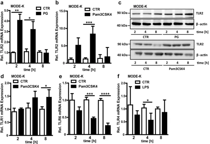
up-regulated upon longer stimulation periods with the TLR2 agonist Pam3CSK4 (Fig. 3d), while TLR6 expression was diminished at longer stimulation periods (Fig. 3e). In contrast to TLR4 expression in the small intestine of colonized mice (Fig. 1e) and to antibiotic decimation of the gut microbiota (Fig. 1j), treatment with LPS for 4h resulted in decreased TLR4 mRNA levels in MODE-K cells (Fig. 3f). Collectively, these results demonstrate an agonist-specific orchestration of the TLR expression profile in small intestinal epithelial cells.
Since cell culture experiments with Clostridium butyricum, a widely used probiotic bacterium, have recently demonstrated the induction of TLR2 expression in the human colon epithelial cell line HT-29 [20], we decided to further explore the effects of an individual microbial colonizer on the orchestration of the TLR2 signaling complex by the use of GF mouse isolator technology. To this end, we colonized GF C57BL/6 mice with theE. colistrain JP 313 for a time period of 14 days. Monocolonization with theE. colistrain JP 313 did not alter mRNA levels of TLR2, TLR1 and TLR4 (Fig. 4a,b,d), but significantly reduced transcript levels of the TLR2 co-receptor TLR6 (Fig. 4c). This implies that colonization with single microbes could specifically affect expres-sion of epithelial pattern recognition receptors.
Since we found that the gut microbiota and even individual colonizers impact on epithelial TLR2/1 or TLR2/6 receptor component expression, we next analyzed if relevant downstream kinase signaling pathways could be activated in epithelial cells by stimulation with PAMPs. In the sterile infection MODE-K model, stimulation with the TLR2 agonists lipoteichoic acid (LTA), Pam3CSK4 and heat-killed Listeria monocytogenes (HKLM) readily activated TLR downstream signaling pathways as shown by phosphorylation of ERK1/2 (mitogen-activated protein kinase 1 and 2) (Fig. 5a). ERK phosphorylation was also increased by stimulation with PG, LPS and MALP-2. The bacterial TLR2-agonists Macrophage-activating lipopeptide-2 (MALP-2), LTA and LPS-induced phosphorylation of the inhibitor of Nuclear Factor kappa-light-chain-enhancer of activated B cells (IkB), the natural inhibitor of the transcription factor NFkB (Fig. 5b). Furthermore, Protein-kinase B (AKT)-phosphorylation could also be triggered by stimulation with Pam3CSK4 in the MODE-K sterile infection model (Fig. 5c). Comparable to the changes in TLR2 expression (Fig. 3a), the increase in phosphorylation was most intense 2 to 4 hours upon stimulation (Fig. 5a–c) and decreased to levels of unstimulated controls at 8 hours upon stimulation (Fig. 5c). Collectively, we show that the small intestinal epithelial cell line MODE-K is reactive to TLR2 Table 1.Primer Table.
Primer Abbreviation Recognized cDNA Oligonucleotide Sequence
mL32_for 60S ribosomal protein L32 TGGCTCCTTCGTTGCTGCTG
mL32_rev 60S ribosomal protein L32 CTGGACGGCTAATGCTGGTG
mCasp3_for murine Caspase 3 TGGTGATGAAGGGGTCATTTATG
mCasp3_rev murine Caspase 3 TTCGGCTTTCCAGTCAGACTC
mTLR1_for murine Toll-like receptor 1 TCAAGCATTTGGACCTCTCCT
mTLR1_rev murine Toll-like receptor 1 TTCTTTGCATATAGGCAGGGC
mTLR2_for murine Toll-like receptor 2 ACAATAGAGGGAGACGCCTTT
mTLR2_rev murine Toll-like receptor 2 AGTGTCTGGTAAGGATTTCCCAT
mTLR4_for murine Toll-like receptor 4 ATGGCATGGCTTACACCACC
mTLR4_rev murine Toll-like receptor 4 GAGGCCATTTTTGTCTCCACA
mTLR6_for murine Toll-like receptor 6 TGAGCCAAGACAGAAAACCCA
mTLR6_rev murine Toll-like receptor 6 GGGACATGAGTAAGGTTCCTGTT
mKi67_for murine antigen identified by
monoclonal antibody Ki-67
CAATGTGCCTCGCAGTAAGA
mKi67_rev murine antigen identified by
monoclonal antibody Ki-67
GCATCTTTGGGGTTTTCTCA
mCyclinD1_for murine Cyclin D1 GCGTACCCTGACACCAATCTC
mCyclinD2_rev murine Cyclin D1 CTCCTCTTCGCACTTCTGCTC
mMyD88_for murine Myeloid differentiation
primary response gene (88)
AGGACAAACGCCGGAACTTTT
mMyD88_rev murine Myeloid differentiation
primary response gene (88)
GCCGATAGTCTGTCTGTTCTAGT
mTrif_for murine TIR-domain-containing
adapter-inducing interferon-b
TTGGGGACATACGTTACACTCC
mTrif_rev murine TIR-domain-containing
adapter-inducing interferon-b
CGGTGTGTTACATAGCTTGCTG
16S UniF_for Universal primer for 16S qPCR GTGSTGCAYGGYYGTCGTCA
16S UniR_rev Universal primer for 16S qPCR ACGTCRTCCMCNCCTTCCTC
8F Universal primer for control PCRs AGAGTTTGATCCTGGCTCAG
338R Universal primer for control PCRs TGCTGCCTCCCGTAGGAGT
activating PAMPs via several protein kinase pathways related to cellular proliferation.
A pivotal role of MyD88-dependent pattern recognition has previously been suggested for proliferation and differentiation of the gut epithelial lining [21] and a mitogenic effect has been demonstrated for TLR2 agonists in a human lung epithelial cell line [22]. However, the induction of the proliferative cell response in intestinal epithelial cells has not been unambiguously assigned to TLR2 in previous studies. The presence of an intestinal microbiota results in increased renewal of the epithelial lineage [3,6,8] as indicated by increased mRNA levels of the proliferation marker Ki67 in CONV-R mice compared with GF controls (Fig. 6a). Increased small intestinal cell turn over was further corroborated by increased Cyclin D1 mRNA levels (Fig. 6b), which drives G1/S phase transition, and increased mRNA levels of the apoptotic marker Caspase-3 (Fig. 6c). Since TLR2 is up-regulated upon microbial colonization, we reasoned that TLR2 signaling could stimulate the proliferation of small intestinal epithelial cells. Indeed, transcript levels of the proliferation marker Ki67 and Cyclin D1 were vastly reduced in Tlr22/2 mice
(Fig. 6d,e). In support of decreased cell turn over in Tlr22/2
mice, mRNA levels of the apoptotic marker Caspase-3 were also decreased (Fig. 6f). Reduced apoptosis in the small intestine of Tlr22/2mice was accompanied by decreased numbers of cleaved
Caspase-3 positive cells in the villus structures (Fig. 6g and
Fig. S1). In addition to TLR2 deficiency, the markers of cell proliferation (Ki67), cell turn over (Cyclin D1) and apoptosis (Caspase-3) were also changed in the ileum of Tlr42/2mice, but
not in Tlr52/2mice, indicating that intestinal homeostasis could
be influenced by diverse PAMPs (Fig. S2). Only Caspase-3 was affected in the Tlr72/2mice, whereas Ki67 and Cyclin D1 were
unchanged (Fig. S2). The role of TLR signaling on the expression of apoptotic markers was in line with the significant increase in TUNEL-positive nuclei in small intestinal villus structures of CONV-R mice compared with GF controls that are devoid of gut microbial TLR activation (Fig. S3a,b). Interestingly, monocolo-nization with theE. colistrain JP 313 was not sufficient to alter small intestinal cell turn over as Ki67, Cyclin D1 and Caspase-3 transcript levels were unchanged (Fig. 6h–j). In accordance with decreased small intestinal tissue renewal observed in GF and Figure 1. Microbial colonization leads to induction of TLR2 receptor expression in the small intestine. a,Relative TLR2 mRNA levels in small intestinal tissues from GF, CONV-D and CONV-R mice (n = 7 Swiss Webster mice per group).b,TLR2 immunoblot of isolated small-intestinal enterocyte lysates from GF and CONV-R mice (n = 6 mice per group; shown is one representative blot).c–e,Relative TLR1, 6 and 4 mRNA levels in small intestinal tissues from GF, CONV-D and CONV-R mice (n = 6–7 mice per group).f,qPCR analyses of feces samples of control mice and mice treated with antibiotics for 7 days. qPCR was performed using universal primers for 16S bacterial sequences and normed toE. colibacterial counts (cfu/ml).g–j,Relative mRNA levels of TLR2, 1, 6 and 4 in small intestinal tissues from CONV-R mice treated with a cocktail (ABX) of ampicillin (1 g/L) and neomycin (0.5 g/L) for 7 days (n = 6–7 mice per group). Female Swiss Webster mice or cells isolated from these mice were analyzed. Results are shown as means6s.e.m. One asterisk, P,0.05; two asterisks, P,0.01; three asterisks, P,0.005.
Tlr22/2mice, we found that 2 h stimulation of MODE-K cells
with the TLR2 agonist PG resulted in a pronounced proliferation response as indicated by elevated Ki67 transcript levels (Fig. 6k). In line, BrdU incorporation of MODE-K cells was increased by 2-fold after 24 hours of PG stimulation (Fig. 6l). We could demonstrate that pattern recognition induced cell proliferation was clearly TLR2 mediated since siRNA silencing of TLR2 expression led to a vast reduction of BrdU incorporation of PG stimulated MODE-K cells (Fig. 6m,n). Our results indicate that the orchestration of TLR2 signals in terminally differentiated small intestinal epithelial cells regulates cell turn over by evoking a pronounced epithelial proliferation response that is accompanied by a modest increase in apoptosis.
Discussion
Here, we report that expression of the epithelial pattern recognition receptor TLR2 and its co-receptor TLR1 in the small intestinal mucosa is tuned by PAMPs of the intestinal microbiota (Fig. 1). Our results with Myd882/2 and Trif2/2 mouse lines suggest that transcriptional regulation of TLRs in the small intestine depends on the adapter molecules MyD88 and TRIF, which also underlay a regulation by the gut microbiota (Fig. 2). Microbiota-driven TLR expression is a reversible process as antibiotic decimation of gut microbial communities led to reduced transcript levels of various TLRs (e.g. TLR1, TLR2, TLR6 and TLR4) (Fig. 1). Both Myd882/2 and Trif2/2 impaired expres-sion of TLR2/1 and TLR2/6 in the ileum (Fig. 2e, g and
Fig. 2i–k). Interestingly, Tlr2-deficiency leads to decreased TLR1 transcripts, but increases TLR6 transcript levels (Fig. 2m,n). Furthermore, we found that Tlr2-deficiency impacts on ileal TLR4 mRNA levels and vice versa (Fig. 2o,p). In accordance with a previous cell culture study [20], we could specifically provoke alterations in TLR transcript levels in the small intestine by monocolonization withE. coliJP 313 (Fig. 4). Up-regulation of the TLR2 signaling complex by TLR2 agonists could be confirmed in a cell culture model using the small intestinal epithelial cell line MODE-K (Fig. 3a–f). Interestingly, this terminally differentiated intestinal epithelial cell line is able to recognize PAMPs. Pam3CSK4 signals via the TLR2/1 complex, while PG triggers diverse signaling pathways [23]. Increased TLR2 transcripts upon Pam3CSK4 stimulation (Fig. 3b) along with modestly increased TLR1 transcripts (Fig. 3d) and de-creased TLR6 transcripts (Fig. 3e) suggest that stimulation with the TLR2/1 agonist Pam3CSK4 augments signaling though its utilized signaling complex TLR2/1, but impairs TLR2/6 signaling. Expression profiling of cell cycle and proliferation markers in small intestinal tissues from GF and CONV-R mice and from Tlr22/2 mice demonstrates a role for TLR2 in small intestinal cell turn over (Fig. 6a–g), but this proliferation response could not be evoked by monocolonization with theE. colistrain JP 313 (Fig. 6h–j). With a sterile infection cell culture model and siRNA silencing of TLR2 we were able to specifically pinpoint the induction of the epithelial proliferation response to TLR2 (Fig. 6k–n). These findings are further corroborated by cell culture experiments which demonstrated a role for TLR2 in activation of diverse protein kinase pathways that are involved in Figure 2. Adapter molecules MyD88 and TRIF alter expression
of TLRs in the ileum – indications for TLR receptor cross-talk in the small intestine. a+b, Relative MyD88 and TRIF mRNA levels in
small intestine from GF, CONV-D and CONV-R mice (n = 6–7 Swiss Webster mice per group).c+d, Relative MyD88 and TRIF mRNA levels in
small intestine from CONV-R mice treated for 7 days with an antibiotic cocktail (ABX) (n = 6–8 Swiss Webster mice per group). e–h, Relative TLR2, 1, 6 and 4 mRNA levels in small intestine from MyD882/2mice
compared to wildtype (WT) controls (n = 6–7 C57BL/6J mice per group). i–l, Relative TLR2, 1, 6 and 4 mRNA levels in small intestine from TRIF2/2
mice compared to WT controls (n = 6–7 mice per group).m–o, Relative
TLR1, 6 and 4 mRNA levels in small intestine from TLR22/2 mice
compared to WT controls (n = 6–7 mice per group).p, Relative TLR2 mRNA levels in small intestine from TLR42/2 mice compared to WT
controls (n = 6–7 mice per group). Female mice were analyzed. Results are shown as means6s.e.m. One asterisk, P,0.05; two asterisks, P, 0.01; three asterisks, P,0.005; four asterisks, P,0.001.
epithelial proliferation of terminally differentiated small intestinal epithelial cells (Fig. 5).
Although it is evident that innate immune sensing augments renewal of the intestinal epithelial lining primarily from the
stem cell niche situated at the base of the crypts of Lieberku¨hn [1,8], we suggest that the TLR2 dependent proliferation response that can be evoked in differentiated small intestinal epithelial cells (MODE-K) might represent an additional route that ensures integrity of the intestinal lining during renewal of the intestinal mucosa (Fig. S4). This proliferative adaption to microbial stimuli could serve to support efficient nutrient uptake in the distal small intestine. A recent study with GF rats has demonstrated a role for the gut microbiota and for E. coli in the augmentation of colonic epithelial cell proliferation as indicated by elevated proliferation markers, but the microbial components that unleash this proliferation response and the activated signaling pathways were not delineated [24,25]. Our data suggest that increased epithelial cell proliferation (Fig. 6) could be due to increased TLR2 signaling resulting in activation of the ERK1/2 and AKT pathways (Fig. 5). These results are in line with previous cell culture studies demonstrating the dependence of intestinal epithelial cell proliferation on ERK1/2 and AKT signaling [26,27]. Further studies are needed to identify which cell cycle stimulatory factors are specifically induced via TLR2 on small intestinal epithelial cells that can act in an autocrine or paracrine manner to induce proliferative and apoptotic pathways in the small intestinal mucosa and to pinpoint the possible role of ROS signaling pathways that are linked to innate immune signaling and renewal of the intestinal epithelium [28].
Figure 3. Agonist-specific orchestration of the TLR expression profile in a mouse small intestinal epithelial cell line. a+b,Relative
mRNA expression of TLR2 in MODE-K cells stimulated with PG (50mg/ml) or Pam3CSK4 (0.5mg/ml) for 2, 4 or, 8 hours (n = 4).c,TLR2 immunoblot of PG or Pam3CSK4 stimulated MODE-K cells. Cells were treated with or without (CTR) PG or Pam3CSK4 for 2, 4, or 8 hours (n = 3, representative blot).d– f,Relative TLR1, 6 and 4 mRNA expression in MODE-K cells stimulated with Pam3CSK4 or LPS for 2, 4, or 8 hours (n = 4). Results are shown as means6 s.e.m. One asterisk, P,0.05; two asterisks, P,0.01; three asterisks, P,0.005; four asterisks, P,0.001.
doi:10.1371/journal.pone.0113080.g003
Figure 4. Monocolonization withE. coliJP313 decreases TLR6 transcript levels. a–d,Relative TLR2, 1, 6, and 4 mRNA levels in small intestine from mice colonized for 14 days withE. coliJP313 (n = 7 male C57BL/6 mice per group). Results are shown as means6s.e.m. Two asterisks, P,0.01.
Interestingly, decimation of the gut microbiota by treatment with broad-spectrum antibiotics diminished transcript levels of various TLRs in the small intestine of CONV-R mice (Fig. 1f–j). This finding implies that eradication of colonizing microbial communities during antibiotic therapy may possibly attenuate epithelial innate immune sensing and hence to some extent limit the development of inflammation. This in some cases might be a double edged sword, since antibiotic treatment may select for resistant germs and at the same time disable protective innate immune responses. Furthermore, there is mounting evidence for that deficiency in pattern recognition could select for colonization with pathobionts that may exert deleterious effects in the absence of an adequate inflammatory response [29]. Our data on small intestinal TLR2 expression imply a crosstalk between MyD88 and TRIF-dependent TLR signaling circuits [30,31], since TLR2 expression is affected in both MyD88 and TRIF-deficient mouse lines (Fig. 2e–l). Further, we found that deficient TLR4 signaling augments TLR2 transcripts and vice versa (Fig. 2m–p). Since it is not sufficiently resolved how stimulation of TLRs with specific agonists or genetic deficiency of individual TLRs affects the expression of other receptors of this family, the context-specific role of the crosstalk between MyD88 and TRIF-dependent signaling pathways in the modulation of TLR expression in the small intestinal epithelium will deserve further investigation. Such efforts will be inevitable to understand the complexity of TLR signaling and its role in small intestinal tissue homeostasis.
While a robust effect of a complex gut microbiota was observed on small intestinal TLR2 expression (Fig. 1a–c), renewal and cell
turn over (Fig. 6a–c), it was not possible to augment mucosal proliferation in the small intestine by colonization with the proteobacterial colonizer E. coli JP 313 (Fig. 6h–j) [32]. This strain of E. coli inoculated alone did not trigger mucosal proliferation possibly due to the fact that colonization with JP 313 alone is not sufficient to counterbalance mucosal atrophy observed in GF mice [5,24]. The situation in monocolonized mice is in contrast to cell culture experiments with PG stimulated MODE-K cells where a pronounced TLR2-dependent prolifera-tion response could be evoked (Fig. 6k–n). Hence, monocoloni-zations with additional gut microbes and importantly with bacterial deletion mutants that target PAMP synthesis will be mandatory to reveal the significance of individual microbes and bacterial membrane components for TLR induction and epithelial cell proliferation.
In addition to the identified role of TLR2 in gut epithelial cell proliferation this pattern recognition receptor has recently been implicated in permeability regulation of the epithelial lining [33]. The regulation of barrier function is one way how TLR2 could affect host metabolism, but there is increasing evidence for additional modes of action [34]. TLR2 has been shown to have profound effects on the composition of gut microbial communities [34,35]. A recent study suggests that alterations in gut microbiota composition as they are found in TLR deficient mice may rather be caused by familiar transmission than by innate deficiency [36]. Alterations in the diversity of the gut microbiota are well known to impact on host metabolism [34]. These recent reports clearly point to roles of TLR2 beyond regulation of small intestinal tissue Figure 5. MODE-K cells are reactive to TLR2 activating PAMPs and LPS. a,Detection of ERK1/2-phosphorylation of TLR-agonist (LTA, 10mg/
ml; Pam3CSK4, 0.5mg/ml; HKLM, 26105cells per well; PG, 50mg/ml; MALP-2, 2mg/ml; LPS, 100 ng/ml) treated MODE-K cells (n = 4–10).b,Detection
of IkB-phosphorylation of TLR-agonist treated MODE-K cells (n = 3). c, Detection of AKT-phosphorylation of Pam3CSK4 treated MODE-K cells (n = 3).One representative immunoblot is shown for each agonist and kinase.
homeostasis. In this context, further experiments including cell-specific targeting of TLR2 are required to differentiate the effects arising from the myeloid lineage from the effects that are originating from epithelial cells.
Previous work has established the protective role of TLR2 and TLR4 and of the gut microbiota to resist to chemical induced epithelial injury [21]. Mice that were deficient in MyD88-dependent signaling or mice subjected to antibiotic treatment resulting in deciminated indigeneous microbiota showed a vastly Figure 6. TLR2 signals in the small intestinal epithelium evoke a proliferation response and increase signs of apoptosis in terminally differentiated epithelium. a–c, Relative Ki67, Cyclin D1 or Caspase-3 mRNA levels in small intestine from GF and CONV-R mice (n = 7 Swiss Webster mice per group).d–f, Relative Ki67, Cyclin D1 or Caspase-3 mRNA levels in small intestine from Tlr22/2mice compared with WT
controls (n = 7 C57BL/6J mice per group).g, Number of Caspase-3 positive cells per 10 villi in small intestinal samples from WT or Tlr22/2mice.h–j,
Relative Ki67, Cyclin D1 and Caspase-3 mRNA levels in small intestine from mice colonized for 14 days withE. coliJP313 (n = 7 C57BL/6 mice per group).k, Relative Ki67 mRNA levels in MODE-K cells stimulated with PG (50mg/ml) for 2, 4, or 8 hours (n = 4).l, Relative proliferation of PG treated
MODE-K cells measured by incorporation of BrdU compared to untreated control cells (n = 3).m, Relative proliferation of MODE-K cells transfected with siRNA against TLR2 or scrambled control RNA measured by incorporation of BrdU (n = 4).n, TLR2 immunoblot of siRNA transfected MODE-K cells. Results are shown as means6s.e.m. One asterisk, P,0.05; two asterisks, P,0.01; three asterisks, P,0.005; four asterisks, P,0.001.
increased lethality during long term DSS treatment. A defect of steady-state intestinal epithelial homeostasis in absence of TLR signaling was suggested to be fundamental for the increased susceptibility to epithelial damage [21]. Indeed, the induction of a proliferation response in terminally differentiated intestinal epithelial cells via TLR2 could be beneficial under conditions where enhanced renewal and mucosal repair is required, e.g. in radiotherapy or chemotherapy-induced intestinal mucositis [37]. With respect to these relevant clinical complications further studies are needed to pinpoint the exact role of innate immune signaling in mucosal repair processes.
Supporting Information
Figure S1 TLR2-deficiency leads to decreased signs of apoptosis in the small intestine. Stainings of Caspase-3 expression in small intestinal tissue sections from WT and Tlr22/2
mice. Sections were embedded in paraffin, cut in 8mm sections and stained with an anti-Caspase-3 antibody. 20x magnifications are shown.
(TIF)
Figure S2 Cell turn over is changed in the ileum of Tlr42/2and Tlr72/2mice, but not in Tlr52/2mice. a–c,
Relative Ki67, Cyclin D1 and Caspase-3 mRNA levels in small intestine from Tlr42/2mice compared with WT controls (n = 7
female mice per group). d–f, Relative Ki67, Cyclin D1 and Caspase-3 mRNA levels in small intestine from Tlr52/2 mice
compared with WT controls (n = 6 male mice per group). g–i,
Relative Ki67, Cyclin D1 and Caspase-3 mRNA levels in small intestine from Tlr72/2mice compared with WT controls (n = 5–8
mice per group). Results are shown as means 6 s.e.m. One asterisk, P,0.05; two asterisks, P,0.01; four asterisks, P,0.001. (TIF)
Figure S3 The gut microbiota increases apoptosis in the small intestine. a, TUNEL-staining of small intestinal tissue sections of GF and CONV-R female Swiss Webster mice.
Paraffin-embedded samples were cut in 8mm sections. Tissues
were deparaffinized, rehydrated and nicks were FITC labeled by the Terminal deoxynucleotidyl Transferase (TdT) reaction. Apoptotic cells are stained with fluorescein (green), nuclei are stained with DAPI (blue). 20x magnifications are shown. b,
Quantitative analysis of TUNEL-positive cells in GF and CONV-R tissue sections. CONV-Results are shown as means 6 s.e.m. Four asterisks, P,0.001.
(TIF)
Figure S4 Model delineating the role of the gut micro-biota on TLR2 agonist stimulated increase in down-stream kinase signaling, TLR2 expression and cell turn over of terminally differentiated enterocytes in the ileum.
(TIF)
Acknowledgments
We are grateful to Klaus-Peter Derreth for expert technical assistance, to Dominique Kaiserlian (INSERM, Cedex, France) for providing the MODE-K epithelial cell line, to Martin Busch (University of Wu¨rzburg, Germany) and Jennifer Hahlbrock (University Medical Center Mainz, Germany) for assisting with qPCR analyses, to Markus Radsak (Mainz, Germany) for providing Tlr42/2, Myd882/2 and Trif2/2 mouse lines, to Kerstin Steinbrink (Mainz, Germany) for providing Tlr72/2 mice, to Stepan Gambaryan (University of Wu¨rzburg, Germany) for antibody reagents, to Evelyne Turlin (Institute Pasteur, Paris, France) for providing E. coli strain JP313, to Fredrik Ba¨ckhed and Carina Arvidsson (University of Gothenburg, Sweden) for their help in establishing germ-free mouse technology in Mainz.
Author Contributions
Conceived and designed the experiments: NH UW CR. Performed the experiments: NH ISB NE SJ ML. Analyzed the data: NH ISB SJ ML CR. Contributed reagents/materials/analysis tools: UW CR. Contributed to the writing of the manuscript: NH ISB SJ ML UW CR.
References
1. Clevers H (2013) The intestinal crypt: a prototype stem cell compartment. Cell 154: 274–84.
2. Johansson ME, Sjo¨vall H, Hansson GC (2013) The gastrointestinal mucus system in health and disease. Nat Rev Gastroenterol Hepatol. 10: 352–61. 3. Abrams GD, Bauer H, Sprinz H (1963) Influence of the normal flora on mucosal
morphology and cellular renewal in the ileum. A comparison of germ-free and conventional mice. Lab Invest. 12: 355–64.
4. Stappenbeck TS, Hooper LV, Gordon JI (2002) Developmental regulation of intestinal angiogenesis by indigenous microbes via Paneth cells. Proc. Natl Acad. Sci. USA 99: 15451–15455.
5. Reinhardt C, Bergentall M, Greiner TU, Schaffner F, O¨ stergren-Lunde´n G, et al. (2012) Tissue factor and PAR1 promote microbiota-induced intestinal vascular remodelling. Nature 483: 627–31.
6. Savage DC, Siegel JE, Snellen JE, Whitt DD (1981) Transit time of epithelial cells in the small intestines of germfree mice and ex-germfree mice associated with indigeneous microorganisms. Appl Environ Microbiol. 42: 996–1001. 7. Abreu MT (2010) Toll-like receptor signalling in the intestinal epithelium: how
bacterial recognition shapes intestinal function. Nat Rev Immunol. 10: 131–143. 8. Nigro G, Rossi R, Commere P-H, Jay P, Sansonetti P (2014) The cytosolic bacterial peptidoglycan sensor Nod2 affords stem cell protection and links microbes to gut epithelial regeneration. Cell Host Microbe. 15: 792–8. 9. Yamamoto M, Sato S, Hemmi H, Hoshino K, Kaisho T, et al. (2003) Role of
adaptor TRIF in the MyD88-independent signalling pathway. Science 301: 640–3.
10. Otte JM, Cario E, Podolsky DK (2004) Mechanisms of cross hyporesponsiveness to Toll-like receptor bacterial ligands in intestinal epithelial cells. Gastroenter-ology 126: 1054–70.
11. Hornef MW, Frisan T, Vanderwalle A, Normark S, Richter-Dahlfors A (2002) Toll-like receptor 4 resides in the Golgi apparatus and colocalizes with internalized lipopolysaccharide in intestinal epithelial cells. J Exp Med. 195: 559–70.
12. Ortega-Cava CF, Ishihara S, Rumi MA, Kawashima K, Ishimura N, et al. (2003) Stratigic compartmentalization of Toll-like receptor 4 in the mouse gut. J Immunol. 170: 3977–85.
13. Cario E, Brown D, McKee M, Lynch-Devaney K, Gerken G, et al. (2002) Commensal-associated molecular patterns induce selective toll-like receptor-trafficking from apical membrane to cytoplasmic compartments in polarized intestinal epithelium. Am J Pathol. 160: 165–73.
14. Lundin A, Bok CM, Aronsson L, Bjo¨rkholm B, Gustafsson JA, et al. (2008) Gut flora, Toll-like receptors and nuclear receptors: a tripartite communicaton that tunes innate immunity in large intestine. Cell Microbiol. 10: 1093–1103. 15. Janssens S, Beyaert R (2003) Functional diversity and regulation of different
interleukin-1 receptor-associated kinase (IRAK) family members. Mol Cell. 11: 293–302.
16. Zhang G, Ghosh S (2002) Negative regulation of Toll-like receptor 2-mediated signaling via Tollip. J Biol Chem. 277: 7059–65.
17. Cani PD, Bibiloni R, Knauf C, Waget A, Neyrinck AM, et al. (2008) Changes in gut microbiota control metabolic endotoxemia –induced inflammation in high-fat diet-induces obesity and diabetes in mice. Diabetes 57: 1470–81. 18. Fuller Z, Louis P, Mihajlovski A, Rungapamestry V, Ratcliffe B, et al. (2007)
Influence of cabbage processing methods and prebiotic manipulation of colonic microflora on glucosinolate breakdown in man. Br J Nutr. 98: 364–72. 19. Vidal K, Grosjean I, Evillard JP, Gespach C, Kaiserlian D (1993)
Immortalization of mouse intestinal epithelial cells by the SV40-large T gene. Phenotypic and immune characterization of the MODE-K cell line. J Immunol Methods. 166: 63–73.
20. Gao Q, Qi L, Wu T, Wang J (2012)Clostridium butyricumactivates TLR2-mediated MyD88-independent signaling pathway in HT-29 cells. Mol Cell Biochem. 361: 31–37.
22. Shaykhiev R, Behr J, Bals R (2008) Microbial patterns signalling via Toll-like receptors 2 and 5 contribute to epithelial repair, growth and survival. PLoS one 1: e1393.
23. Ozinsky A, Underhill DM, Fontenot JD, Haijar AM, Smith KD, et al. (2000) The repertoire for pattern recognition of pathogens by the innate immune system is defined by cooperation between toll-like receptors. Proc Natl Acad Sci USA. 97: 13766–71.
24. Tomas J (2014) Early colonizing E. coli elicits remodeling of rat colonic epithelium shifting toward a new homeostatic state. ISME J.: doi: 10.1038/ ismej.2014.111.
25. Cherbuy C, Honvo-Houeto E, Bruneau A, Bridonneau C, Mayeur C, et al. (2010) Microbiota matures colonic epithelium through a coordinated induction of cell cycle-related proteins in gnotobiotic rat. Am J Physiol Liver Physiol. 299: G348–357.
26. Go¨ke M, Kanai M, Lynch-Devaney K, Podolsky DK (1998) Rapid mitogen-activated protein kinase activation by transforming growth factor alpha in wounded rat intestinal epithelial cells. Gastroenterology 114: 697–705. 27. Sheng H, Shao J, Townsend CM Jr, Evers BM (2003) Phosphatidylinositol
3-kinase mediates proliferative signals in intestinal epithelial cells. Gut 52: 1472–8. 28. Sommer F, Ba¨ckhed F (2014) The gut microbiota engages different signaling pathways to induce Duox2 expression in the ileum and colon epithelium. Mucosal Immunol. doi: 10.1038/mi.2014.74.
29. Ji Y, Sun S, Goodrich JK, Kim H, Poole AC, et al. (2014) Diet-induced alterations in gut microbiota contribute to lethal pulmonary damage in TLR2/ TLR4-deficient mice. Cell Rep. 8: 137–49.
30. Petnicki-Ocwieja T, Chung E, Acosta DI, Ramos LT, Shin OS, et al. (2013) TRIF mediates Toll-like receptor 2-dependent inflammatory responses to
Borrelia burgdorferi. Infect Immun. 81: 402–10.
31. Ouyang X, Negishi H, Takeda R, Fujita Y, Taniguchi T, et al. (2007) Cooperation between MyD88 and TRIF pathways in TLR synergy via IRF5 activation. Biochem Biophys Res Commun. 354: 1045–51.
32. Kamada N, Chen GY, Inohara N, Nunez G (2013) Control of pathogens and pathobionts by the gut microbiota. Nat Immunol. 14: 685–690.
33. Hanson PJ, Moran AP, Butler K (2011) Paracellular permeability is increased by basal lipopolysaccharide in a primary culture of colonic epithelial cells; an effect prevented by an activator of Toll-like receptor-2. Innate Immun. 17: 269–82. 34. Caricilli AM, Picardi PK, de Abreu LL, Ueno M, Prada PO, et al. (2011) Gut
microbiota is a key modulator of insulin resistance in TLR2 knock out mice. PLoS Biol. 9: e1001212.
35. Round JL, Lee SM, Li J, Tran G, Jabri B, et al. (2011) The Toll-like receptor 2 pathway establishes colonization by a commensal of the human microbiota. Science 332: 974–7.
36. Ubeda C, Lipuma L, Gobourne A, Viale A, Leiner I, et al. (2012) Familial transmission rather than defective innate immunity shapes the distinct intestinal microbiota of TLR-deficient mice. J Exp Med. 209: 1445–56.