ISSN 0100-879X
BIOMEDICAL SCIENCES
AND
CLINICAL INVESTIGATION
www.bjournal.com.br
www.bjournal.com.br
Volume 43 (04) 325-408 April 2010
Braz J Med Biol Res, March 2010, Volume 43(4) 330-337
Tumor necrosis factor alpha increases epithelial barrier
permeability by disrupting tight junctions in Caco-2 cells
W. Cui, L.X. Li, C.M. Sun, Y. Wen, Y. Zhou, Y.L. Dong and P. Liu
Institutional Sponsors
Tumor necrosis factor alpha increases
epithelial barrier permeability by
disrupting tight junctions in Caco-2 cells
W. Cui
1, L.X. Li
2, C.M. Sun
1, Y. Wen
1, Y. Zhou
1, Y.L. Dong
1and P. Liu
11Department of Infectious Diseases, the First Affiliated Hospital, China Medical University,
Shenyang, Liaoning, China
2Department of Infectious Diseases, The First Hospital of Xiamen Affiliated to the Fujian Medical University,
Xiamen, Fujian, China
Abstract
The objectives of this studywere to determine the effect of tumor necrosis factor alpha (TNF-α) on intestinal epithelial cell permeability and the expression of tight junction proteins. Caco-2 cells were plated onto Transwell® microporous filters and treated with TNF-α (10 or 100 ng/mL) for 0, 4, 8, 16, or 24 h. The transepithelial electrical resistance and the mucosal-to-serosal flux ratesof the established paracellular marker Lucifer yellow were measured in filter-grown monolayers of Caco-2intestinal cells. The localization and expression of the tight junction protein occludin were detected by immunofluorescence and West-ern blot analysis,respectively. SYBR-Green-based real-time PCR was used to measure the expression of occludin mRNA. TNF-α treatment produced concentration-and time-dependent decreases in Caco-2 transepithelial resistance and increasesin transepithelial permeability to the paracellular marker Lucifer yellow.Western blot results indicated that TNF-α decreased the expression of phosphorylated occludin in detergent-insolublefractions but did not affect the expression of non-phosphorylated occludin protein. Real-time RT-PCR data showed that TNF-α did not affect the expression of occludin mRNA. Taken together, our data demonstrate that TNF-α increases Caco-2 monolayer permeability, decreasesoccludin protein expression and disturbs intercellular junctions.
Key words: TNF-α; Intestinal epithelial barrier; Occludin; Caco-2 cells; Transepithelial permeability
Introduction
Correspondence: P. Liu, Department of Infectious Diseases, the First Affiliated Hospital, China Medical University, Shenyang, Liaoning, China. Fax: +86-24-3153-5053. E-mail: [email protected]
Received June 17, 2009. Accepted March 3, 2010. Available online March 19, 2010. Published April 12, 2010. The intestinal mucosal barrier plays an important role in
the body’s protection against luminal pathogens and antigenic molecules. The intestinal barrier includes secreted mucus
and the epithelial cell itself, which serves as a selective bar -rier permitting the uptake of nutrients, ions and other desired solutes, thus helping to maintain homeostasis of the internal
environment. Pathological events such as intestinal inflam
-mation, sepsis, burn, end-stage liver diseases, and severe
pancreatitis cause impairment of the intestinal epithelial barrier. Therefore, bacteria and their lysis products, such as lipopolysaccharide, gain access to the portal and systemic
circulations inducing systemic inflammatory response syn
-drome or even multiple organ dysfunction syn-drome.
Increased intestinal epithelial barrier permeability is caused not only by exogenous factors such as infection and ischemia but also by the local immune system. Tumor
necrosis factor alpha (TNF-α), which is increased in many disease states, is a proinflammatory cytokine. Both in vivo
and in vitro studies (1-5) have demonstrated that TNF-α can
increase the permeability of the intestinal epithelium and
that anti-TNF-α antibody can restore the barrier’s function (6,7). The precise mechanism by which TNF-α increases intestinal permeability is unclear. However, epithelial cell
apoptosis (3,4,8), protein kinase C, nuclear factor-kappaB,
myosin light chain kinase, and mitogen-activated protein kinase are thought to be involved (9-12).
In intestinal epithelial cells, tight junctions (TJ) are the most luminal cell-to-cell junctions. They seal the
paracel-lular space between individual epithelial cells against pas
-sive solute flux. The transmembrane proteins, occludins,
claudins and zonular occludens proteins, form the TJ.
TNF alpha disrupts intestinal tight junctions 331
protein of the TJ, is considered to be the major regulatory
protein of TJs (13-15). Post-translational modifications of the occludin protein involve phosphorylation events, and multiple phosphorylation sites have been identified on oc
-cludin serine and threonine residues (16). Several proteins
such as Rho kinase, protein kinase C, protein phosphatase 2A, and casein kinase 2 may regulate occludin phospho-rylation (17,18). The phosphophospho-rylation state of occludin mediates its association with the cell membrane and barrier permeability. Sakakibara et al. (19) demonstrated that TJ assembly induced by a calcium switch is paralleledby oc-cludin phosphorylation, and highly phosphorylatedoccludin
molecules are selectively concentrated at TJs, whilenon- or less phosphorylated occludin is localized in the cytoplasm. It has also been shown that thephosphorylation of occludin might regulate TJ permeabilityin response to histamine and
lysophosphatidic acid (16).
Thefindings cited above lead to the conclusion that
TNF-α can cause elevation of intestinal epithelial perme -ability, and the phosphorylation state of occludin may
regulate TJ permeability. However, it is unclear whether the elevation of intestinal epithelial permeability induced by TNF-α correlates with changes in occludin phosphorylation.
Therefore, in the present study, we employed Caco-2 cells as an in vitro intestinal epithelial model, observing changes in barrier permeability, in TJs and in the expression of the
highly or non-phosphorylated occludin induced by TNF-α
to characterize the mechanism of the disruption of the
epithelial barrier in response to TNF-α.
Material and Methods
Cell culture
The Caco-2 cell line (passage 23) was obtained from the American TypeCulture Collection (Manassas, USA), and stock cultures weregrown in culture medium consisting of DMEM
(Gibco, USA) with 4.5 mg/mL glucose,10 g/L non-essential amino acids (Gibco), 50 U/mL penicillin, 50 U/mL streptomycin,
4 mM glutamine, and20% FBS (20). The culture medium was
changed every 1 to 2days. The cells were subcultured by
diges-tion with 0.25%trypsin and 0.9 mM ethylenediamine-tetraacetic acid in Ca2+-freeand Mg2+-free PBS. For growth on filters,
Caco-2 cells wereplated onto Transwell microporous filters
(Corning-Costar, USA) and monitored regularlyby measuring
transepithelial electrical resistance (TER). For experimental
purposes, only Caco-2 cellsbetween passages 24 and 30 were used.Cells were cultured for about 21 days before treat-ment.Next, cells were fed serum-free culture medium, and the medium was supplemented with the indicatedconcentration
of TNF-α (10 or 100 ng/mL; R&D Systems, USA) for various times (0, 4, 8, 16, or 24 h).
Determination of Caco-2 intestinal monolayer resistance and paracellular permeability
The TER of the filter-grown Caco-2intestinal monolayers
was measured with an epithelial voltameter(World
Preci-sion Instruments, USA) as previously described (21). For
resistance measurements, both the apical and basolateral sides of the epithelia were bathed in cell culture medium.
Electrical resistance was measured until similar values were recorded for three consecutive measurements.
The effect of TNF-α on Caco-2 monolayer paracellular
permeabilitywas determined by using the paracellular marker Lucifer yellow (Sigma-Aldrich,USA) as previously
described (22). DMEM, pH 7.4, was usedas the
incuba-tion soluincuba-tion during the experiments. HBSS, pH 7.4, buffer
solution (100 µL) was added to the apical compartment,
while 600 µL was added to the basolateral compartment
toensure equal hydrostatic pressure, as recommended by the manufacturer.Known concentrations of Lucifer yellow,
the permeability marker (40 μg/L), were added to the apical
solution. Basolateralsamples were taken at 60 min, and
fluorescence intensitywas determined with a fluorescent
plate reader. The amount of Lucifer yellow transported into the basolateralcompartment was extrapolated from a
stan-dard curve. The apical-to-basolateral transport of Lucifer
yellow was reported as the percentage of basolateral Lucifer yellow concentration compared with the original Lucifer yellow concentration. Lowconcentrations of permeability markers were used to ensure that a negligible osmotic or concentration gradient was introduced.All flux studies were carried out at 37°C. All experimentswere repeated three to six times in triplicate to ensure reproducibility.
Transmission electron microscopy
After dehydration through a graded series of ethanol, the membrane-intact Transwell insert was embedded in Araldite
epoxy resin. Areas selected for ultrastructural observations
andultrathin sections (~80 nm in thickness) were cut using a diamondknife and stained with saturated uranyl acetate and Reynold’slead citrate. Sections were viewed on a Hitachi
H7000 transmissionelectron microscope, and images were
captured digitally. Ultrastructureobservations were made
from multiple sites (>10) of junctionalcomplexes that were
clearly identified. Three examiners analyzed at least three
images from each treatment group in a blind fashion.
Preparation of detergent-soluble and -insoluble protein fractions
As previously described (19), Caco-2 cells were washed
three times with ice-cold PBS, immediatelyincubated in
Nonidet P-40 extraction buffer (25 mM HEPES, pH7.4,
150 mM NaCl, 4 mM EDTA, 1% Nonidet P-40, 25 mM
NaF, 1 mMNa3VO4, 10 mM sodium pyrophosphate, and
protease inhibitors)on ice for 30 min and centrifuged at 20,000 g for 30 min at4°C. The detergent-soluble frac-tion was transferred to amicrocentrifuge tube and the insoluble fraction was collected inSDS extraction buffer
(25 mM HEPES, pH 7.4, 4 mM EDTA, 1% SDS,25 mM
and sonicated. Supernatants (cytoskeleton-associated fractions)were then obtained after centrifugation at 20,000
g for 30 min. Protein concentrationswere measured using a Bradford Protein Assay Kit (Santa Cruz Biotechnology,
USA) according to manufacturer instructions using bovine
serum albumin as standard.
Assessment of occludin protein expression by Western blot analysis
Equal amounts of proteins (50 µg) from each sample
wereseparated by SDS-PAGE (8% polyacrylamide gel) and transferred to Immobilon™ transfer polyvinylidene
difluoride membranes (Millipore,USA). Blots were blocked with 5% nonfat milk and incubated sequentially with a rabbit polyclonal occludin antibody (1:1000; Cat. #711500,
Zymed, USA) and a horseradish peroxidase-conjugated
secondary antibody (1:2000) (Santa Cruz Biotechnology).
Antibody binding was detected with ECL plus (Amersham Pharmacia Biotech, USA).
RNA isolation and reverse transcription
The TRIZOL® reagent (Invitrogen Life Technology,
USA) was used to homogenize cells and to isolate total
RNA according to manufacturer instructions. The RNA
was purified by treatment with DNase I and deprotein
-ated by PI-PCI-ECHO. The concentration of total RNA
was determined by absorbanceat 260/280 nm. Reverse transcription was carried out using the SYBR® RT-PCR kit
(Takara Bio, Japan). Fromeach sample, 500 ng total RNA
was reverse-transcribed into cDNA in a 10-µL reaction
containing 5X M-MLV buffer,250 µM of each dNTP, 100 µM random hexamer, 10 units of RNase inhibitor, and 50 units of M-MLV RTase. Reactions were performed at 25°C
for 10 min, 42°C for10 min, and 95°C for 2 min.
Quantification of occludin mRNA expression using real-time PCR
SYBR-Green-based real-time PCR was used to mea
-sure relative gene expression in each sample. First, we
constructed the RNA standards for the gene of interest
(occludin) and for the housekeeping gene (GAPDH). To start
this process, PCR primers were created. The T7 promoter
was then added to a 5’-upstream primer, while twenty T’s
were added to a 3’-downstream primer. For the Caco-2
sample, the total RNA was reverse-transcribed and ampli
-fied by PCR (Takara Bio). For the RNA standards, the PCR
products were transcribed in vitro. The RNA was treated
with DNase I (Takara Bio) and purified by PI-PCI-ECHO. Samples were annealed using a gradient and amplified by real-time PCR. Then, standard curves were constructed for
the gene of interest (occludin) and for the housekeeping
gene (GAPDH).
The reactions for real-time PCR were carried out using the Line-gene 3310 real-time PCR detection system and the TaKaRa SYBR® RT-PCR kit (Takara). Each real-time
PCR assay contained 2 µL cDNA template, 12.5 µL SYBR Premix Ex Taq, and 0.25 µL of each forward and reverse primer (10 µM) in a 25-µL reaction. Primer design for the real-time PCR was performed using Primer Express version
2 from Takara Bio. The primers used in this study wereas
follows: Ocln-F: 5’-AAGAGTTGACAGTCCCATGGCATA C-3’, Ocln-R: 5’-ATCCACAGGCGAAGTTAATGGAAG-3’; GAPDH-F: 5’-AAATGGTGAAGGTCGGTGTG-3’, GAPDH-R: 5’-TGAAGGGGTCGTTGATGG-3’). All runs were performed
according to the PCRprotocol (95°C for 30 min, and 45 cyclesof 95°C for 5 s and 60°C for 20 s. For each sample, real-time PCR was performed in triplicate, and theaverage threshold cycle (Ct) was calculated. A standard curvewas
generated to convert the Ct into copy number. Expression
of occludin mRNA was normalized to GAPDH mRNA ex -pression. Theaverage copy number of occludin mRNAwas
set to 1.0 in the control samples. The relative expression of
occludin mRNA in the treatedsamples was determined as a fold increase compared with controlsamples.
Statistical analysis
Data are reported as means ± SD. Statistical analysis was performed by the Student t-test for unpaired data and by
one-way ANOVA with the LSDpost hoc test for comparison
with the control. A P value of ≤ 0.05 was used toindicate
statistical significance. All experiments were repeateda minimum of three times to ensure reproducibility.
Results
Effect of TNF-α on Caco-2 intestinal epithelial TJ permeability
In the following studies, the effect of TNF-α on Caco-2
TJ permeabilitywas determined by measuring TER and epithelial permeabilityto the paracellular marker Lucifer yellow. Increasing concentrations ofTNF-α (0-100 ng/mL) caused a dose-dependent decrease inCaco-2 TER after a 24-h period of treatment (Figure 1A). The cell monolayer
TER decreased from 124.5 ± 0.3 to 36 ± 0.5 Ω/cm2 after 10
ng/mL TNF-α treatment, and the maximaldrop in Caco-2
TER occurred when the concentration of TNF-α was 100 ng/mL (28 ± 0.7 Ω/cm2). The time course of the TNF-α (100
ng/mL)effect on Caco-2 TER is shown in Figure 1B. TNF-α did not havea significant effect on Caco-2 TER during the first 4 h of treatment (115.5 ± 0.6 vs 128.4 ± 1.0 Ω/cm2).
There was a sharp time-dependent drop in Caco-2 TER
between8 and 24 h (8 h: 65.1 ± 0.8, 16 h: 45.9 ± 0.4, 24
h: 23.05 ± 1.7 Ω/cm2) and the maximal drop in TER was
reached by 24 hof TNF-α treatment.
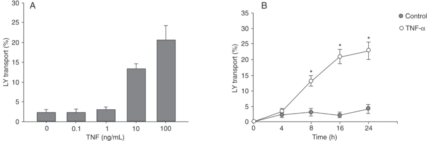
Conversely, TNF-α (100 ng/mL) caused a dose- and
time-dependentincrease in Caco-2 paracellular permeability to Lucifer yellow, and the maximal increase in paracellular permeability was also reached by 24 h of treatment with 100
ng/mL TNF-α (Figure 2). These results indicate that TNF-α
TNF alpha disrupts intestinal tight junctions 333
Effects of TNF-α on the ultrastructure of TJ
Since epithelial TJs regulate paracellular permeability,
the ultrastructure of Caco-2 cells, treated as previously
described,was analyzed by transmission electron micro-copy (Figure 3). The presenceof electron-dense material in the space between the cells and near thebrush border
reflected the TJs. In cells lacking TNF-α (Figure 3A), the
TJ had an intact structure. Incells treated with 100 ng/
mL TNF-α in the medium for 24 h, the TJs were reduced
and containedless electron-dense material and the space between cells was enlarged (Figure 3B). These results
demonstrate that TNF-α treatment resulted indisruption of normal TJ morphology.
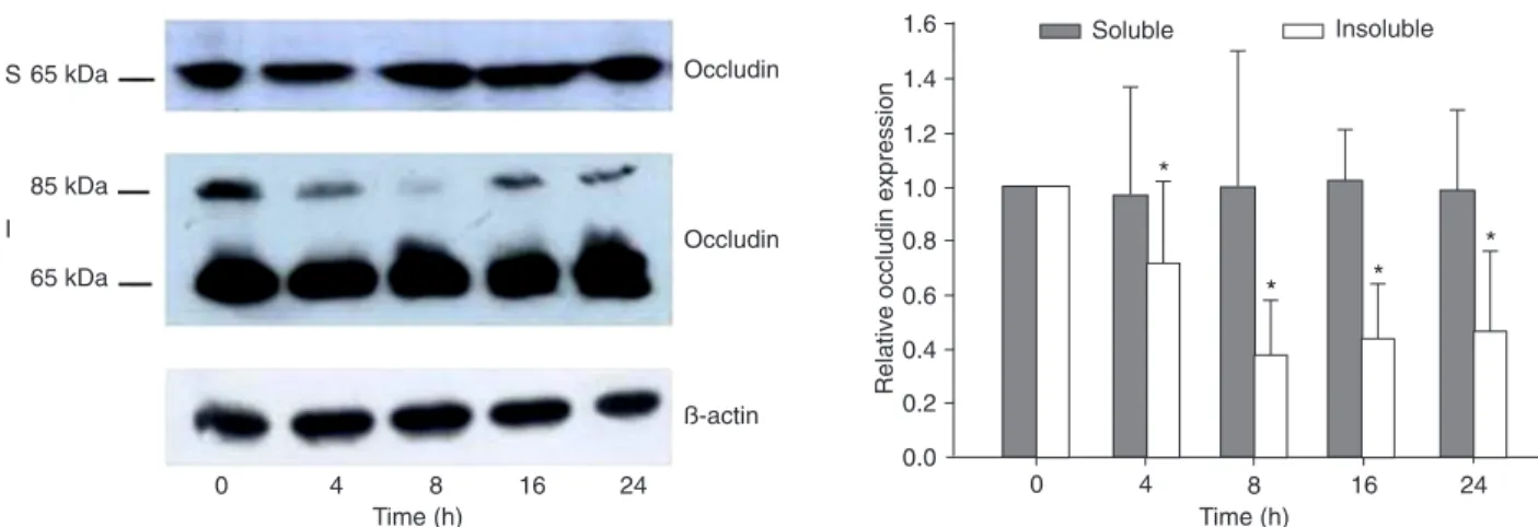
TNF-α induces a protein-specific down-regulation of occludin protein
Western blot using an antibody specific for occludin demonstrated that both 85- and 65-kDa forms were seen in
detergent-insolubleprotein extracts. However, only the 65-kDa form wasseen in the detergent-soluble fraction. TNF-α
did not affect the expression of the 65-kDa occludin in either
detergent-soluble or insolublefractions. However, it reduced
the 85-kDa occludin expression in the detergent-insoluble
fractions (Figure 4). According to Sakakibara et al. (19),
occludin from Madin-Darby canine kidney cells resolved as several bands between 62 and 82 kDa by SDS-PAGE
analysis. Among these bands, the lower predominant bands
Figure 1. Effect of tumor necrosis factor alpha (TNF-α) on transepithelial electrical resistance(TER). TNF-α produced a concentration- and time-dependent decrease in Caco-2 TER. A, Confluent Caco-2 monolayers were treated with increasing concentrations of TNF-α
applied to the basolateral compartment and TER was measured after 24 h. Data are reported as mean percent of control ± SD, N = 3, in triplicate. *P < 0.01 for TER compared with control (one-way ANOVA). B, Confluent Caco-2 monolayers were treated with TNF-α
(100 ng/mL) and TER was measured at the times indicated. Data are reported as the mean percent of control ± SD, N = 3, in triplicate. *P < 0.01 for TER compared with control (one-way ANOVA).
Figure 2. Effect of tumor necrosis factor alpha (TNF-α) on Caco-2 paracellular permeability. The effect of TNF-α on Caco-2 mucosal-to-serosal flux of the paracellular marker, luminal yellow (LY) was monitored as described in Material and Methods. A, Effect of increasing
concentrations of TNF-α on Caco-2 transepithelial LY flux. LY flux was measured after 24 h. B, Time-course of the effect of TNF-α (100
Figure 3. Effect of tumor necrosis factor alpha (TNF-α) on the ultrastructure of tight junctions. Caco-2 cells were treated with (B) or without (A) 100 ng/mL TNF-α for 24 h. Cells were observed by transmission electron microscopy. The junction structures between the adjoining cells are indicated by arrows. TNF-α produced disruption of normal TJ morphology. Bars = 200 nM.
Figure 4. Time-course of the effect of tumor necrosis factor alpha (TNF-α) on the expression of the tight junction protein occludin in Caco-2 monolayers. Caco-2 monolayers were treated with TNF-α (100 ng/mL) for increasing times (0-24 h). A, Protein expression was determined by Western blot analysis with rabbit anti-occludin antibody as described in Material and Methods. The 85-kDa protein was phosphorylated and the 65-kDa protein was less or not at all phosphorylated occludin protein. TNF-α did not cause any changes in detergent-soluble (S) occludin protein expression, but it did facilitate a progressive down-regulation in detergent-insoluble (I) oc -cludin protein expression. B, Densitometric analysis of Western blot. Data are reported as means ± SD. *P < 0.05 for occludin protein
TNF alpha disrupts intestinal tight junctions 335
were essentially extracted with 1% NP-40, whereas the other
higher Mr bands were selectively recovered in the NP-40-insoluble fraction. Phosphoamino acid analyses identified
phosphoserines in the higher Mr bands of occludin, and this insolubilization correlated temporally with TJ formation
detected by immunofluorescence microscopy. Therefore, our results indicated that TNF-α reduced the expression of the phosphorylated and active occludin protein.
Effect of TNF-α on occludin mRNA expression To examine whether the TNF-α-induced decreasein occludin protein expression was associated with changes
in occludin mRNA transcription, the effect of TNF-α on oc -cludin transcriptexpression was determined. We set the
mRNA relative expression of controls as 1; the results from
the real-time quantitative analyses showed that there were no significant differences in occludin expression at different
concentrations of TNF-α (10 ng/mL TNF-α: 0.97 ± 0.06; 100
ng/mL TNF-α: 0.95 ± 0.07; P > 0.05). Similar results were shown at different times of 100 ng/mL TNF-α (4 h: 1.02 ± 0.6; 8 h: 1.01 ± 0.4; 16 h: 0.98 ± 0.3; 24 h: 1.03 ± 0.6; P >
0.05). These findings suggest that TNF-α had no effect on occludin mRNA expression.
Discussion
The intestinal barrier is primarily formed by the intestinal epithelial cells and the TJs between them. Fluxes through the intestinal epithelium mainly proceed through a
transcel-lular route, with specific membrane pumps and channels,
as well as through a paracellular route controlled by TJs.
Evidence has shown that cultured epithelial monolayers lose the barrier function after exposure to TNF-α (1-4,8-10,12,23-25). Our study also supports this viewpoint. The TER of the Caco-2 monolayers decreased in a dose- and time-dependent manner after TNF-α treatment. Further
studies to detect paracellular permeability using Lucifer
yellow, which is specifically transported through the para
-cellular space (26), yielded similar TER results.
There have been many hypotheses regarding the mechanism of TNF-α-induced barrier dysfunction, includ -ing epithelial apoptosis, a nonapoptotic mechanism and
reduced transcription of TJ proteins (3,4,9-12). However,
the exact mechanism remains unclear. Claude (27) has
proven that the number of TJs logarithmically correlates with the TER, indicating that TJs play an indispensable role in maintaining epithelial barrier function. To confirm whether TNF-α increases paracellular permeability by disrupting the structure of TJs, we observed the morpho
-logical changes of Caco-2 monolayers caused by TNF-α
under transmission electron microscopy and found that the
TJs were incontinuous after exposure to TNF-α (100 ng/ mL) for 24 h. Similar observations have been reported in a study of the human intestinal cell line HT-29/B6 (1). These phenomena suggest that TNF-α-induced TJ disruption was
partially related to the elevation of paracellular permeability
in Caco-2 monolayers.
A TJ is a macromolecular assembly of proteins that circumscribe the apical region of polarized epithelial cells. TJs are formed by different kinds of junctional proteins including transmembrane proteins such as occludin and
claudins, junctional adhesion molecules, AF6, 7H6, and zonula occludens ZO-1, ZO-2, ZO-3. Occludin was the first identified transmembrane protein and is critical in maintain
-ing the function of a TJ barrier. Studies have shown that the introduction of occludin into cultured L fibroblasts formed a well-developed network of TJ strands along the cell-to-cell borders (28). The overexpression of occludin in MDCK cells increased the number of TJs and the TER of epithelial cells
(29,30). In addition, the transfection of Xenopus embryos with truncated occludins impaired TJ permeability (31).
These observations indicate that occludin is a key compo -nent of TJs, structurally as well as functionally.
Occludins have phosphorylated and nonphosphorylated forms. The phosphorylated occludin is active and has been
demonstrated to be necessary for TJ assembly (32,33). Sakakibara et al. (19), using phosphoamino acid analyses,
showed that phosphorylated occludin protein was selectively recovered in the NP-40-insoluble fraction and appeared in
the higher Mr bands, whereas non-phosphorylated occludin protein was predominantly in the NP-40-soluble fraction and appeared in the lower Mr bands. Therefore, we prepared the NP-40-soluble and NP-40-insoluble protein fractions to
hybridize with occludin antibody. Our results demonstrated that the protein within the NP-40 soluble fraction was 65 kDa, and its expression was not affected by TNF-α. In the
NP-40-insoluble fraction, there were two molecular forms
of the occludin protein, 65 and 85 kDa. Expression of the 85-kDa occludin protein was down-regulated by TNF-α, and a significant decrease occurred 4 h after TNF-α treatment,
ahead of the increase in paracellular permeability induced
by TNF-α. These results indicate that TNF-α might change
the action of the occludin protein to cause the abnormal TJ and epithelial barrier.
Regarding the effect of TNF-α on the expression of oc
-cludin, the results from various studies are controversial. Most studies showed that TNF-α had no effect on the expres
-sion of the occludin protein (34-36), whereas others found that TNF-α or a mixture of cytokines (TNF-α, interferon-γ, interleukin-1β) reduced the amount and expression of occlu
-din (30,37,38). In our study, TNF-α changed the expression
of the special phosphorylated occludin protein in Caco-2
cells, supporting the conclusion that TNF-α can affect the
expression of TJ proteins, disrupt the TJs and increase the permeability of the intestinal barrier.
Studies have demonstrated that gene expression in the epithelial HT-29/B6 cell line mediated by promoters was down-regulated by TNF-α (39), which indicated that the
expression of occludin was also regulated at the
mRNA by real-time PCR, and the results showed that TNF-α,
regardless of concentration and time, had no effect on the
level of occludin mRNA. These findings indicate that TNF-α
modulated occludin in Caco-2 monolayers at the protein
level rather than at the transcription level.
Our results suggest that TNF-α increased the per -meability of Caco-2 monolayers by decreasing occludin phosphorylation. The change in occludin phosphorylation
caused the occludin protein to detach from TJs and caused TJ disruption.
Acknowledgments
Research supported by a National Science Foundation
of China (#30670947) and the National Ministry of Health of the People’s Republic of China (#97100252).
References
1. Schmitz H, Fromm M, Bentzel CJ, Scholz P, Detjen K, Mankertz J, et al. Tumor necrosis factor-alpha (TNF-alpha) regulates the epithelial barrier in the human intestinal cell line HT-29/B6. J Cell Sci 1999; 112 (Part 1): 137-146.
2. Gitter AH, Bendfeldt K, Schmitz H, Schulzke JD, Bentzel CJ, Fromm M. Epithelial barrier defects in HT-29/B6 colonic cell monolayers induced by tumor necrosis factor-alpha. Ann N Y Acad Sci 2000; 915: 193-203.
3. Bojarski C, Gitter AH, Bendfeldt K, Mankertz J, Schmitz H, Wagner S, et al. Permeability of human HT-29/B6 colonic epithelium as a function of apoptosis. J Physiol 2001; 535:
541-552.
4. Ma TY, Boivin MA, Ye D, Pedram A, Said HM. Mechanism of TNF-{alpha} modulation of Caco-2 intestinal epithelial tight junction barrier: role of myosin light-chain kinase protein expression. Am J Physiol Gastrointest Liver Physiol 2005;
288: G422-G430.
5. Ma TY, Iwamoto GK, Hoa NT, Akotia V, Pedram A, Boivin MA, et al. TNF-alpha-induced increase in intestinal epithelial tight junction permeability requires NF-kappa B activation. Am J Physiol Gastrointest Liver Physiol 2004; 286: G367-G376.
6. Suenaert P, Bulteel V, Lemmens L, Noman M, Geypens B, Van Assche G, et al. Anti-tumor necrosis factor treatment re-stores the gut barrier in Crohn’s disease. Am J Gastroenterol
2002; 97: 2000-2004.
7. Rutgeerts P, Feagan BG, Lichtenstein GR, Mayer LF, Schreiber S, Colombel JF, et al. Comparison of scheduled and episodic treatment strategies of infliximab in Crohn’s disease. Gastroenterology 2004; 126: 402-413.
8. Bruewer M, Luegering A, Kucharzik T, Parkos CA, Madara JL, Hopkins AM, et al. Proinflammatory cytokines disrupt epithelial barrier function by apoptosis-independent mecha-nisms. J Immunol 2003; 171: 6164-6172.
9. Ye D, Ma I, Ma TY. Molecular mechanism of tumor necrosis factor-alpha modulation of intestinal epithelial tight junction barrier. Am J Physiol Gastrointest Liver Physiol 2006; 290:
G496-G504.
10. Wang F, Graham WV, Wang Y, Witkowski ED, Schwarz BT, Turner JR. Interferon-gamma and tumor necrosis factor-al-pha synergize to induce intestinal epithelial barrier dysfunc-tion by up-regulating myosin light chain kinase expression.
Am J Pathol 2005; 166: 409-419.
11. Patrick DM, Leone AK, Shellenberger JJ, Dudowicz KA, King JM. Proinflammatory cytokines tumor necrosis factor-alpha and interferon-gamma modulate epithelial barrier function in Madin-Darby canine kidney cells through mitogen activated protein kinase signaling. BMC Physiol 2006; 6: 2.
12. Clark EC, Patel SD, Chadwick PR, Warhurst G, Curry A,
Carlson GL. Glutamine deprivation facilitates tumour necro-sis factor induced bacterial translocation in Caco-2 cells by depletion of enterocyte fuel substrate. Gut 2003; 52:
224-230.
13. Furuse M, Hirase T, Itoh M, Nagafuchi A, Yonemura S, Tsukita S, et al. Occludin: a novel integral membrane protein localizing at tight junctions. J Cell Biol 1993; 123:
1777-1788.
14. Luabeya MK, Dallasta LM, Achim CL, Pauza CD, Hamilton RL. Blood-brain barrier disruption in Simian immunodefi-ciency virus encephalitis. Neuropathol Appl Neurobiol 2000;
26: 454-462.
15. Papadopoulos MC, Saadoun S, Davies DC, Bell BA. Emerg-ing molecular mechanisms of brain tumour oedema. Br J Neurosurg 2001; 15: 101-108.
16. Hirase T, Kawashima S, Wong EY, Ueyama T, Rikitake Y, Tsukita S, et al. Regulation of tight junction permeability and occludin phosphorylation by Rhoa-p160ROCK-dependent and -independent mechanisms. J Biol Chem 2001; 276:
10423-10431.
17. Andreeva AY, Krause E, Muller EC, Blasig IE, Utepbergenov DI. Protein kinase C regulates the phosphorylation and cel-lular localization of occludin. J Biol Chem 2001; 276:
38480-38486.
18. Seth A, Sheth P, Elias BC, Rao R. Protein phosphatases 2A and 1 interact with occludin and negatively regulate the assembly of tight junctions in the CACO-2 cell monolayer. J Biol Chem 2007; 282: 11487-11498.
19. Sakakibara A, Furuse M, Saitou M, Ando-Akatsuka Y, Tsukita S. Possible involvement of phosphorylation of occludin in tight junction formation. J Cell Biol 1997; 137: 1393-1401.
20. Ma TY, Nguyen D, Bui V, Nguyen H, Hoa N. Ethanol modula-tion of intestinal epithelial tight juncmodula-tion barrier. Am J Physiol
1999; 276: G965-G974.
21. Ma TY, Hollander D, Riga R, Bhalla D. Autoradiographic determination of permeation pathway of permeability probes across intestinal and tracheal epithelia. J Lab Clin Med 1993;
122: 590-600.
22. Hidalgo IJ, Raub TJ, Borchardt RT. Characterization of the human colon carcinoma cell line (Caco-2) as a model system for intestinal epithelial permeability. Gastroenterology 1989;
96: 736-749.
23. Wang Q, Guo XL, Noel G, Ogle C. Heat shock stress ame-liorates cytokine mixture-induced permeability by downregu-lating the nitric oxide and signal transducer and activator of transcription pathways in Caco-2 cells. Shock 2007; 27:
179-185.
TNF alpha disrupts intestinal tight junctions 337
tight junctions. Annu Rev Physiol 1998; 60: 143-159.
25. Van Itallie C, Rahner C, Anderson JM. Regulated expression of claudin-4 decreases paracellular conductance through a selective decrease in sodium permeability. J Clin Invest
2001; 107: 1319-1327.
26. Fish SM, Proujansky R, Reenstra WW. Synergistic effects of interferon gamma and tumour necrosis factor alpha on T84 cell function. Gut 1999; 45: 191-198.
27. Claude P. Morphological factors influencing transepithelial permeability: a model for the resistance of the zonula oc-cludens. J Membr Biol 1978; 39: 219-232.
28. Furuse M, Fujita K, Hiiragi T, Fujimoto K, Tsukita S. Clau-din-1 and -2: novel integral membrane proteins localizing at tight junctions with no sequence similarity to occludin. J Cell Biol 1998; 141: 1539-1550.
29. McCarthy KM, Skare IB, Stankewich MC, Furuse M, Tsukita S, Rogers RA, et al. Occludin is a functional component of the tight junction. J Cell Sci 1996; 109 (Part 9): 2287-2298.
30. Balda MS, Whitney JA, Flores C, Gonzalez S, Cereijido M, Matter K. Functional dissociation of paracellular permeability and transepithelial electrical resistance and disruption of the apical-basolateral intramembrane diffusion barrier by expression of a mutant tight junction membrane protein. J Cell Biol 1996; 134: 1031-1049.
31. Chen Y, Merzdorf C, Paul DL, Goodenough DA. COOH ter-minus of occludin is required for tight junction barrier function in early Xenopus embryos. J Cell Biol 1997; 138: 891-899.
32. Tsukamoto T, Nigam SK. Role of tyrosine phosphorylation in the reassembly of occludin and other tight junction proteins.
Am J Physiol 1999; 276: F737-F750.
33. Chen YH, Lu Q, Goodenough DA, Jeansonne B. Nonrecep-tor tyrosine kinase c-Yes interacts with occludin during tight junction formation in canine kidney epithelial cells. Mol Biol Cell 2002; 13: 1227-1237.
34. Vietor I, Bader T, Paiha K, Huber LA. Perturbation of the tight junction permeability barrier by occludin loop peptides acti-vates beta-catenin/TCF/LEF-mediated transcription. EMBO Rep 2001; 2: 306-312.
35. Poritz LS, Garver KI, Tilberg AF, Koltun WA. Tumor necrosis factor alpha disrupts tight junction assembly. J Surg Res
2004; 116: 14-18.
36. Quesnell RR, Erickson J, Schultz BD. Apical electrolyte concentration modulates barrier function and tight junction protein localization in bovine mammary epithelium. Am J Physiol Cell Physiol 2007; 292: C305-C318.
37. Han X, Fink MP, Delude RL. Proinflammatory cytokines cause NO*-dependent and -independent changes in expres-sion and localization of tight junction proteins in intestinal epithelial cells. Shock 2003; 19: 229-237.
38. Sappington PL, Han X, Yang R, Delude RL, Fink MP. Ethyl pyruvate ameliorates intestinal epithelial barrier dysfunc-tion in endotoxemic mice and immunostimulated caco-2 enterocytic monolayers. J Pharmacol Exp Ther 2003; 304:
464-476.
39. Mankertz J, Waller JS, Hillenbrand B, Tavalali S, Florian P, Schoneberg T, et al. Gene expression of the tight junction protein occludin includes differential splicing and alternative promoter usage. Biochem Biophys Res Commun 2002; 298: