Protective Effects in Cardiac Myocytes
Christiane Neuber1,2, June Uebeler1,2, Thomas Schulze1,2, Hannieh Sotoud1,2, Ali El-Armouche3,4,5,
Thomas Eschenhagen1,2*
1Department of Experimental Pharmacology and Toxicology, University Medical Center Hamburg-Eppendorf, Hamburg, Germany, 2DZHK (German Center for Cardiovascular Research), partner site, Hamburg/Kiel/Luebeck, Germany,3Department of Pharmacology, University Medical Center Goettingen, Goettingen, Germany, 4DZHK (German Center for Cardiovascular Research), partner site Goettingen, Germany,5Department of Pharmacology, University of Technology Dresden, Dresden, Germany
Abstract
Endoplasmic reticulum (ER) stress has been implicated in a variety of cardiovascular diseases. During ER stress, disruption of the complex of protein phosphatase 1 regulatory subunit 15A and catalytic subunit of protein phosphatase 1 by the small molecule guanabenz (antihypertensive, a2-adrenoceptor agonist) and subsequent inhibition of stress-induced dephos-phorylation of eukaryotic translation initiation factor 2a (eIF2a) results in prolonged eIF2a phosphorylation, inhibition of protein synthesis and protection from ER stress. In this study we assessed whether guanabenz protects against ER stress in cardiac myocytes and affects the function of 3 dimensional engineered heart tissue (EHT). We utilized neonatal rat cardiac myocytes for the assessment of cell viability and activation of ER stress-signalling pathways and EHT for functional analysis. (i) Tunicamycin induced ER stress as measured by increased mRNA and protein levels of glucose-regulated protein 78 kDa, P-eIF2a, activating transcription factor 4, C/EBP homologous protein, and cell death. (ii) Guanabenz had no measurable effect alone, but antagonized the effects of tunicamycin on ER stress markers. (iii) Tunicamycin and other known inducers of ER stress (hydrogen peroxide, doxorubicin, thapsigargin) induced cardiac myocyte death, and this was antagonized by guanabenz in a concentration- and time-dependent manner. (iv) ER stressors also induced acute or delayed contractile dysfunction in spontaneously beating EHTs and this was, with the notable exception of relaxation deficits under thapsigargin, not significantly affected by guanabenz. The data confirm that guanabenz interferes with ER stress-signalling and has protective effects on cell survival. Data show for the first time that this concept extends to cardiac myocytes. The modest protection in EHTs points to more complex mechanisms of force regulation in intact functional heart muscle.
Citation:Neuber C, Uebeler J, Schulze T, Sotoud H, El-Armouche A, et al. (2014) Guanabenz Interferes with ER Stress and Exerts Protective Effects in Cardiac Myocytes. PLoS ONE 9(6): e98893. doi:10.1371/journal.pone.0098893
Editor:Emilio Hirsch, University of Torino, Italy
ReceivedFebruary 7, 2014;AcceptedMay 8, 2014;PublishedJune 3, 2014
Copyright:ß2014 Neuber et al. This is an open-access article distributed under the terms of the Creative Commons Attribution License, which permits unrestricted use, distribution, and reproduction in any medium, provided the original author and source are credited.
Funding:The study was supported by the European Union (FP7 Angioscaff). The funders had no role in study design, data collection and analysis, decision to publish, or preparation of the manuscript.
Competing Interests:The authors have declared that no competing interests exist. * E-mail: [email protected]
Introduction
Proper endoplasmic reticulum (ER) function is essential for the maintenance of cellular processes such as protein assembling, calcium homeostasis and lipid biosynthesis [1]. The ER is highly sensitive to changing environmental conditions. Alterations of ER homeostasis disrupt correct protein folding and lead to accumu-lation of unfolded and misfolded proteins within the cell. To cope with ER stress conditions the cell initiates several signaling cascades known as the unfolded protein response (UPR). Three transmembrane proteins sense the protein folding status in the ER lumen: inositol-requiring protein-1 (IRE1), activating transcription factor-6 (ATF6) and protein kinase RNA (PKR)-like ER kinase (PERK). These UPR sensors are in an inactive state while they are coupled to glucose-regulated protein 78 kDa (GRP78). In case of ER stress and accumulation of unfolded proteins, GRP78 is released allowing oligomerization and subsequent activation of the typical sensors. This results in immediate (i) transcriptional up-regulation of ER-chaperones and folding enzymes to correct protein folding, (ii) translational attenuation to reduce total protein load of the ER, and (iii) activation of the ER-associated protein
degradation (ERAD) pathway to protect from misfolded proteins [1]. In particular, activation of PERK decreases global protein synthesis rate by phosphorylating the a-subunit of eukaryotic
translation initiation factor-2 (eIF2a). PERK simultaneously
activates the transcription factor ATF4, which in turn controls expression of another transcription factor C/EBP homologous protein (CHOP) and consequently the further expression of GADD34 [2–4]. The growth arrest and DNA damage gene (GADD)34 is also known as PPPP1R15A, a regulatory subunit of the catalytic subunit of protein phosphatase 1 (PP1c). It participates in a negative feedback loop that terminates UPR signaling. Accordingly, stress-induced expression of GADD34 mediates dephosphorylation of eIF2avia recruitment of PP1c and
thereby allows recovery of protein synthesis [5–7].
Pharmacological inhibition of GADD34 by guanabenz, ana2
Cardiomyocyte culture and assessment of cell viability
Neonatal rat cardiac myocytes (NRCM) were isolated from 1–3 day old neonates (Wistar and Lewis rats) as described earlier [16]. Experimental procedures were reviewed and approved by Ethics Committee, University of Hamburg. NRCM were cultured in Minimal Essential Medium (MEM) supplemented with 10% fetal calf serum (FCS), 1% penicillin/streptomycin and 1 mM 59 -brom-29-deoxyuridin (BrdU). For assessment of cell viability cells were plated in 96-well plates at a density of 30,000 cells/well and for molecular biological analysis in 12-well plates at a density of 330,000 cells/well. Cells were maintained at 37 uC in 7% CO2
atmosphere and were in minimum cultured for one week before starting an experiment. One day before each experiment medium was replaced with fresh medium.
To assess cell viability ER stress was elicited by addition of fresh media containing tunicamycin (Sigma-Aldrich), hydrogen perox-ide (H2O2, Merck), thapsigargin (Sigma-Aldrich) or doxorubicin
(Sigma-Aldrich) in the specified concentrations. Guanabenz (Tocris) was always freshly dissolved in water and added as indicated. Cell viability was determined by using Cell viability Counting Kit-8 (Dojindo) according to the manufacturer’s protocol. The water-soluble tetrazolium salt WST-8 [2-(2-methoxy-4-nitro-phenyl)-3-(4-nitrophenyl)-5-(2.4-disulfophenyl)-2H-tetrazolium] is reduced by dehydrogenase activities in living cells into yellow-colored formazan, which is soluble in culture media. Absorbance was measured at 450 nm using a microplate reader (Tecan Safire 2). The amount of generated formazan is directly proportional to the number of living cells.
Engineered heart tissue and analysis
Fibrin-based engineered heart tissues (EHT) from neonatal rat heart cells were generated and cultured as previously described [17]. Briefly, for each EHT, a 100ml-reconstitution mix contain-ing 4.16105cells/EHT, bovine fibrinogen, aprotinin and Dul-becco’s Modified Eagle Medium (DMEM) was mixed with 3ml thrombin and pipetted around two elastic silicone posts. EHTs were cultured up to 21 days in culture medium (DMEM, 10% horse serum, 2% chick embryo extract, 1% penicillin/streptomy-cin, insulin 10mg/ml, 33mg/ml aprotinin). Contraction measure-ments were performed by video optical recording as previously described [17]. Average force, frequency, contraction and relaxation times were calculated from the recorded contractions by an algorithm that takes into account the elastic properties of the silicone posts. EHTs represent 3-dimensional heart tissue-like structures that exhibit spontaneous, regular, and synchronous beating and allow measurement of contractile force under isometric conditions.
CCT TGC AAA TTC-3’, transcript size: 161 bp), GADD34 (forward: 5’- CTG AAG GGT AGA AAG GTG CAC-3’, reverse: 5’-CTT CGA TCT CGT GCA AAC TG-3’, transcript size: 118 bp), GRP78 (forward: 5’-CAT CAA TGA GCC AAC AGC AG-3’, reverse: 5’-CAT TAG TGG CCA CCA CTT CA-3’, transcript size: 152 bp) and Maxima SYBR Green/ROX qPCR Master Mix (Thermo Scientific) with the ABIPrism 7900 Sequence Detection System (Applied Biosystems).
Western Blot Analysis
Immunoblot analyses were performed as described earlier [16]. Membranes were incubated with the following primary antibodies against ATF4 (1:200, Santa Cruz, pAb), CHOP (1:1000, Cell Signaling, mAb), eIF2a(1:1000, Cell Signaling, pAb),
phoshpho-eIF2a (1:1000, Cell Signaling, pAb), GRP78 (1:1000, Cell
Signaling, pAb), eIF4 (1:1000, Cell Signaling, pAb), IRE1a
(1:1000, Cell Signaling, pAb),a-actinin (1:1000, Sigma-Aldrich,
mAb), calsequestrin (1:2500, Dianova, pAb) and with secondary antibodies rabbit IgG POX (1:5000, Sigma-Aldrich) and anti-mouse IgG POX (1:5000, Sigma-Aldrich).
Statistical analysis
All statistical tests were performed in GraphPad Prism version 5.02. In detail, one-way ANOVA and Dunnett’s multiple comparison post-test (to compare to control condition) or two-way ANOVA and Bonferroni’s multiple comparison post-test (to compare all groups) were used for more than two groups. Average data are presented as mean6SEM. Differences were considered significant when p,0.05. P-values are displayed graphically as follows: *P,0.05, **P,0.01, ***P,0.001.
Results
Treatment of neonatal rat cardiomyocytes with tunicamycin or guanabenz
To induce ER stress in neonatal rat cardiomyocytes (NRCM) we treated cells with increasing concentrations of tunicamycin (0.1–2.5mg/ml) and measured cell survival after 24 hours. As
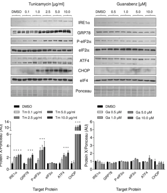
expected, tunicamycin reduced cell viability in a concentration-dependent manner (Figure 1A), whereas guanabenz (0.5–50mM) did not affect cell survival (Figure 1B). Additionally, we measured the effect of the treatment on different branches of UPR on mRNA and protein level. Tunicamycin strongly upregulated mRNA concentrations of the ER-chaperone GRP78 and activated the PERK pathway, which is reflected by the increase of the typical UPR targets ATF4, GADD34 and CHOP in a concen-tration- and time-dependent manner (Figure 2A and B). Accord-ingly, protein amounts of GRP78, ATF4 and CHOP were elevated as well (Figure 3 left panel). Increased eIF2a
Figure 1. Effects of tunicamycin and guanabenz on cell viability of neonatal rat cardiomyocytes (NRCM).(A) Concentration-dependent reduction of cell viability by tunicamycin treatment for 24 hours. (B) Treatment with increasing concentrations of guanabenz for 24 hours did not affect cell viability. Data are means6SEM (n = 8); ***P,0.0001.
doi:10.1371/journal.pone.0098893.g001
Figure 2. Effects of tunicamycin and guanabenz on different branches of the UPR in NRCM on the mRNA level.(A) Tunicamycin (Tm) treatment of NRCM for 12 hours increased levels of the UPR targets GRP78, GADD34, ATF4, and CHOP. (B) Time-dependent increase of UPR targets after treatment with tunicamycin (2.5mg/ml). (C) Guanabenz (Ga) treatment of NRCM for 24 hours did not affect levels of UPR targets. Data are means
6 SEM (n = 3–5); Changes of mRNA expression of$1.6 or #0.5 compared to DMSO control and a corrected P-value,0.05 were considered significant.
machinery. Besides activation of the PERK pathway, we could detect activation of a second UPR pathway that is initiated by IRE1a resulting in concomitant elevation of its protein amount
(Figure 3 left panel). The third UPR pathway that is reflected by initiation of ATF6 was not activated by tunicamycin (data not shown).
As expected, guanabenz alone (0.5–50mM) did not affect these UPR targets, neither on mRNA (Figure 2C) or protein level nor the phosphorylation status of eIF2a(Figure 3 right panel). It also
did not induce its proposed pharmacological target GADD34 or the constitutively active form CReP (Figure 2C). Taken together, the glycosylation inhibitor tunicamycin induced ER stress in neonatal rat cardiomyocytes that activated the different branches of the UPR while guanabenz alone had no effect.
Treatment of neonatal rat cardiomyocytes with ER stressors and guanabenz
To examine whether guanabenz affected the different branches of the UPR in stressed cells, NRCM were treated with tunicamycin 2.5mg/ml with or without guanabenz 2.5mM.
Protein levels of IRE1a, GRP78, CHOP as well as eIF2a
phosphorylation were analysed over 36 hours (Figure 4). As seen before, tunicamycin induced the expression of the ER stress markers IRE1a, GRP78 and the pro-apoptotic protein CHOP. In
addition, it increased phosphorylation of eIF2awith a maximum
between 6 and 12 h and a non-significant trend for a decrease at later time points. Treatment with guanabenz markedly reduced the effect of tunicamycin on IRE1a, GRP78, CHOP levels, and
significantly prolonged eIF2a phosphorylation at 24 and 36 h,
compatible with the proposed mechanism of action of guanabenz.
Figure 3. Effects of tunicamycin and guanabenz on different branches of the UPR in NRCM on the protein level.Upper panel – representative immunoblots of lysates of NRCMs treated with tunicamycin (left) or guanabenz (right) for 12 and 24 hours, respectively. Lower panel – concentration-dependent increase of UPR targets proteins after treatment with tunicamycin (Tm) or guanabenz (Ga) compared to DMSO control. Please note that the intensity of the upper unspecific band for GRP78 is caused by differences in blotting conditions (10% acrylamide/bisacrylamide gel). Data are means6SEM (n = 3); *P,0.05.
Next we assessed whether the effects of guanabenz on tunicamycin-induced ER stress extends to protection against cytotoxicity exerted by the known ER stressors tunicamycin (Figure 5A and B), hydrogen peroxide (H2O2; Figure 5C),
doxorubicin (Figure 5D) or thapsigargin (Figure 5E). Like tunicamycin, these drugs are known to activate the PERK pathway in the given concentrations and have an impact on cardiomyocyte function [19–21]. In accordance with these data, we saw a comparable induction of UPR by hydrogen peroxide treatment (data not shown). Cytotoxicity was determined as the reduction in formazan production. Different protocols of treat-ment with the cytotoxic drugs were tested. All 4 compounds reduced the viability of NRCM by 25–75% compared to vehicle control. The strongest toxicity was observed with a short (4 h) pulse of a high concentration of H2O2 (80mM). Preincubation
with guanabenz promoted the survival of NRCM (Figure 5) at
least at certain time points. The concentration-response relation of guanabenz was bell-shaped with a maximally effective concentra-tion around 2–3mM.
Treatment of engineered heart tissue with ER stressors and guanabenz
The above results indicated a protective guanabenz effect against drug-induced ER stress and cell death in cardiac myocytes. To evaluate whether this effect also translates into preserved contractile function we employed the model of engineered heart tissue (EHT), 3-dimensional, spontaneously beating and force-developing muscle strips [17]. We incubated EHTs with equiv-alent concentrations of tunicamycin, thapsigargin, doxorubicin or hydrogen peroxide with and without guanabenz and measured force development over time. Preincubation with guanabenz had no effect on EHT contractility and is indicated in the respective
Figure 4. Representative immunoblots of ER stress marker proteins related to the loading control calsequestrin (CSQ) in lysates of NRCM treated with tunicamycin (Tm; 2.5mg/ml) in the presence or absence of guanabenz (Ga; 2.5mM) for the indicated times (0, 6,
12, 24 and 36 h).Bar graphs show densitometric and statistical analysis of the immunoblots. Data are means6SEM (n = 6); *P,0.05 compared to the respective time points.
figures as time zero (0 h). In all cases exposure to cytotoxic agents resulted in a reduction of force or in an abnormal beating pattern (Figure 6). Compared to NRCM in 2D culture the toxic effect of tunicamycin was delayed in EHTs, and higher concentrations of thapsigargin (500 nM) and hydrogen peroxide (600mM) were
needed to induce acute toxicity (reduction of peak force), indicating higher resistance to stress in the 3D culture model. Addition of guanabenz (2.5–10mM) did not affect peak force significantly in any of these experiments, but was associated with a trend towards improved contractile function in each experiment and at each time point. A short pulse of hydrogen peroxide treatment led to a strong reduction in peak force, but EHTs fully recovered after 20 hours washout, arguing against a major role of cell death in this experiment. Accordingly, measurement of serum creatine kinase (CK) and lactate dehydrogenase (LDH) activities as markers of cardiomyocyte death showed no evidence for cell death in the EHT model (Figure S1). The SR Ca2+
ATPase (SERCA) inhibitor thapsigargin induced phases of very fast beating and lack of complete relaxation (Figure 6C), compatible with an inhibitory effect of the compound on Ca2+
reuptake into the SR in diastole. Of note, the onset of effect of thapsigargin was delayed (data not
shown) and was reduced by guanabenz at 3mM and almost completely abolished at 10mM. This was unexpected, because guanabenz probably does not directly interfere with SR Ca2+
handling. It indicates that ER stress is a consequence of SERCA inhibition and participates in the disturbation of SR Ca2+
handling.
Discussion
ER stress is increasingly recognized as a contributor to the pathophysiology of cardiovascular diseases, suggesting therapeutic interventions in its mechanisms as a new target for drug therapy. Yet, knowledge about exact molecular mechanisms of the UPR and signalling pathways leading to cardioprotection remains limited [14]. In principle, activation of the different UPR signalling branches is considered to protect cells from ER stress. For example, increased phosphorylation of eIF2a causes a
transient reduction of global protein synthesis and thereby of the amount of new proteins that require proper folding. Prolonged eIF2a phosphorylation was associated with attenuated toxicity
[9,22]. It has been proposed that selective inhibition of GADD34,
Figure 5. Guanabenz protected NRCM from deleterious endoplasmic reticulum stress.Values are normalized to vehicle control (100%). Data are means6SEM. (A) Assessment of cell viability after treatment with tunicamycin (Tm; 2.5mg/ml) and indicated concentrations of guanabenz
for 10 h. (n = 8); **P,0.001; ***P,0.0001. (B) Protective effect of guanabenz (Ga; 2.5mM) in cells treated with tunicamycin (0.1mg/ml) for 12, 24, and
54 hours. (n = 4); ***P,0.0001. (C) Concentration-dependent protection by guanabenz against cellular stress induced by exposure to H2O2(80mM)
for 4 hours (n = 7–14); **P,0.001; ***P,0.0001. Cell viability of NRCM treated with doxorubicin (Dox; 100 nM) for 48 hours (D) or with thapsigargin (Tg; 100 nM) for 72 hours (E), with or without indicated concentrations of guanabenz. (n = 6–7); *P,0.05 **P,0.001.
a stress-induced regulatory subunit of protein phosphatase 1 (PP1), causes selective attenuation of eIF2adephosphorylation by PP1 in
stressed cells [10,23–25]. Indeed, guanabenz, a small molecule interfering with PP1-GADD34 interaction, protected cells from lethal ER stress without affecting cell function alone [9]. Here we tested whether this concept can be expanded to cardiac myocytes. The main findings were: (i) Tunicamycin induced ER stress and consequently activated several UPR signalling pathways. (ii) Guanabenz did not increase the levels of UPR targets, but antagonized the effects of tunicamycin on markers of ER stress in cardiac myocytes. (iii) Tunicamycin and other known inducers of ER stress led to cardiac myocyte death and this was concentration-and time-dependently antagonized by guanabenz. (iv) ER stressors also caused acute or delayed contractile dysfunction in EHTs, which was not significantly affected by guanabenz, with the notable exception of relaxation deficits under thapsigargin. Thus, our results in single cardiac myocytes principally support those in cultured cell lines, but modest protection in our EHT model point
to a more complex mechanism in intact functional heart muscle, which may be critical for developing this approach for therapeutic purposes.
The beneficial effects of guanabenz on cell survival showed a bell-shaped concentration-dependency. For example, guanabenz in the presence of 80mM hydrogen peroxide had no effect at 1,
tripled cell survival at 2, 3 and 4mM (,70% compared to 23%)
and had no effect at 10mM. Similar results have been described
before by Tsaytler et al. [9]. This narrow ‘‘therapeutic window’’ could be related to the proposed mechanism of action, but also to the interference with various targets, e.g. IRE1a or even other
unknown targets. Tunicamycin treatment of cardiac myocytes induced upregulation of several UPR targets (GRP78, IRE1a,
p-eIF2a, ATF4 and CHOP) including activation of GADD34 on the
mRNA level, pointing indeed to activation of the PERK pathway. In our hands, guanabenz markedly reduced the effects of tunicamycin on IRE1a, GRP78, CHOP, and prolonged eIF2a
phosphorylation, even though the statistics of the latter effect were
Figure 6. Impact of guanabenz on EHT contractility during exposure to ER stress.(A) Force development of EHTs after treatment with tunicamycin (Tm; 2.5mg/ml) for 12, 24, 36 and 48 hours, with or without guanabenz (Ga; 2.5mM). Pre-drug values Tm: 0.1760.01 vs. Ga: 0.1760.02.
(B) Force development of EHTs with or without guanabenz (2.5mM) during 30 min acute exposure to H2O2(600mM) and during following 20 hours
recovery time. Pre-drug values Tm: 0.1660.01 vs. Ga: 0.1660.01. (C) Representative beating pattern of an EHT treated with thapsigargin (Tg; 500 nM) for 1 hour, with or without guanabenz (3mM, 10mM) compared to an untreated control EHT. (D) Force development of EHTs after treatment with
doxorubicin (Dox; 100 nM) for 12, 24 and 48 hours, with or without guanabenz (2.5mM). Pre-drug values Tm: 0.1560.03 vs. Ga: 0.1660.02. Data are
means6SEM (n = 4–6). All drugs showed a time-dependent effect on EHT contractility (Two-way ANOVA, row factor, P,0.05), but differences between the groups were not statistically significant.
distinct pathways promoting either cell survival or cell death: (i) The intrinsic endoribonuclease activity of IRE1a results in
production of the transcription factor X-box binding protein (XBP)1 [1,12,14] that induces the expression of genes involved in protein folding and degradation of unfolded proteins to restore protein homeostasis. (ii) IRE1a mediates cell death and
inflam-matory signaling [26] via activation of the apoptosis signal-regulating kinase (ASK)1 [27,28]. Both, the increase of spliced XBP1 [15] and activation of ASK1 [29] potentially contribute to the pathogenesis of heart diseases. Guanabenz clearly reduced the effect of tunicamycin on IRE1afor yet unknown reasons. It might
be a direct interaction of the small molecule with elements of the IRE1a signaling pathway or a secondary effect resulting from
translational repression and slowdown of global protein synthesis rate through activation of the PERK pathway. Interpretation of the data is complicated by the fact that the underlying mechanisms are not yet fully understood. On the one hand, activation of IRE1aand subsequent XBP1 splicing were shown to reduce ER
stress and promote cell survival [30–32]. On the other hand, also inhibition of the IRE1a apoptotic downstream target ASK1
reduced cell death induced by ER stress [12,33,34]. For detailed information see [12,35]. Our data show that the IRE1apathway is
activated during ER stress conditions in cardiac myocytes and reduced by concomitant guanabenz treatment. If IRE1a
activa-tion was protective, its downregulaactiva-tion by guanabenz may attenuate a cytoprotective effect of the drug and explain why it had only mild protective effects in cardiac myocytes and especially in EHT.
In addition to the concentration-dependency, the cytoprotective effects in cardiac myoyctes were time-dependent and varied between 4 and 72 h depending on the cytotoxic agent. This transient effect is consistent with the results obtained by Tsaytler et al. [9] and with a temporary reduction of protein synthesis by the proposed mechanism. Balanced feedback inhibition of the UPR by GADD34-mediated dephosphorylation of eIF2a seems to be
essential for cell survival. Thus, sustained eIF2aphosphorylation is
lethal to cells in vitro [36] and in vivo [37] as is the complete loss of eIF2aphosphorylation [38,39]. Therapeutic interference with this
process therefore likely needs to be mild and transient.
The impact of guanabenz treatment on EHTs during exposure to different ER stressors was modest and did not reach statistical significance. The only clear-cut effect was an almost complete prevention of the marked relaxation deficits induced by a high concentration of the SERCA inhibitor thapsigargin by guanabenz
and millimolar [40]. In our hands protective effects were seen around 2.5mM. At higher concentrations (30mM) it exerted direct negative inotropic and lusitropic effects in engineered heart tissue (Figure S2), but also protected against adverse effects of high concentrations of epinephrine (data not shown). This observation may be related to yet another function of this small molecule, namely agonistic activity at the trace amine-associated receptor 1 [46] which mediates various cardiac effects of trace amines [48-51].
In summary, our data are principally consistent with the proposed protective effect of guanabenz via inhibition of PPP1R15A and ER stress, but also point to various other modes of action and possible targets in heart cells. This issue needs consideration when developing ER stress-directed concepts based on guanabenz for therapeutic purposes.
Supporting Information
Figure S1 Impact of tunicamcin treatment (Tm; 1.0mg/
ml) on EHTs compared to non-treated controls.(A) Force development of EHTs under tunicamycin treatment after 0, 24, 48, 72, and 96 hours. (B) Measurement of lactate dehydrogenase (LDH) activity during tunicamycin treatment after 0, 48, and 96 hours. Serum creatine kinase (CK) activity was below detection level. Data are means 6 SEM (n = 3); ***P,0.001 (Two-way ANOVA, Bonferroni post-test).
(TIF)
Figure S2 Long-term effect of guanabenz treatment for 14 days (Ga; 3, 10, 30mM) on EHT contractility compared to non-treated controls. (A) Force development of EHTs under guanabenz treatment with the indicated concentrations. (B) Contraction time T180% (time from 20% to
100% of peak height of the force peak) and (C) relaxation time T280%(time from 100% to 20% of peak height of the force peak).
Data are means6SEM (n = 4); **P,0.01, ***P,0.001 (One-way ANOVA, Dunnett post-test).
(TIF)
Author Contributions
Conceived and designed the experiments: CN TE. Performed the experiments: CN JU TS HS. Analyzed the data: CN HS TE. Wrote the paper: CN AEA TE.
References
1. Ron D, Walter P (2007) Signal integration in the endoplasmic reticulum unfolded protein response. Nat Rev Mol Cell Biol 8: 519–529.
2. Harding HP, Zhang Y, Ron D (1999) Protein translation and folding are coupled by an endoplasmic-reticulum-resident kinase. Nature 397: 271–274.
4. Rutkowski DT, Kaufman RJ (2004) A trip to the ER: coping with stress. Trends Cell Biol 14: 20–28.
5. Novoa I, Zeng H, Harding HP, Ron D (2001) Feedback inhibition of the unfolded protein response by GADD34-mediated dephosphorylation of eIF2alpha. J Cell Biol 153: 1011–1022.
6. Ma Y, Hendershot LM (2003) Delineation of a negative feedback regulatory loop that controls protein translation during endoplasmic reticulum stress. J Biol Chem 278: 34864–34873.
7. Harding HP, Zhang Y, Scheuner D, Chen JJ, Kaufman RJ, et al. (2009) Ppp1r15 gene knockout reveals an essential role for translation initiation factor 2 alpha (eIF2alpha) dephosphorylation in mammalian development. Proc Natl Acad Sci U S A 106: 1832–1837.
8. Holmes B, Brogden RN, Heel RC, Speight TM, Avery GS (1983) Guanabenz. A review of its pharmacodynamic properties and therapeutic efficacy in hypertension. Drugs 26: 212–229.
9. Tsaytler P, Harding HP, Ron D, Bertolotti A (2011) Selective inhibition of a regulatory subunit of protein phosphatase 1 restores proteostasis. Science 332: 91–94.
10. Tsaytler P, Bertolotti A (2013) Exploiting the selectivity of protein phosphatase 1 for pharmacological intervention. FEBS J 280: 766–770.
11. Lin JH, Walter P, Yen TS (2008) Endoplasmic reticulum stress in disease pathogenesis. Annu Rev Pathol 3: 399–425.
12. Kim I, Xu W, Reed JC (2008) Cell death and endoplasmic reticulum stress: disease relevance and therapeutic opportunities. Nat Rev Drug Discov 7: 1013– 1030.
13. Kitakaze M, Tsukamoto O (2010) What is the role of ER stress in the heart? Introduction and series overview. Circ Res 107: 15–18.
14. Minamino T, Kitakaze M (2010) ER stress in cardiovascular disease. J Mol Cell Cardiol 48: 1105–1110.
15. Okada K, Minamino T, Tsukamoto Y, Liao Y, Tsukamoto O, et al. (2004) Prolonged endoplasmic reticulum stress in hypertrophic and failing heart after aortic constriction: possible contribution of endoplasmic reticulum stress to cardiac myocyte apoptosis. Circulation 110: 705–712.
16. El-Armouche A, Rau T, Zolk O, Ditz D, Pamminger T, et al. (2003) Evidence for protein phosphatase inhibitor-1 playing an amplifier role in beta-adrenergic signaling in cardiac myocytes. FASEB J 17: 437–439.
17. Hansen A, Eder A, Bonstrup M, Flato M, Mewe M, et al. (2010) Development of a drug screening platform based on engineered heart tissue. Circ Res 107: 35– 44.
18. El-Armouche A, Wittkopper K, Degenhardt F, Weinberger F, Didie M, et al. (2008) Phosphatase inhibitor-1-deficient mice are protected from catecholamine-induced arrhythmias and myocardial hypertrophy. Cardiovasc Res 80: 396–406. 19. Liu L, Wise DR, Diehl JA, Simon MC (2008) Hypoxic reactive oxygen species regulate the integrated stress response and cell survival. J Biol Chem 283: 31153–31162.
20. Zhang Y, Ren J (2011) Thapsigargin triggers cardiac contractile dysfunction via NADPH oxidase-mediated mitochondrial dysfunction: Role of Akt dephosphor-ylation. Free radical biology & medicine 51: 2172–2184.
21. Zhang YW, Shi J, Li YJ, Wei L (2009) Cardiomyocyte death in doxorubicin-induced cardiotoxicity. Arch Immunol Ther Exp (Warsz) 57: 435–445. 22. Fu HY, Okada K-i, Liao Y, Tsukamoto O, Isomura T, et al. (2010) Ablation of
C/EBP homologous protein attenuates endoplasmic reticulum-mediated apoptosis and cardiac dysfunction induced by pressure overload. Circulation 122: 361–369.
23. Boyce M, Bryant KF, Jousse C, Long K, Harding HP, et al. (2005) A selective inhibitor of eIF2alpha dephosphorylation protects cells from ER stress. Science 307: 935–939.
24. Boyce M, Py BF, Ryazanov AG, Minden JS, Long K, et al. (2008) A pharmacoproteomic approach implicates eukaryotic elongation factor 2 kinase in ER stress-induced cell death. Cell Death Differ 15: 589–599.
25. Cnop M, Ladriere L, Hekerman P, Ortis F, Cardozo AK, et al. (2007) Selective inhibition of eukaryotic translation initiation factor 2 alpha dephosphorylation potentiates fatty acid-induced endoplasmic reticulum stress and causes pancreatic beta-cell dysfunction and apoptosis. J Biol Chem 282: 3989–3997. 26. Minamino T, Komuro I, Kitakaze M (2010) Endoplasmic reticulum stress as a
therapeutic target in cardiovascular disease. Circ Res 107: 1071–1082. 27. Ron D, Hubbard SR (2008) How IRE1 reacts to ER stress. Cell 132: 24–26. 28. Urano F, Wang X, Bertolotti A, Zhang Y, Chung P, et al. (2000) Coupling of
stress in the ER to activation of JNK protein kinases by transmembrane protein kinase IRE1. Science 287: 664–666.
29. Yamaguchi O, Higuchi Y, Hirotani S, Kashiwase K, Nakayama H, et al. (2003) Targeted deletion of apoptosis signal-regulating kinase 1 attenuates left ventricular remodeling. Proc Natl Acad Sci U S A 100: 15883–15888. 30. Korennykh AV, Egea PF, Korostelev AA, Finer-Moore J, Zhang C, et al. (2009)
The unfolded protein response signals through high-order assembly of Ire1. Nature 457: 687–693.
31. Lin JH, Li H, Yasumura D, Cohen HR, Zhang C, et al. (2007) IRE1 signaling affects cell fate during the unfolded protein response. Science 318: 944–949. 32. Han D, Upton JP, Hagen A, Callahan J, Oakes SA, et al. (2008) A kinase
inhibitor activates the IRE1alpha RNase to confer cytoprotection against ER stress. Biochemical and biophysical research communications 365: 777–783. 33. Kim I, Shu CW, Xu W, Shiau CW, Grant D, et al. (2009) Chemical biology
investigation of cell death pathways activated by endoplasmic reticulum stress reveals cytoprotective modulators of ASK1. J Biol Chem 284: 1593–1603. 34. Nishitoh H, Matsuzawa A, Tobiume K, Saegusa K, Takeda K, et al. (2002)
ASK1 is essential for endoplasmic reticulum stress-induced neuronal cell death triggered by expanded polyglutamine repeats. Genes Dev 16: 1345–1355. 35. Matsukawa J, Matsuzawa A, Takeda K, Ichijo H (2004) The ASK1-MAP kinase
cascades in mammalian stress response. J Biochem 136: 261–265.
36. Srivastava SP, Kumar KU, Kaufman RJ (1998) Phosphorylation of eukaryotic translation initiation factor 2 mediates apoptosis in response to activation of the double-stranded RNA-dependent protein kinase. J Biol Chem 273: 2416–2423. 37. DeGracia DJ, Sullivan JM, Neumar RW, Alousi SS, Hikade KR, et al. (1997) Effect of brain ischemia and reperfusion on the localization of phosphorylated eukaryotic initiation factor 2 alpha. J Cereb Blood Flow Metab 17: 1291–1302. 38. Harding HP, Zhang Y, Bertolotti A, Zeng H, Ron D (2000) Perk is essential for translational regulation and cell survival during the unfolded protein response. Mol Cell 5: 897–904.
39. Scheuner D, Song B, McEwen E, Liu C, Laybutt R, et al. (2001) Translational control is required for the unfolded protein response and in vivo glucose homeostasis. Mol Cell 7: 1165–1176.
40. Barbezier N, Chartier A, Bidet Y, Buttstedt A, Voisset C, et al. (2011) Antiprion drugs 6-aminophenanthridine and guanabenz reduce PABPN1 toxicity and aggregation in oculopharyngeal muscular dystrophy. EMBO Mol Med 3: 35–49. 41. Tribouillard-Tanvier D, Beringue V, Desban N, Gug F, Bach S, et al. (2008) Antihypertensive drug guanabenz is active in vivo against both yeast and mammalian prions. PLoS One 3: e1981.
42. Ernsberger P, Meeley MP, Mann JJ, Reis DJ (1987) Clonidine binds to imidazole binding sites as well as alpha 2-adrenoceptors in the ventrolateral medulla. Eur J Pharmacol 134: 1–13.
43. Lachaud-Pettiti V, Podevin RA, Chretien Y, Parini A (1991) Imidazoline-guanidinium and alpha 2-adrenergic binding sites in basolateral membranes from human kidney. Eur J Pharmacol 206: 23–31.
44. Bognar IT, Enero MA (1984) alpha 1-Antagonist activity of the alpha 2-adrenoceptor agonist guanabenz in perfused mesenteric artery of the rat. Eur J Pharmacol 103: 173–175.
45. Ozaita A, Olmos G, Boronat MA, Lizcano JM, Unzeta M, et al. (1997) Inhibition of monoamine oxidase A and B activities by imidazol(ine)/guanidine drugs, nature of the interaction and distinction from I2-imidazoline receptors in rat liver. Br J Pharmacol 121: 901–912.
46. Hu LA, Zhou T, Ahn J, Wang S, Zhou J, et al. (2009) Human and mouse trace amine-associated receptor 1 have distinct pharmacology towards endogenous monoamines and imidazoline receptor ligands. Biochem J 424: 39–45. 47. Norez C, Vandebrouck C, Antigny F, Dannhoffer L, Blondel M, et al. (2008)
Guanabenz, an alpha2-selective adrenergic agonist, activates Ca2+-dependent chloride currents in cystic fibrosis human airway epithelial cells. Eur J Pharmacol 592: 33–40.
48. Frascarelli S, Ghelardoni S, Chiellini G, Galli E, Ronca F, et al. (2011) Cardioprotective effect of 3-iodothyronamine in perfused rat heart subjected to ischemia and reperfusion. Cardiovasc Drugs Ther 25: 307–313.
49. Frascarelli S, Ghelardoni S, Chiellini G, Vargiu R, Ronca-Testoni S, et al. (2008) Cardiac effects of trace amines: pharmacological characterization of trace amine-associated receptors. Eur J Pharmacol 587: 231–236.
50. Ghelardoni S, Suffredini S, Frascarelli S, Brogioni S, Chiellini G, et al. (2009) Modulation of cardiac ionic homeostasis by 3-iodothyronamine. J Cell Mol Med 13: 3082–3090.