Serotonin regulates osteoblast proliferation
and function
in vitro
S.Q. Dai
1*, L.P. Yu
1*, X. Shi
2, H. Wu
3, P. Shao
1, G.Y. Yin
1and Y.Z. Wei
1 1Department of Orthopedic Surgery, The First Affiliated Hospital, Nanjing Medical University, Nanjing, Jiangsu, China
2
Department of Obstetrics and Gynecology, The First Affiliated Hospital, Nanjing Medical University, Nanjing, Jiangsu, China
3
Emergency Department, The First Affiliated Hospital, Soochow University, Suzhou, China
Abstract
The monoamine serotonin (5-hydroxytryptamine, 5-HT), a well-known neurotransmitter, also has important functions outside the central nervous system. The objective of this study was to investigate the role of 5-HT in the proliferation, differentiation, and function of osteoblastsin vitro. We treated rat primary calvarial osteoblasts with various concentrations of 5-HT (1 nM to 10mM) and assessed the rate of osteoblast proliferation, expression levels of osteoblast-specific proteins and genes, and the
ability to form mineralized nodules. Next, we detected which 5-HT receptor subtypes were expressed in rat osteoblasts at different stages of osteoblast differentiation. We found that 5-HT could inhibit osteoblast proliferation, differentiation, and mineralization at low concentrations, but this inhibitory effect was mitigated at relatively high concentrations. Six of the 5-HT receptor subtypes (5-HT1A, 5-HT1B, 5-HT1D, 5-HT2A, 5-HT2B, and 5-HT2C) were found to exist in rat osteoblasts. Of these,
5-HT2Aand 5-HT1Breceptors had the highest expression levels, at both early and late stages of differentiation. Our results
indicated that 5-HT can regulate osteoblast proliferation and functionin vitro.
Key words: Serotonin; Osteoblast; Proliferation; Differentiation; 5-HT receptors; 5-HT
Introduction
Bone remodeling is a highly regulated process that requires a tight coupling of bone formation with resorption to maintain skeletal mass. Imbalances in bone formation and resorption lead to pathological conditions such as osteope-nia, osteoporosis, and osteomalacia. Bone mass and osteoblast activity, as well as proliferation and differentiation of osteoblast precursors, are regulated by many factors, including hormones and locally produced growth factors and cytokines, which respond to hormonal activation (1-4).
In recent years, the neurotransmitter serotonin (also known as 5-hydroxytryptamine, 5-HT) was discovered to be involved in bone metabolism. Clinical observations suggest that 5-HT might be associated with bone mass. Selective 5-HT reuptake inhibitors (SSRIs) are routinely used to treat depression in adults (5), children, and adolescents (6-8). SSRIs hinder the 5-HT transporter from taking up 5-HT from the synaptic space, thus increasing extracellular levels of 5-HT. It has been reported that patients taking the SSRI fluoxetine appear to have an elevated risk of fracture (9-16). Consistent with this finding, serum 5-HT was reported to be inversely correlated with
femoral neck total and trabecular volumetric bone mineral density (7). Functional serotonergic pathways in bones (17-19) may enable 5-HT to influence skeletal biology. These observations suggest that there may be an important relationship between 5-HT and bone remodeling. Reports of the effects of 5-HT on bone are conflicting. Yadav et al. (20) reported that 5-HT acted on osteoblasts via the 5-HT1Breceptor to inhibit their proliferation, while many other researchers found that 5-HT had an opposite effect (18,19,21-26). The reason for this inconsistency is unknown. The purpose of the present study was to explore the possible physiological roles of 5-HT in bone metabolism. Our data suggest that 5-HT plays a significant role in the regulation of bone biologyin vitro.
Material and Methods
Ethics statement
All experimental procedures involving animals were performed in accordance with the protocols approved by the Experimental Animal Ethics Committee of Nanjing
Correspondence: G.Y. Yin and/or Y.Z. Wei, Department of Orthopedic Surgery, The First Affiliated Hospital, Nanjing Medical University, 300 Guangzhou Road, Nanjing, Jiangsu 210029, China. E-mail: guoyongyin001@sina.com and/or wyzljm@163.com
*These authors contributed equally to this study.
Medical University, China, and conformed to the Guide for the Care and Use of Laboratory Animals of the National Institutes of Health (USA). All efforts were made to minimize suffering.
Isolation, culture, and preparation of rat calvarial osteoblasts
Primary osteoblasts were isolated by collagenase digestion from calvariae of Sprague-Dawley rats that were 1-2 days old. Osteoblasts were grown in complete medium, i.e., a-minimal essential medium (HyClone, USA) supplemented with 10% fetal bovine serum (FBS; HyClone), 100 U/mL penicillin, and 0.1 mg/mL strepto-mycin (Gibco, USA). Because FBS is known to contain relatively high levels of 5-HT from platelet lysis (approxi-mately 300 ng/mL by enzyme-linked immunosorbent assay, ELISA) (27), 5-HT was stripped from the FBS by incubation with dextran-coated charcoal (Sigma, USA). The concentration after treatment was confirmed by high performance liquid chromatography to be below 1 pM in the medium containing 10% FBS. All the operations involving 5-HT needed to be protected from light, because 5-HT is an unstable compound and decomposes quickly. Osteoblasts at passage 3 were used to perform all cell studies and were divided into six groups, which were cultured in the presence of various concentrations of 5-HT (Sigma): 0 M (control group) and 1 nM, 10 nM, 100 nM, 1mM, and 10mM (experimental groups). After attach-ment, the osteoblasts were serum starved for 12 h prior to experiments. 5-HT was then added, beginning on day 1. After the cells reached confluence, at approximately day 5, the complete medium was replaced with a differentia-tion medium (complete medium containing 50mg/mL ascorbic acid, and 10 mM b-glycerophosphate, Sigma) for appropriate mineralization.
Cell proliferation assay
Cell proliferation was determined using the Cell Counting Kit-8 (CCK-8; Dojindo, Japan) as described elsewhere (28). Osteoblasts of passage 3 were cultured for 2 days with 5-HT prior to CCK-8 assay. Absorbance (optical density, OD) at 450 nm was measured with a microplate spectrophotometer (BioTek, USA). Cell num-ber was correlated with OD values. The cell proliferation rate was calculated as a percentage as follows: (ODserotonin–ODblank)/(ODcontrol–ODblank)6100.
Quantitative real-time RT-PCR analysis
The level of type I collagen (col1a1) mRNA was examined by quantitative real-time reverse transcription polymerase chain reaction (qRT-PCR) analysis on day 5. RNA isolated from cells cultured with 5-HT was concen-trated using a NanoDrop 2000 microvolume spectro-photometer (Thermo Scientific, USA), and cDNA was synthesized using M-MuLV reverse transcriptase (Fermentas, USA). PCR was performed using a Power
SYBR Green PCR Master Mix (Applied Biosystems, USA) on a Real-Time Thermal Cycler apparatus (Mastercycler ep realplex; Eppendorf, Germany). The relative level of expression for the target gene was normalized by the housekeeping gene GAPDH and calculated using the 2–DDCt relative quantification method, as described previously (29). Primer sequences for each gene are listed in Table 1.
For assessment of 5-HT receptor mRNA expression, we selected osteoblasts from the control group at day 5 and day 15 to represent early and late stages of osteoblast differentiation, respectively. When the program was completed, we analyzed the real-time PCR products of 5-HT receptors by electrophoresis on a 1.5% agarose/ Tris-acetate-EDTA (TAE) gel and stained them with ethidium bromide to further confirm amplification specifi-city and amplicon size.
Western blot analysis
To assess alkaline phosphatase (ALP) protein expres-sion, we performed Western blot analysis, as described elsewhere (30). In brief, proteins were extracted from different experimental groups at day 10 and quantified. Twenty micrograms of supernatant protein samples were subjected to sodium dodecylsulfate-polyacrylamide gel electrophoresis and transferred to Immobilon-P polyvi-nylidene fluoride (PVDF) membranes (Millipore, USA). Following blocking, immunoblots were incubated with anti-ALP monoclonal antibody (1:10,000; Abcam, UK) overnight at 46C. A GAPDH antibody (Sigma) was used as a protein loading control. Blots were then incubated with horseradish peroxidase-conjugated secondary anti-body (1:10,000; Bioworld, USA) at 376C for 1 h and visualized using a SuperSignal West Pico chemilumines-cence substrate kit (Pierce, USA). The membranes were scanned using a Molecular Imager (Bio-Rad, USA), followed by data analysis using the Image Lab software (Bio-Rad). Data are reported as the protein-to-GAPDH ratio to correct for variations in protein loading.
Alkaline phosphatase activity assay
ALP activity was assessed at day 10 using a phosphate assay kit (BioAssay Systems, USA), and the assessment was based on the cleavage of p-nitrophenyl phosphate, as described elsewhere (31). The product of the enzyme reaction, p-nitrophenol, was assessed by measuring the absorbance at 405 nm. The protein concentration of each sample was measured using a bicinchoninic acid protein assay reagent kit (Pierce). ALP activity was expressed as the ratio of OD to protein content.
ELISA
manufacturer’s instructions. The ELISA plates were analyzed at 450 nm with a microplate reader (BioTek). The OCN concentration of each sample was calculated according to the standard curve.
Detection and quantification of mineralization At day 15, we used Alizarin Red S (AR-S; Sigma) stain (32,33) to determine the extent of mineralized matrix in the plates. In brief, cells were fixed in ice-cold 70% (v/v) ethanol and then stained with 40 mM AR-S, pH 4.2. The plates were incubated for 10 min at room temperature with gentle shaking. Stained monolayers were visualized by means of phase microscopy with an inverted micro-scope (Nikon, Japan). AR-S was released from the cell matrix by incubation in 10% (w/v) cetylpyridinium chloride in 10 mM Na2PO4, pH 7.0, for 15 min. The released dye was transferred to a 96-well plate and assessed at 562 nm using a microplate reader (BioTek).
Statistical analysis
All experiments were performed in triplicate, and the data are reported as means±SE. Statistical analyses
were performed using the SPSS 13.0 software package (SPSS, USA). We performed one-way analysis of variance followed by the Dunnettpost hoctest for multiple comparisons between groups. In all cases, P values less than 0.05 were considered to be statistically significant.
Results
5-HT inhibited proliferation of primary osteoblasts Primary osteoblasts were incubated with 5-HT for 2 days, and the proliferation rate was measured as shown in Figure 1. Compared to the growth of control cells, that
Table 1. Primer sets used in real-time RT-PCR.
Primer FW/RV sequence (59R39) Product
length (bp)
Accession No. (NCBI)
Col1a1 CTGCCCCTCGCAGGGGTTTG/GCCTGCACATGTGTGGCCGA 72 NM_053304.1
GAPDH GCTCTCTGCTCCTCCCTGTTCT/CAGGCGTCCGATACGGCCAAA 117 NM_017008.3
5-HT1AR CCGCTGCGCTGATCTCGCTC/GATCGGTCTTCCGGGGTGCG 88 NM_012585.1
5-HT1BR GCGAGTCTCAGACGCCCTGC/GGGTCTTGGTGGCTTTGCGCT 71 NM_022225.1
5-HT1DR TCACGCGGCGGCCATGATTG/CTGCCGCCAGAAGAGCGGTG 76 NM_012852.1
5-HT2AR AGGCTCCTACGCAGGCCGAA/CCCAGCACCTTGCACGCCTT 69 NM_017254.1
5-HT2BR AATGTCCTTGGCGGTGGCTGA/GCCAGTGGGAGGGGCCATGTA 99 NM_017250.1
5-HT2CR GCGATTGCAGCCGAGTCCGT/AACCCGCTAGCGTCCGGGAG 80 NM_012765.3
FW: forward; RV: reverse; bp: base pairs; GAPDH: glyceraldehyde-3-phosphate dehydrogenase; Col1a1: type 1 collagen.
Figure 1.Serotonin inhibited proliferation of primary osteoblasts. Growth of osteoblasts treated with serotonin (1 nM-10mM) was inhibited compared to controls. This influence was alleviated in the 10mM and 1mM groups (relatively high concentrations). Data are reported as means±SE. *P,0.05 vs control (ctrl) group (Dunnett test).
of osteoblasts treated with 5-HT was inhibited. The inhibitory effect increased gradually in a dose-dependent manner as the 5-HT concentration increased (1-100 nM), but this effect was alleviated in the 10mM and 1mM groups (relatively high concentrations) and a reverse trend was shown.
5-HT affected the differentiation of primary osteoblasts
The effect of 5-HT on osteoblast differentiation was determined by measuring the expression of col1a1 mRNA, ALP, and OCN proteins after exposure to 5-HT-containing media. Expression of col1a1 mRNA was significantly reduced (P,0.05) by the addition of 10 nM to 10mM 5-HT. The 100 nM 5-HT group had the lowest levels of col1a1 gene expression (Figure 2A).
Activity of ALP, a marker of bone formation, and expression of ALP protein were measured to assess the effect of 5-HT on osteoblast differentiation. ALP was expressed in osteoblasts during long-term cultivation, with maximum expression at day 10. ALP protein expression of the experimental groups (,1-100 nM) decreased gradually, and 100 nM 5-HT reduced protein expression most significantly (P,0.05). However, this inhibitory effect was attenuated when the concentration reached ,1-10mM (Figure 2B). ALP enzyme activity (Figure 2C) in all groups showed a pattern that was similar to ALP protein expression.
OCN was expressed at a later stage of osteoblast differentiation and represented the beginning of bone matrix mineralization. We found that OCN content in the cultured supernatant of all groups, which reflects the
amount of OCN synthesis of the osteoblasts, showed a ‘‘V’’ pattern, with the lowest level in the 100 nM group (Figure 2D). Interestingly, neither immunofluorescence nor Western blot analysis detected OCN protein expres-sion in the osteoblasts (data not shown).
5-HT suppressed mineralization of primary osteoblasts
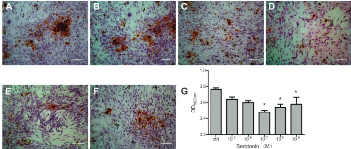
The effects of 5-HT on osteoblast mineralization were investigated at day 15 by using AR-S staining, which identifies calcium content within the bone matrix. Decreased mineralization relative to controls was observed in cultures treated with 5-HT (Figure 3). Mineralized nodule formation was reduced in all groups of 5-HT-treated osteoblasts, but this finding was statisti-cally significant only at 100 nM to 10mM 5-HT (P,0.05). Interestingly, the inhibitory effect rebounded with the increase in concentration from 1mM to 10mM 5-HT, similar to the pattern observed in results of proliferation and differentiation assays.
5-HT receptor mRNA in primary osteoblasts
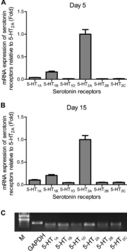
Next, we detected which 5-HT receptor subtypes were expressed in rat primary osteoblasts. We harvested cells from the control group at days 5 and 15 to assess the relative mRNA expression of 5-HT1A, 5-HT1B, 5-HT1D, 5-HT2A, 5-HT2B, and 5-HT2C, which are reported to exist in bones (19,20,25). Our results indicated that all six subtypes of 5-HT receptors were present in rat osteo-blasts, as shown in Figure 4. Of these, 5-HT1Band 5-HT2A had the highest expression, at both early and late stages of osteoblast differentiation. Electrophoresis on agarose/
TAE gel further confirmed the presence of 5-HT receptors (Figure 4C).
Discussion
Our preliminary experiment characterized the differ-entiation of rat primary osteoblast cultures. Changes in ALP activity and differential gene expression characterize the following three distinct stages (34): growth (prolifera-tion), up to 5-6 days; matrix maturation (or differentia(prolifera-tion), up to 10-11 days; and mineralization, up to 15-16 days. Each of the three osteoblast-specific proteins that we analyzed reaches peak expression during a different stage (34), which explains why we detected col1a1 at day 5, ALP at day 10, and OCN at day 15. Because there is a significant effect of cell density on the rate of osteoblast
proliferation and differentiation, cytometry was used to ensure that the number of cells in each group was equal. Our results showed that 5-HT at low concentrations resulted in a decrease in the proliferation rate of rat osteoblasts in a dose-dependent manner (in low-dosage groups). The presented results are consistent with, and also contradictory to, those of some previous studies. Our results were similar to those reported by Yadav et al. (20). They found that proliferation of wild-type osteoblasts decreased when they were treated with 5-HT for 24 h.
Col1a1, ALP, and OCN, all synthesized by osteo-blasts, are important components of the extracellular matrix and are indispensable for the onset of mineraliza-tion and bone formamineraliza-tion. Therefore, the expression levels of these three proteins could reflect, to a large extent, the ability of osteoblasts to generate bones. Our results indicated that the addition of 5-HT did affect their expression profiles as well as the mineralization capability of osteoblasts. The dominant effect was inhibitory, especially in low-dosage 5-HT groups. However, 5-HT has also been reported to decrease expression of CycD1, CycD2, and CycE1 without affecting expression ofcol1a1
or of other genes characteristic of the osteoblast phenotype (20).
However, at relatively high concentrations, this inhibi-tion effect was significantly attenuated, and showed a trend to promote the proliferation and function of osteoblasts. In order to clarify this strange phenomenon, we next tried to detect which 5-HT receptor subtypes were expressed in rat primary osteoblasts, because the activation of different 5-HT receptors might cause different effects on osteoblasts.
We found all six of the 5-HT receptor subtypes (5-HT1A, 5-HT1B, 5-HT1D, 5-HT2A, 5-HT2B, and 5-HT2C) were found in rat osteoblasts. Of these, 5-HT2A and 5-HT1B receptors had the highest expression levels, at both early and late stages of differentiation. Consistent with our findings in rats, 5-HT receptors, including 5-HT1A, 5-HT1B, 5-HT1D, 5-HT2A, 5-HT2B, and 5-HT2C, have been reported to be widely distributed in murine tissues (20,35). It has been reported that 5-HT promotes the growth of cells of various origins via the 5-HT2A receptor (36,37). Expression of 5-HT2Breceptor mRNA was demonstrated in fetal chicken bone cells (19). Occupancy of the 5-HT2B receptor pharmacologically stimulated the proliferation of periosteal fibroblasts (19). 5-HT may also facilitate osteoblast proliferation and differentiation, via the 5-HT2B receptor (22). Yadav and colleagues (20) have demonstrated that, among the known 5-HT receptors, only three are significantly expressed in osteoblasts: 5-HT1B(the most highly expressed), 5-HT2A, and 5-HT2B. Subsequently, they confirmed that only the 5-HT1B receptor was functional in osteoblasts and was critical to the signal transduction of 5-HT. 5-HT bound to the 5-HT1B receptor caused a decrease in cyclin expression and osteoblast proliferation (20).
Figure 4. Expression profiles of 5-HT receptors in primary osteoblasts. Cells from the control group were harvested at day 5 (A) and day 15 (B) to assess the relative levels of mRNA expression of 5-HT1A, 5-HT1B, 5-HT1D, 5-HT2A, 5-HT2B, and
5-HT2C receptors. Electrophoresis on agarose/TAE gel further
Based on the observations of these researchers and our data that 5-HT1Band 5-HT2A5-HT receptor subtypes were primarily detected in osteoblasts in our experiments, we speculate that the cause of dual effects of 5-HT on bone metabolism may rely on the activation of different receptor subtypes. The 5-HT1Breceptor belongs to the Gai-protein coupled receptor (GPCR) and suppresses the activity of cAMP protein kinase A (PKA) after activation, thereby inhibiting bone formation (20). Meanwhile, the inhibition of PKA leads to phosphorylation of activating transcription factor 4, stimulating the differentiation of osteoclasts (38). However, the 5-HT2A/Breceptor belongs to the Ga q/11-GPCR, which transducts signals through the phospholi-pase C-inositol phosphate 3/diacylglycerol-protein kinase C (PLC-IP3/DAG-PKC) signaling pathway. Activation of this signaling pathway can promote the proliferation of osteoblasts and promote bone formation (24).
The signaling pathways of 5-HT1B and 5-HT2A/B receptor subtypes are remarkably similar to those of the parathyroid hormone (PTH) receptor PTH1R. PTH1R, which also belongs to GPCR, regulates osteoblast proliferation, differentiation, and function through the cAMP-PKA and PLC-IP3/DAG-PKC signaling pathways (39). PTH can produce both anabolic and catabolic effects by activating different signaling pathways, depending on its administration method. PTH, at relatively high concentrations, is required for efficient activation of the PLC-PKC pathway; this is in contrast to activation of the cAMP-PKA pathway, which occurs at low concentrations in the same cell host (39).
Given all that, we speculate that 5-HT at low concentrations activates the 5-HT1Breceptor, which leads to the antiproliferation of osteoblasts. At relatively high concentrations, it may activate the 5-HT2A/B receptor, resulting in osteoblast proliferation. Because osteoblast
number and cell viability at the end of the growth period will have a sustained impact on the subsequent differ-entiation progress, the effects of 5-HT on osteoblast differentiation and mineralization might also be secondary to its proliferation-regulating effect. This might explain the dual and perplexing effects of 5-HT on osteoblast proliferation and function. Additionally, GPCRs are well known for their ability to become desensitized upon exposure to excess ligand. 5-HT at concentrations of 1-10mM might have led to the desensitization of 5-HT2A and 5-HT1B receptors, which might also be the cause of this phenomenon. To elucidate this mechanism, further studies are warranted, and selective agonists and antagonists specific to 5-HT receptor subtypes will be required, as well as an analysis of receptor-binding activities of each receptor subtype.
In summary, our study confirmed that 5-HT could impair the proliferation of osteoblasts at low concentrations, leading to decreased differentiation and matrix deposition. However, at high concentrations, this inhibition was significantly attenuated, and showed a trend to promote bone formation. The receptors 5-HT1A, 5-HT1B, 5-HT1D, 5-HT2A, 5-HT2B, and 5-HT2Cwere all present at early and late stages of osteoblast differentiation, while receptors 5-HT2A and 5-HT1Bwere most expressed. These data suggest that 5-HT plays a significant role in the modulation of bone metabolism. The cause for the dual effects of 5-HT on bone metabolism may rely on the different signaling pathways of these two receptor subtypes.
Acknowledgments
Research supported by a grant from the National Natural Science Foundation of China to L.P. Yu (#30600626).
References
1. Anastasilakis AD, Polyzos SA, Delaroudis S, Bisbinas I, Sakellariou GT, Gkiomisi A, et al. The role of cytokines and adipocytokines in zoledronate-induced acute phase reaction in postmenopausal women with low bone mass.Clin Endocrinol 2012; 77: 816-822, doi: 10.1111/j.1365-2265.2012.04459.x. 2. Cutler GB Jr. The role of estrogen in bone growth and
maturation during childhood and adolescence. J Steroid Biochem Mol Biol1997; 61: 141-144, doi: 10.1016/S0960-0760(97)80005-2.
3. Somjen D. Vitamin D modulation of the activity of estrogenic compounds in bone cells in vitro and in vivo. Crit Rev Eukaryot Gene Expr 2007; 17: 115-147, doi: 10.1615/ CritRevEukarGeneExpr.v17.i2.30.
4. Vescini F, Grimaldi F. PTH 1-84: bone rebuilding as a target for the therapy of severe osteoporosis. Clin Cases Miner Bone Metab2012; 9: 31-36.
5. Vaswani M, Linda FK, Ramesh S. Role of selective serotonin reuptake inhibitors in psychiatric disorders: a comprehensive review. Prog Neuropsychopharmacol Biol
Psychiatry 2003; 27: 85-102, doi: 10.1016/S0278-5846(02)00338-X.
6. Ambrosini PJ. A review of pharmacotherapy of major depression in children and adolescents. Psychiatr Serv 2000; 51: 627-633, doi: 10.1176/appi.ps.51.5.627. 7. Kastelic EA, Labellarte MJ, Riddle MA. Selective serotonin
reuptake inhibitors for children and adolescents. Curr Psychiatry Rep 2000; 2: 117-123, doi: 10.1007/s11920-000-0055-x.
8. Ryan ND. Medication treatment for depression in children and adolescents.CNS Spectr2003; 8: 283-287.
9. Bolton JM, Metge C, Lix L, Prior H, Sareen J, Leslie WD. Fracture risk from psychotropic medications: a population-based analysis.J Clin Psychopharmacol2008; 28: 384-391, doi: 10.1097/JCP.0b013e31817d5943.
10.4088/JCP.08m04595gre.
11. Diem SJ, Blackwell TL, Stone KL, Yaffe K, Haney EM, Bliziotes MM, et al. Use of antidepressants and rates of hip bone loss in older women: the study of osteoporotic fractures. Arch Intern Med 2007; 167: 1240-1245, doi: 10.1001/archinte.167.12.1240.
12. Lewis CE, Ewing SK, Taylor BC, Shikany JM, Fink HA, Ensrud KE, et al. Predictors of non-spine fracture in elderly men: the MrOS study.J Bone Miner Res2007; 22: 211-219, doi: 10.1359/jbmr.061017.
13. Liu B, Anderson G, Mittmann N, To T, Axcell T, Shear N. Use of selective serotonin-reuptake inhibitors or tricyclic antide-pressants and risk of hip fractures in elderly people.Lancet 1998; 351: 1303-1307, doi: 10.1016/S0140-6736(97)09528-7. 14. Richards JB, Papaioannou A, Adachi JD, Joseph L, Whitson HE, Prior JC, et al. Effect of selective serotonin reuptake inhibitors on the risk of fracture.Arch Intern Med2007; 167: 188-194, doi: 10.1001/archinte.167.2.188.
15. Vestergaard P, Rejnmark L, Mosekilde L. Selective ser-otonin reuptake inhibitors and other antidepressants and risk of fracture. Calcif Tissue Int 2008; 82: 92-101, doi: 10.1007/s00223-007-9099-9.
16. Ziere G, Dieleman JP, van der Cammen TJ, Hofman A, Pols HA, Stricker BH. Selective serotonin reuptake inhibiting antidepressants are associated with an increased risk of nonvertebral fractures.J Clin Psychopharmacol2008; 28: 411-417, doi: 10.1097/JCP.0b013e31817e0ecb.
17. Battaglino R, Fu J, Spate U, Ersoy U, Joe M, Sedaghat L, et al. Serotonin regulates osteoclast differentiation through its transporter.J Bone Miner Res2004; 19: 1420-1431, doi: 10.1359/JBMR.040606.
18. Bliziotes MM, Eshleman AJ, Zhang XW, Wiren KM. Neurotransmitter action in osteoblasts: expression of a functional system for serotonin receptor activation and reuptake. Bone 2001; 29: 477-486, doi: 10.1016/S8756-3282(01)00593-2.
19. Westbroek I, van der Plas A, de Rooij KE, Klein-Nulend J, Nijweide PJ. Expression of serotonin receptors in bone. J Biol Chem 2001; 276: 28961-28968, doi: 10.1074/ jbc.M101824200.
20. Yadav VK, Ryu JH, Suda N, Tanaka KF, Gingrich JA, Schutz G, et al. Lrp5 controls bone formation by inhibiting serotonin synthesis in the duodenum.Cell2008; 135: 825-837, doi: 10.1016/j.cell.2008.09.059.
21. Baudry A, Bitard J, Mouillet-Richard S, Locker M, Poliard A, Launay JM, et al. Serotonergic 5-HT(2B) receptor controls tissue-nonspecific alkaline phosphatase activity in osteo-blasts via eicosanoids and phosphatidylinositol-specific phospholipase C. J Biol Chem 2010; 285: 26066-26073, doi: 10.1074/jbc.M109.073791.
22. Collet C, Schiltz C, Geoffroy V, Maroteaux L, Launay JM, de Vernejoul MC. The serotonin 5-HT2B receptor controls bone mass via osteoblast recruitment and proliferation. FASEB J2008; 22: 418-427, doi: 10.1096/fj.07-9209com. 23. Cui Y, Niziolek PJ, MacDonald BT, Zylstra CR, Alenina N,
Robinson DR, et al. Lrp5 functions in bone to regulate bone mass.Nat Med2011; 17: 684-691, doi: 10.1038/nm.2388. 24. Gustafsson BI, Westbroek I, Waarsing JH, Waldum H,
Solligard E, Brunsvik A, et al. Long-term serotonin adminis-tration leads to higher bone mineral density, affects bone architecture, and leads to higher femoral bone stiffness in rats.
J Cell Biochem2006; 97: 1283-1291, doi: 10.1002/jcb.20733. 25. Hirai T, Tokumo K, Tsuchiya D, Nishio H. Expression of mRNA for 5-HT2 receptors and proteins related to inactiva-tion of 5-HT in mouse osteoblasts.J Pharmacol Sci2009; 109: 319-323, doi: 10.1254/jphs.08243SC.
26. Locker M, Bitard J, Collet C, Poliard A, Mutel V, Launay JM, et al. Stepwise control of osteogenic differentiation by 5-HT(2B) receptor signaling: nitric oxide production and phospholipase A2 activation. Cell Signal 2006; 18: 628-639, doi: 10.1016/j.cellsig.2005.06.006.
27. Modder UI, Achenbach SJ, Amin S, Riggs BL, Melton LJ III, Khosla S. Relation of serum serotonin levels to bone density and structural parameters in women. J Bone Miner Res 2010; 25: 415-422, doi: 10.1359/jbmr.090721.
28. Yin Z, Chen X, Chen JL, Shen WL, Hieu Nguyen TM, Gao L, et al. The regulation of tendon stem cell differentiation by the alignment of nanofibers.Biomaterials2010; 31: 2163-2175, doi: 10.1016/j.biomaterials.2009.11.083.
29. Schmittgen TD, Zakrajsek BA. Effect of experimental treatment on housekeeping gene expression: validation by real-time, quantitative RT-PCR. J Biochem Biophys Methods 2000; 46: 69-81, doi: 10.1016/S0165-022X(00) 00129-9.
30. Bliziotes M, Eshleman A, Burt-Pichat B, Zhang XW, Hashimoto J, Wiren K, et al. Serotonin transporter and receptor expression in osteocytic MLO-Y4 cells.Bone2006; 39: 1313-1321, doi: 10.1016/j.bone.2006.06.009.
31. Nakano Y. Novel function of DUSP14/MKP6 (dual specific phosphatase 14) as a nonspecific regulatory molecule for delayed-type hypersensitivity. Br J Dermatol 2007; 156: 848-860, doi: 10.1111/j.1365-2133.2006.07708.x.
32. Maeda T, Kawane T, Horiuchi N. Statins augment vascular endothelial growth factor expression in osteoblastic cells via inhibition of protein prenylation.Endocrinology2003; 144: 681-692, doi: 10.1210/en.2002-220682.
33. Maeda T, Matsunuma A, Kawane T, Horiuchi N. Simvastatin promotes osteoblast differentiation and mineralization in MC3T3-E1 cells. Biochem Biophys Res Commun 2001; 280: 874-877, doi: 10.1006/bbrc.2000.4232.
34. Lian JB, Stein GS. Concepts of osteoblast growth and differentiation: basis for modulation of bone cell develop-ment and tissue formation.Crit Rev Oral Biol Med1992; 3: 269-305.
35. Lauder JM, Wilkie MB, Wu C, Singh S. Expression of 5-HT(2A), 5-HT(2B) and 5-HT(2C) receptors in the mouse embryo. Int J Dev Neurosci 2000; 18: 653-662, doi: 10.1016/S0736-5748(00)00032-0.
36. Pakala R, Willerson JT, Benedict CR. Mitogenic effect of serotonin on vascular endothelial cells.Circulation1994; 90: 1919-1926, doi: 10.1161/01.CIR.90.4.1919.
37. Stroebel M, Goppelt-Struebe M. Signal transduction path-ways responsible for serotonin-mediated prostaglandin G/H synthase expression in rat mesangial cells. J Biol Chem 1994; 269: 22952-22957.
38. Kode A, Mosialou I, Silva BC, Rached MT, Zhou B, Wang J, et al. FOXO1 orchestrates the bone-suppressing function of gut-derived serotonin.J Clin Invest2012; 122: 3490-3503, doi: 10.1172/JCI64906.


