RESEARCH ARTICLE
Laboratory Investigations of African Pouched
Rats (
Cricetomys gambianus
) as a Potential
Reservoir Host Species for Monkeypox Virus
Christina L. Hutson1*, Yoshinori J. Nakazawa1, Joshua Self1, Victoria A. Olson1, Russell L. Regnery1, Zachary Braden1, Sonja Weiss1, Jean Malekani2, Eddie Jackson3,
Mallory Tate3, Kevin L. Karem1, Tonie E. Rocke4, Jorge E. Osorio5, Inger K. Damon1, Darin S. Carroll1
1Poxvirus and Rabies Branch, Centers for Disease Control and Prevention, Atlanta, Georgia, United States of America,2Department of Biology, University of Kinshasa, Kinshasa, Democratic Republic of Congo, 3Animal Resources Branch, Centers for Disease Control and Prevention, Atlanta, Georgia, United States of America,4U.S. Geological Survey-National Wildlife Health Center, Madison, Wisconsin, United States of America,5Department of Pathobiological Science, School of Veterinary Medicine, University of Wisconsin, Madison, Wisconsin, United States of America
*zuu6@cdc.gov
Abstract
Monkeypox is a zoonotic disease endemic to central and western Africa, where it is a major public health concern. AlthoughMonkeypox virus(MPXV) and monkeypox disease in humans have been well characterized, little is known about its natural history, or its mainte-nance in animal populations of sylvatic reservoir(s). In 2003, several species of rodents imported from Ghana were involved in a monkeypox outbreak in the United States with indi-viduals of three African rodent genera (Cricetomys,Graphiurus,Funisciurus) shown to be infected with MPXV. Here, we examine the course of MPXV infection inCricetomys gam-bianus(pouched Gambian rats) and this rodent species’competence as a host for the virus. We obtained ten Gambian rats from an introduced colony in Grassy Key, Florida and infected eight of these via scarification with a challenge dose of 4X10⁴plaque forming units (pfu) from either of the two primary clades of MPXV: Congo Basin (C-MPXV: n = 4) or West African (W-MPXV: n = 4); an additional 2 animals served as PBS controls. Viral shedding and the effect of infection on activity and physiological aspects of the animals were mea-sured. MPXV challenged animals had significantly higher core body temperatures, reduced activity and increased weight loss than PBS controls. Viable virus was found in samples taken from animals in both experimental groups (C-MPXV and W-MPXV) between 3 and 27 days post infection (p.i.) (up to 1X10⁸pfu/ml), with viral DNA found until day 56 p.i. The results from this work show thatCricetomys gambianus(and by inference, probably the closely related species,Cricetomys emini) can be infected with MPXV and shed viable virus particles; thus suggesting that these animals may be involved in the maintenance of MPXV in wildlife mammalian populations. More research is needed to elucidate the epidemiology of MPXV and the role of Gambian rats and other species.
PLOS Neglected Tropical Diseases | DOI:10.1371/journal.pntd.0004013 October 30, 2015 1 / 20
OPEN ACCESS
Citation:Hutson CL, Nakazawa YJ, Self J, Olson VA, Regnery RL, Braden Z, et al. (2015) Laboratory Investigations of African Pouched Rats (Cricetomys gambianus) as a Potential Reservoir Host Species for Monkeypox Virus. PLoS Negl Trop Dis 9(10): e0004013. doi:10.1371/journal.pntd.0004013
Editor:David Joseph Diemert, The George Washington University School of Medicine and Health Sciences, UNITED STATES
Received:February 24, 2015
Accepted:July 28, 2015
Published:October 30, 2015
Copyright:This is an open access article, free of all copyright, and may be freely reproduced, distributed, transmitted, modified, built upon, or otherwise used by anyone for any lawful purpose. The work is made available under theCreative Commons CC0public domain dedication.
Data Availability Statement:All relevant data are within the paper and its Supporting Information files.
Funding:This research was supported by the National Institutes of Health (NIH) grant
1R01TW008859-01 (“Sylvatic Reservoirs of Human Monkeypox”). The NIH funders had no role in our study design, data collection and analysis, decision to publish, or preparation of the manuscript.
Author Summary
Post smallpox eradication,Monkeypox virus(MPXV) has emerged as the most important human health threat within theOrthopoxvirusgenus. Sporadic outbreaks of monkeypox within Africa, concern over the potential of the virus to move outside of its natural range, as well as the increasing proportion of unvaccinated people now susceptible to MPXV (due to cessation of smallpox vaccination), makes it important to understand how the virus is transmitted to humans within Africa. Thus far the natural reservoir(s) of MXPV has eluded identification; however several rodent species including African pouched rats (Cricetomysspp.) have been implicated as possible reservoirs.Cricetomysare often found living in close proximity to humans (and invading homes) and additionally serve as a food source within Africa. Therefore, it is important to utilize laboratory methods to examine the course of MPXV infection inCricetomysand thus determine this rodent species’ com-petence as a host for the virus. We challenged eight animals with MPXV (4X10⁴pfu) and assessed clinical symptoms and molecular markers of disease. Our results showCricetomys
can be infected with MPXV and shed high loads of virus via multiple routes, supporting the hypothesis that they may be involved in the maintenance and transmission of the virus within Africa.
Introduction
Currently, 10 species are known in the genusOrthopoxvirus(OPXV); 6 of them (Ectromelia,
Cowpox,Volepox,Taterapox,MonkeypoxandVaccinia virus) have been shown to circulate in rodent species [1–5]. With the eradication ofVariola virus(the causative agent of smallpox),
Monkeypox virus(MPXV) is the OPXV that is most problematic with respect to global public health concerns. Through genotyping techniques, prior studies have identified 2 distinct MPXV clades, termed West African and Congo Basin due to their geographic location [6,7]. Congo Basin MPXV has been shown to be more virulent within both animal models as well as humans [8–12]. Currently, the reservoir host(s) and the ecological parameters surrounding transmission of this zoonotic disease from native African species into local human populations is/are uncertain.
Historically, MPXV was thought to have a relatively narrow range of permissive animal hosts, but subsequent outbreaks in zoological gardens and captive primate colonies expanded our knowledge of animal species that suffer acute MPXV infections [13–15]. Since the early 1970’s, field researchers have conducted ecological investigations that involved the collection of vertebrate animals living in proximity to humans suffering from monkeypox disease. During these investigations, a variety of assays were used to identify several species with serological evi-dence (anti-OPXV antibodies) of past OPXV infection, including a wide variety of mammalian taxa such as ungulates (hoofed animals), non-human primates, and rodents (including squir-rels of various genera,Cricetomys,Graphiurus, and shrews to name a few) [16–20]. However, little success was achieved in determining which of these species or groups of species was responsible for maintaining MPXV in its native range.
sooty mangabey (Cercocebus atys) that was found dead during a long-term monitoring pro-gram in Taï National Park, Cote d’Ivoire, which represents the second MPXV isolate obtained from a wild animal [22].
In 2003, several species of imported African rodents from Ghana were involved in the intro-duction of MPXV into the United States and its spread into captive North American prairie dogs (Cynomys ludovicianus), and subsequently to humans. Specifically, viable virus was found in the tissues of individuals of three African rodent genera (Cricetomys,Graphiurus,Funisciurus) and additionally,Graphiurusshowed evidence of maintaining a persistent infection [23]. Unfortu-nately, it could not be determined at what point during the outbreak these three genera became infected; therefore it is not possible to confidently determine if any of these genera served as the index case species, only that they are susceptible to MPXV infection. Subsequent field research in Ghana Africa of wild-caught rodents revealed anti-OPXV antibodies in four genera and evidence of OPXV DNA in three genera. In only two of the genera studied (CricetomysandGraphiurus) was it possible to detect anti-OPXV antibodies and OPXV DNA [20].
Members of the genusCricetomysare native to the savannahs and rain forests of tropical Africa, they dig burrows for shelter and food storage; and can reach body lengths of>67cm
and weights of>730 grams [24]. These species are commonly exploited as bushmeat [25,26].
A recent taxonomic revision of the genus divided the previously recognized species and identi-fied three new species; with this, the distribution ofC.gambianusis limited to the savannah region south of the Sahel from the coast of Gambia and Senegal east through Guinea, Côte d’Ivoire, Ghana, Togo, Benin, Nigeria Cameroon and Central African Republic [27].C. gam-bianuswere introduced to the Grassy Key, Florida due to activities related to the exotic pet trade [28].
Thus far, virtually every short-term ecological study targeting animals in MPXV endemic areas and elsewhere have had a high degree of success in collecting mammals with evidence of OPXV infection, but the potential cross reactivity of OPXV assays and the difficulty in obtain-ing wild-caught viremic animals has confounded searches for the true reservoir(s) [16,17,29]. However, based on data from the US and African outbreaks of MPXV,Cricetomyshas emerged as a rodent genus of interest in the search for the sylvatic source of the disease [16,20,23].
In the current study, we conducted a laboratory challenge study to examine the effects of MPXV infection inCricetomys gambianusto assess its capability to maintain prolonged infec-tions with MPXV and to shed infectious virus at levels that could lead to transmission within and between other rodent species or humans. Additionally, we compared the effects of the infection with West African (W-MPX) and Congo Basin (C-MPX) clades of MPXV in this native African rodent in terms of difference in disease presentation (if any) as is the case with human monkeypox disease.
This work complements previous studies which have similarly examined African squirrels and dormice [30,31] as well as studies of non-African species such as prairie dog, ground squir-rels and inbred mice [11,32–38] as potential models for human monkeypox disease. The inves-tigations, including the one described herein, involving African rodents represent laboratory based ecological investigations which complement the early and current field efforts of several research groups meant to elucidate elements surrounding the maintenance and ecology of MPXV in its endemic (African) range.
Materials and Methods
Ethics Statement
Permission was obtained from the Food and Drug Administration (FDA) to capture, transport and use these animals in an experimental study at the Centers for Disease Control and
Cricetomys(Pouched Rats): Potential Reservoir of Monkeypox Virus
Prevention (CDC). All animal handling followed an existing CDC Institutional Animal Care and Use Committee (IACUC)-approved protocol (1376REGRATC). Animals were fully anes-thetized in their cage using 5% inhalant isoflourane prior to any manipulation or sampling procedures.
Study Animals
We followed standard procedures regarding sampling animals potentially infected with viral zoonoses in field collections [39]. All animal handling followed an existing Centers for Disease Control and Prevention (CDC) Institutional Animal Care and Use Committee (IACUC)-approved protocol (1376REGRATC).Cricetomysused in this study were caught in Grassy Key, Florida; where a population of this species became established after its introduction to the area via the exotic pet industry (originally purchased from West African populations) described in more detail elsewhere [28]. Serum was collected from the study animals to confirm the absence of anti-OPXV antibodies via ELISA) Described below in Serology.
Biotelemetry
Two weeks prior to the start of the study, animals were completely anesthetized with 5% inhal-ant isoflourane and implinhal-anted with vital-view mini-mitter IP biotelemetry G2 transmitters fol-lowing the manufacturer’s guidelines. The telemetry systems were set up to record the activity level of each rat as the number of position changes, and core body temperature measurements beginning one week prior to inoculation and continued at 30 minute intervals throughout the study. For analysis, the temperature and activity for each animal on each sampling day was cal-culated by averaging all measures (collected every 30 minutes) for each variable so that one average value per sampling day per animal could be compared. Additionally, averages and standard deviations for these variables were calculated for the different groups (experimental and control groups) per sampling day.
Study Design
Animals were cared for in accordance with CDC IACUC guidelines under an approved proto-col (1376REGRATC). TenCricetomyswere divided into two experimental groups of four ani-mals each (C-MPX and W-MPX) and one PBS control group with two animal. Aniani-mals were individually housed in cages with wire tops and aerosol barrier lids and received fresh food and water daily. The cages were placed on a metal shelving unit inside a (negative pressure)“ hold-ing”Bioclean unit.
Animals were inoculated by a sub-dermal“scarification”route with the appropriate virus (C-MPX or W-MPX) by placing 10l of viral preparation on shaved skin between the scapulae, and lightly pricking the skin 10 times with a 28 gauge needle. The control animals were sham infected using 10l of sterile PBS and housed in adjacent cages on 2 shelves within a Bioclean unit inside of a BSL-3 animal suite. The West African and Congo Basin MPXV strains used in this study were isolated during the 2003 U.S. outbreak (MPXV-2003-044; West African) and an outbreak in the Republic of the Congo in 2003 (MPXV-2003-358; Congo Basin) [7,40]. Both viruses had undergone two passages in African green monkey kidney cells (BSC-40; origi-nally purchased from ATCC and currently maintained by CDC Biologics Information and Ordering System) prior to seed pool production. Sucrose-cushion purified preparations [41] of virus were used for animal challenges. Inocula titers were immediately re-confirmed by stan-dard plaque assay and found to be 4X10⁴pfu (in a total volume of 10l) for both MPXV clades.
5% inhalant isoflourane prior to their manipulation. Once the animal was unconscious, it was weighed and placed on a stainless steel heating surface to maintain normal body temperature and the face was placed in a“nose-cone”to maintain complete anesthesia during sampling. Blood, oral, nasal and rectal swabs were collected on days 0, and every third day through day 21 p.i. (8 samples). After day 21 p.i. sampling intervals were increased to one sample day per week. All animals were euthanized on day 70 p.i., except for one experimental animal that died on day 13 p.i.. Tissue samples were collected from all animals including the following: liver, lung, heart, kidney, spleen, skin, primary lesion and mesenteric lymph node.
These challenge experiments were conducted in a Biological Safety Level 3+ laboratory at the CDC in Atlanta, Georgia. All individual cages were housed in a negative pressure Bioclean unit within a BSL-3 animal suite.
Sample Preparation
DNA from blood samples was extracted using the EZ-1 DNA extraction robot (Qiagen) from 200l of blood after one hour incubation at 55°C to inactivate virus. To recover DNA from swabs (oral, nasal, rectal, scarification site lesion), 400l of PBS was added to each swab and the swab extraction tube systems (SETS; Roche) protocol was followed to recover a homogenate. DNA from swab samples was obtained from 100l of the swab homogenate (after the homoge-nate was incubated with Proteinase K and Buffer G2 at 55°C to inactivate virus) using EZ-1 DNA extraction robot (Qiagen); the remaining swab homogenate was used for virus isolation (see below). Tissue samples were placed in disposable dounce homogenizers with 1 ml of PBS and ground thoroughly to create a slurry. Genomic DNA was obtained form 100l of tissue slurry (after the slurry was incubated with Proteinase K and Buffer G2 at 55°C to inactivate virus) with EZ-1 DNA extraction robot (Qiagen) and the remaining slurries were used for virus isolation (see below). All sample processing and testing was performed under BSL-2 con-ditions with BSL-3 work practices.
Polymerase Chain Reaction
DNA samples prepared from sampled tissues, blood and swabs were tested for the presence of OPXV DNA by PCR using forward and reverse primers and probe designed to be complimen-tary to regions of the E9L (DNA polymerase) gene that are able to detect all Eurasian OPXVs, except for variola [42]. MPXV DNA (50fg–5pg) was used as a standard curve to allow for DNA quantification, and six wells with water were used as negative controls. Reactions were placed in either an ABI 7900 or ABI ViiA7 real-time PCR system and subjected to the following ther-mal cycle parameters: 95°C for 10 minutes; then 95°C for 15 seconds and 63°C for 1 minute for 45 cycles.
Representative samples that tested positive in the generic E9L OPX assay were used for an additional clade specific MPXV- real-time PCR assay to add specificity to the OPXV diagnosis. Forward and reverse primers, and probe for this assay were specific to the West African MPXV clade (GSR_WA) or to the Congo Basin MXPV clade (C3L_assay); PCR reactions were as described within Li et al. [43] and were run on an ABI 7500 PCR platform.
Viable Virus Loads
Specimens testing positive for OPXV DNA by PCR were evaluated for viable virus by tissue culture propagation. Each swab or tissue sample was titrated using 10 fold dilutions of swab eluent or tissue slurry on BSC-40 cell monolayers, incubated at 36oC and 6% CO2for 72
hours, and subsequently stained with crystal violet and formalin to reveal plaques.
Cricetomys(Pouched Rats): Potential Reservoir of Monkeypox Virus
Serology
We used a modified ELISA assay to screen animal samples for presence of anti-OPXV immu-noglobulin types A and G as described in Hutsonet al. [11]. One half of each Microtitreplate (Immulon II; Dynatech) was coated with 0.01 g crudeVaccinia virus(Dryvax grown in BSC-40 cells) in carbonate buffer; the other half was coated with an equal volume of BSC-40 cell lysate diluted in carbonate buffer. After an overnight incubation at 4°C, 10 % formalin was applied for 10 min for inactivation. Plates were blocked for 30 min at room temperature with assay dil-uent [PBS, 0.01 M, pH 7.4 (Gibco)+0.05% Tween-20, 5% dried skim milk, 2% normal goat serum and 2% BSA] followed by three PBST (0.05% Tween-20) washes. Pouched rat sera were diluted in assay diluent (1:100) and added to both sides of the plates. After a 1hr incubation (37°C) they were washed and then a 1:30000 dilution of ImmunoPure A/G conjugate (Pierce) was added to the plates. After incubation (1h at 37°C) and wash, peroxidase substrate was added followed by addition of stop solution (5–15 min later) (Kirkegaard & Perry Laborato-ries). Absorbance was read on a spectrophotometer at 450nm. Both positive and negative human anti-vaccinia sera were used as assay controls. The mean value of all the BSC-40 cell lysate portion of each plate was used to generate a cut-off value (COV) by two standard devia-tions. Specimens with values above the COV were considered positive.
Statistical Analyses
Change of animal weight was calculated as the percent weight difference at each sample day using day 3 p.i. as the base value. We compared core body temperature, weight change and activity between groups (C-MPX vs. Control, W-MPX vs. Control and C-MPX vs. W-MPX) via the Wilcoxon rank-sum test for each sampling day. Additionally, group means of core body temperature, weight loss and activity level were calculated per sample day and a paired com-parison between groups was performed using the Wilcoxon signed-rank test to assess differ-ences in these variables throughout the study. Maximum levels of viable virus and average anti-OPXV antibody were obtained from samples collected from animals of the same group and compared using the Wilcoxon rank-sum test to evaluate differences in viable virus shedding between MPXV strains and type of sample (oral, nasal, rectal, primary lesion and secondary lesion swabs). All statistical analyses were performed using the MASS package [44] in R 2.15.1 [45].
Accession Numbers
The West African and Congo Basin MPXV strains used in this study were isolated during the 2003 U.S. outbreak (MPXV-2003-044; West African) and an outbreak in the Republic of the Congo in 2003 (MPXV-2003-358; Congo Basin), respectively; and have been fully sequenced in previous works (Accession numbers: DQ011157 and DQ011154) [7,40].
Results
SeeTable 1.
Observed Illness
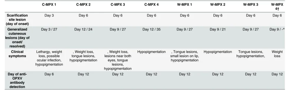
Table 1. Summary of clinical symptoms, day of primary and generalized lesions onset, and day of first detection of immune response in each experimental animal.
C-MPX 1 C-MPX 2 C-MPX 3 C-MPX 4 W-MPX 1 W-MPX 2 W-MPX 3 W-MPX
4†
Scarification site lesion (day of onset)
Day 3 Day 6 Day 6 Day 6 Day 6 Day 6 Day 6 Day 6
Generalized cutaneous lesions (day of
onset/ resolved)
Day 3 / 27 Day 12 / 24 Day 9 / 27 Day 12 / 35 Day 9 / 27 Day 9 / 21 Day 9 / 27 Day 9 / -*
Clinical symptoms Lethargy, weight loss, possible ocular infection, hypopigmentation
, Weight loss, tongue lesions, hypopigmentation
, Weight loss, lesions near both
eyes, tongue lesions, hypopigmentation
Hypopigmentation , Tongue lesions, small lesion on lip,
hypopigmentation
Hypopigmentation Tongue lesions, hypopigmentation,
Weight loss
Day of anti-OPXV antibody detection
Day 6 Day 12 Day 12 Day 12 Day 12 Day 12 Day 12 Day 12
*W-MPX 4 died on day 13 p.i., thus, no information is available for this animal after this day.
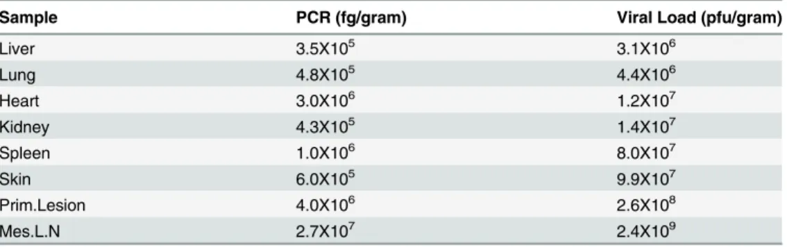
hypopigmented scarring left) by day 27 p.i. All other animals in both experimental groups had distinct primary lesions at the site of scarification by day 6 and distinguishable secondary lesions between days 9–12 p.i. Lesions began to resolve by day 15 and were fully resolved for most animals by day 27, with the exception of one C-MPX challenged animal in which lesions did not completely resolve until day 35 p.i. Two animals in each experimental group developed tongue lesions on day 12 (C-MPX 2 and 3, W-MPX 1 and 3), that resolved by day 18 (C-MPX animals) and 21 p.i. (W-MPX) respectively. Although the gross number of lesions was not noticeably different between viral strains, based on our observations the severity of lesion pre-sentation (size/appearance) seemed more pronounced in animals from group W-MPX than animals from group C-MPX (although this was not a quantifiable measurement). Additionally, animal W-MPX 4 died on day 13 although the overt illness in this animal was not noticeably different from the other animals in its experimental group; tissues samples from this animal were collected during necropsy and tested for OPXV DNA and viable virus. Viral loads in har-vested tissues ranged from 3.1X10⁶(liver) to 2.4X10⁹(mesenteric lymph node) (Table 2).
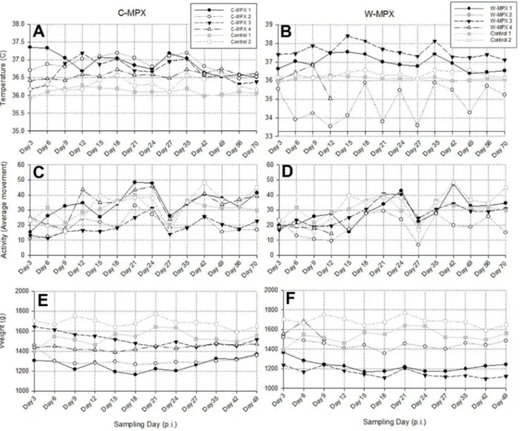
Biotelemetry
There was no significant difference between core body temperatures, activity levels or weight change of experimental animals compared to control animals when compared on a day by day basis (S1 Table). Three animals of group C-MPX and two in group W-MPX showed an increase in temperature following viral challenge (Fig 2). Additionally, when using Wilcoxon signed-rank tests to compare daily average temperatures per group throughout the study, the C-MPX group was significantly higher (p<0.001) than control groups, and significantly higher
than the W-MPX group (p = 0.003). The W-MPX average temperatures were marginally sig-nificantly higher compared to the control group (p = 0.04).
Animals C-MPX 1 and W-MPX 1 increased their activity after being challenged with MPXV; animals C-MPX 4 and W-MPX 3 showed reduced activity immediately after viral chal-lenge but their activity increased after days 9 and 12 p.i., respectively; the rest of the experimen-tal animals were less active than the control animals throughout the entire study (Fig 2). Comparisons of average activity levels per day throughout the study using Wilcoxon signed-rank test were highly significant between each experimental group and the control group with reduced averages throughout the study for both clades (C-MPX: p = 0.004 and W-MPX: p = 0.001), but no significant difference was found between C-MPX and W-MPX (p = 0.593).
Individual weights and activity were measured throughout the study. All experimental ani-mals lost weight after MPXV challenge and started recovering weight between days 21 and 27 p.i. (Fig 2). Weight loss was statistically higher for both experimental groups compared to the control group (p<0.001 for both groups) and the difference between groups was marginally
significant (p = 0.041) with greater weight loss in the C-MPX challenged group.
Live Viral Particles and Viral DNA (Table 3)
animals from a subset of samples (oral, scarification site, nasal swabs) out to day 56 p.i. in both experimental groups (Table 3).
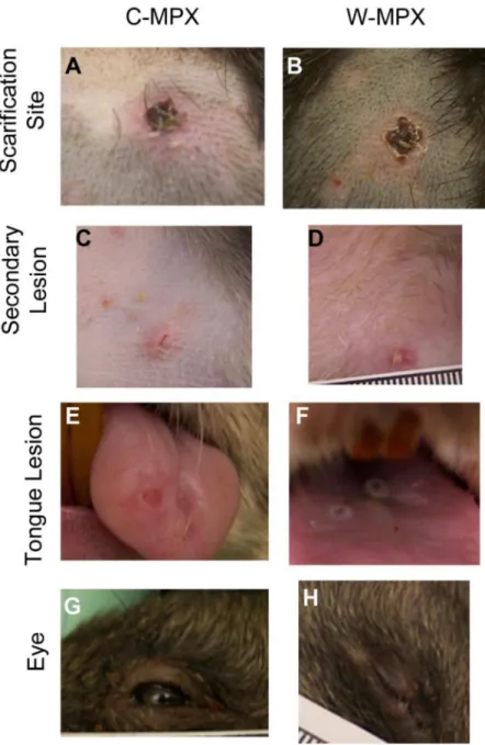
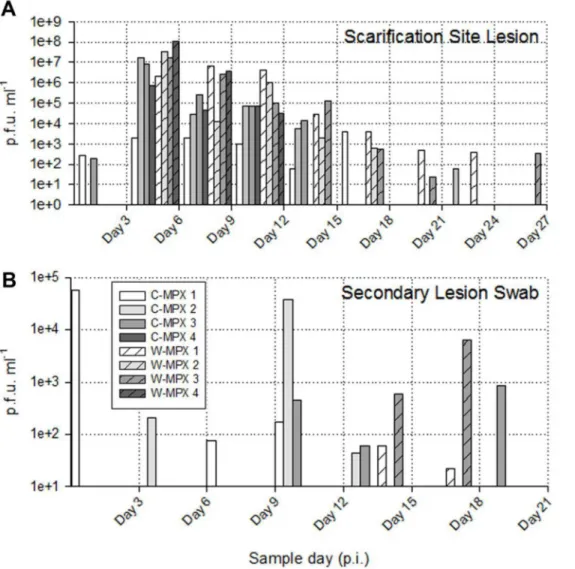
The length of viral shedding was similar to viral DNA results, with some animals/samples having a delay in time between viral DNA positivity and viable virus detection. Infectious viral particles from at least one sample were first obtained from one C-MPX animals (C-MPX 1) and one W-MPX animals (W-MPX 1) on day 3 p.i. and from all experimental animals by day 6 p.i. (for at least one sample;Fig 3andFig 4). Viable virus could not be detected from one rec-tal swab that was positive for viral DNA (C-MPX 2). Additionally, viable virus could not be Fig 1. Representative images of cutaneous lesions after experimental challenge with Congo Basin (A, C, E and G) or West African (B, D, F and H) clades of MPXV.(A and B) Pictures of scarification site lesions, (C and D) secondary lesions C and D, (E and F) tongue lesions; and (G and H) eyelid lesions for animals in each experimental group.
doi:10.1371/journal.pntd.0004013.g001
Cricetomys(Pouched Rats): Potential Reservoir of Monkeypox Virus
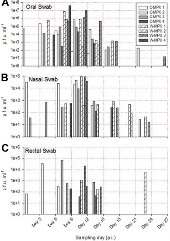
detected from secondary lesion swabs from three animals that were positive for viral DNA (C-MPX 4, W-MPX 2 and W-MPX 4). Cessation of viral shedding from all animals/samples had occurred by day 27 p.i. The mean number of shedding days was not significantly different between viral clades from the oral, nasal, lesions and rectal swabs and time of shedding in days for all samples can be seen inTable 3.
The shedding of viable virus was most prevalent in swabs of the inoculation site on day 6 p.i. (108pfu/ml) followed closely in magnitude by oral and nasal swabs 107and 105pfu/ml for C-MPX and W-MPX challenged animals, respectively; with peak levels seen on days 9 and 12 p.i. then decreasing steadily until the last detection on day 24 p.i. for C-MPX and day 27 p.i. for W-MPX (Figs3and4). Loads of virus from rectal and scarification swabs are also depicted in
Fig 3andFig 4.
Fig 5summarizes the maximum and average viable virus obtained from each sample type by individual sample (Fig 5A) and by experimental group (Fig 5B). Statistical comparison, using the Wilcoxon rank sum test, of maximum viable virus found in blood samples (p = 0.4), oral (p = 1), scarification site (p = 0.8), secondary lesion (p = 0.66), rectal (p = 0.8) and nasal (p = 0.67) swabs between experimental groups showed no significant differences.
Serology
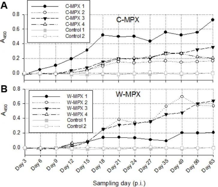
OPXV generic Ig antibodies were detected earliest in C-MPX 1 on day 6 p.i., (this was also the animal that first developed generalized cutaneous lesions on day 3 p.i.) (1). This individual also developed the highest level of antibody response of the C-MPX group with levels increasing most rapidly until day 18 p.i. and continuing to increase through day 63 p.i. (Fig 6). All other MPXV challenged animals developed a detectable antibody response by day 12 p.i.. Anti-body levels varied between individual animals but differences were not significantly different between viral strains. Ig antibody levels fluctuated but increased abruptly for most animals until day 21 p.i.; after this initial increase there was a slower but continued increase in antibody titer or plateau at study end. The W-MPX animal that died on day 13 had Ig antibody levels similar to the other animals in its experimental group at time of death.
Discussion
MPXV continues to be an important human health threat, causing sporadic outbreaks within Africa as well as the potential to spread outside its endemic range, as evidenced by the 2003 US outbreak. Identification of MPXV reservoir(s) is important so the public can be informed of associated risks with handling and consuming those species of animals and to improve infec-tion control measures. Addiinfec-tionally the search to identify a small animal model that closely Table 2. PCR results and viral load for sampled tissues obtained from animal W-MPXV 4 during necropsy.
Sample PCR (fg/gram) Viral Load (pfu/gram)
Liver 3.5X10⁵ 3.1X10⁶
Lung 4.8X10⁵ 4.4X10⁶
Heart 3.0X10⁶ 1.2X10⁷
Kidney 4.3X10⁵ 1.4X10⁷
Spleen 1.0X10⁶ 8.0X10⁷
Skin 6.0X10⁵ 9.9X10⁷
Prim.Lesion 4.0X10⁶ 2.6X10⁸
Mes.L.N 2.7X10⁷ 2.4X10⁹
resembles human monkeypox disease progression continues in order to allow evaluation of vaccines and therapeutics against systemic Orthopoxvirus infection [38]. Up until the relatively recent development of the prairie dog and CAST/Eij MPXV models, there has been a paucity of small-animal models for the study of MPXV; specifically a small-animal model that mimics systemic disease in humans including the development of the characteristic cutaneous lesions. Due to the FDA’s animal rule, having numerous animal models that can be used to test efficacy of therapeutics and anti-virals against a MPXV challenge, especially at time of rash onset, would be beneficial.
Through our experimental infection ofCricetomys, we have shown that these animals may well serve as a natural reservoir of the virus and additionally could be utilized as a relevant Fig 2. Biotelemetry measurements of temperature (A and B) and activity (C and D); and weight (E and F) of Cricetomys challenged with West African or Congo Basin MPXV. (A and B)Recorded core body temperature for experimental and control groups with higher temperatures for animals in C-MPX compared to controls. (C and D) Activity levels showing reduced activity for most experimental animals until day 9 p.i. (C-MPX) and day 12 p.i. (W-MPX), and becoming more active afterwards. (E and F) Weight loss was observed in experimental animals but not in the control group. W-MPX 4 died on day 13 p.i., thus, no information is available for this animal after this day.
doi:10.1371/journal.pntd.0004013.g002
Cricetomys(Pouched Rats): Potential Reservoir of Monkeypox Virus
animal model for the study of MPXV. With the exception of the prairie dog MPXV model, no other small animal model develops the cutaneous lesions that characterize human disease. Through the current study, we have shown thatCricetomysdevelop these skin lesions after MPXV infection and therefore may be utilized for the study of therapeutics at time of rash onset. The time until secondary lesions developed after animal inoculation was 9–12 days with the exception of one animal which developed lesions at 3 days p.i. The 9–12 day incubation before lesion onset is similar to the prairie dog MPXV model as well as the time-course believed to occur after a human is infected. Additionally, we were able to infect the animals with a plau-sible amount of virus to that which likely occurs in a natural transmission setting via scarifica-tion (to mimic a bite/scratch from an infected animal) and infected animals shed large amounts of virus from multiple secretions. During the 2003 US outbreak, people became infected with MPXV due to bites/scratches from infected prairie dogs [40,46]. Although we do not have the data from human exposures in Africa, we can hypothesize that a bite or scratch from an infected animal can lead to infection in people and in other animals. Additionally, it is widely accepted that MPXV is less transmissible via an aerosol route than smallpox in people as well as in an animal model of MPXV [47,48]; therefore a route other than intranasal is prob-ably the most relevant when studying MPXV transmission in potential reservoirs. Animals may be infecting other animals when fighting with each other as occurs during transmission of other viruses such as Hanta virus [49], or perhaps when sharing the same food source and/or bedding and therefore oral excreta is shared. Follow-up studies with additional challenge routes would be worthwhile to explore differences in disease progression in MPXV infected Cricet-omysdue to infection route. However, the results from the current study provide evidence thatCricetomys gambianus (and by inference, probably the closely related species,Cricetomys
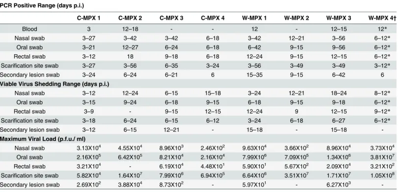
Table 3. Range of days p.i. in which collected samples were PCR positive (top section), contained viable virus (middle section), and the maximum viral load found for each individual.
PCR Positive Range (days p.i.)
C-MPX 1 C-MPX 2 C-MPX 3 C-MPX 4 W-MPX 1 W-MPX 2 W-MPX 3 W-MPX 4†
Blood 3 12–18 - - 12 - 12–15 12*
Nasal swab 3–27 3–42 3–42 6–18 3–42 12–21 3–56 6–12*
Oral swab 3–21 12–27 6–24 6–18 6–42 9–15 9–56 6–12*
Rectal swab 3–12 18 9–18 6–18 12–24 9–15 12–15 6–12*
Scarification site swab 3–27 3–56 6–35 3–24 3–56 3–49 3–49 3–12*
Secondary lesion swab 3–24 6–24 6–21 6 15–35 9–15 6–42 6
Viable Virus Shedding Range (days p.i.)
Nasal swab 3–12 12–24 6–15 15–18 3–24 12–21 18–24 8–12*
Oral swab 3–15 9–24 6–18 9–15 6–18 9–15 9–18 6–12*
Rectal swab 3–9 - 9–15 12–15 12–24 9 12–15 9–12*
Scarification site swab 3–18 6–24 6–15 6–12 3–24 6–18 6–27 6–12*
Secondary lesion swab 3–12 6–15 12–21 - 15–18 - 15–18
-Maximum Viral Load (p.f.u./ ml)
Nasal swab 3.13X10⁴ 4.55X10⁴ 8.96X10³ 2.46X10² 9.63X10⁴ 3.66X10² 8.96X10⁴ 3.73X10⁴
Oral swab 2.16X10⁵ 6.42X10⁵ 8.21X10⁴ 2.16X10⁴ 7.99X10⁶ 7.09X10⁵ 1.34X10⁶ 3.81X10⁷
Rectal swab 3.21X10⁴ - 6.19X10⁴ 4.48X101 5.90X101 5.67X10² 2.09X10⁴ 3.21X10²
Scarification site swab 5.82X10⁴ 1.64X10⁷ 7.99X10⁶ 6.94X10⁵ 6.64X10⁶ 3.51X10⁷ 1.71X10⁷ 1.05X10⁸
Secondary lesion swab 2.69X10² 3.88X10⁴ 8.73X10² - 5.97X101 - 6.27X10³
-*W-MPX 4 died on day 13 p.i., thus, no information is available for this animal after this day.
emini)should be further considered as a likely MPXV reservoir species as well as potential ani-mal model of monkeypox disease.
Both MPXV strains caused an overt rash illness inCricetomys, although the morbidity and mortality were slightly lower than that reported in non-African rodents; particularly when comparing animals challenged with C-MPX [50]. Although observational data noted more pronounced lesions in the W-MPX animals, decreased activity and weight loss in both experi-mental groups suggest that infection with either clade of MPXV produces a systemic infection that affects the normal behavior of the animals (i.e., they became more stationary and con-sumed less food), but infectedCricetomysdid not become moribund during the periods when they were shedding infectious virus of either MPXV strain. Slight differences were observed in body temperatures of MPXV infectedCricetomys, with C-MPX animals having a more marked febrile period and temperature difference compared to the W-MPX groups. This could suggest Fig 3. Viable viral load (pfu/ml) for each experimental animal found in scarification site (A) and secondary lesion swabs (B) throughout the study. (A) The maximum viral load from the scarification site was 1.05X10⁸in an animal challenged with West African MPXV and 1.64X10⁷for an animal challenged
with Congo Basin MPXV. No viable virus was found in sample days after day 27 post infection (p.i.) (B) Lower loads of viable virus were recovered from secondary lesion swabs during a shorter period of time (day 3 p.i. to 21 p.i.); the maximum viral load was 3.88X10⁴for an animal infected with Congo Basin
MPXV and 6.27X10³for an animal infected with West Africa MPXV.
doi:10.1371/journal.pntd.0004013.g003
Cricetomys(Pouched Rats): Potential Reservoir of Monkeypox Virus
a more robust immune response in the C-MPX animals; however no significant differences were seen in anti-OPXV antibody levels between groups, and the course of illness was similar.
Because these were wild-caught, genetically heterogeneous animals, it is not surprising that some differences in disease presentation and mortality were observed. One animal challenged with C-MPX had an earlier disease time-course with both primary and secondary lesions evi-dent by day 3 p.i. The antibody response was also expedited in this animal with antibody detec-tion occurring 6 days earlier than all other animals. Viral DNA and viable virus were first detected earlier in all samples compared to the otherCricetomys; however the peak loads seen were similar. There was only one animal that succumbed to disease, an animal challenged with W-MPX. Although the disease presentation until the animal perished on day 13 was similar to other animals, necropsy results revealed extremely high loads of virus within all tissues tested (106–109p.f.u./g). It is possible that the animal suffered from multi-organ failure and resulting death due to these high loads of virus. It is interesting that only an animal challenged with W-MPX succumbed to disease, as it is the less virulent clade within people as well as other ani-mal models. We have previously genotyped theCricetomysused within this study and shown that these animals originated from West Africa [Mauldin et al. manuscript in draft]. Generally viruses are believed to become attenuated within an animal host after circulation within that Fig 4. Viable viral load (p.f.u./ml) for each experimental animal found in oral (A), nasal (B) and rectal swabs (C) throughout the study.Higher viral loads were obtained from oral swabs than from rectal or nasal swabs. No viable virus was found in sample days after Day 27 post infection (p.i.).
hosts’geographic range [51,52], therefore we believe that this one animal’s death was most likely not reflective of the W-MPX virulence potential withinCricetomys, but specific to this individual animal.
The viral loads from infectedCricetomysare similar to that seen in MPXV infected non-African rodent species such as prairie dogs and ground squirrels in other laboratory challenge investigations, as well as during the 2003 MPX outbreak in the United States [11,23,31,53]. This level of virus should be more than sufficient to achieve transmission to other rodents or humans that might come into contact with the infected animals (especially through a bite) or their excretions. For example, the maximum amount of virus shed from the lesions, nose, and mouth of infectedCricetomys(1.05X10⁸, 9.63X10⁴and 3.8X10⁷pfu/ml, respectively) exceeds Fig 5. Average and maximum viral loads from MPXV challengedCricetomys.(A) Average viral load per individual sample type and experimental group at each sampling day; no viable virus was found in sample days after Day 27 post infection (p.i.). (B) Maximum and average viral load for each sample type and experimental group throughout the entire study. Error bars represent one standard deviation.
doi:10.1371/journal.pntd.0004013.g005
Cricetomys(Pouched Rats): Potential Reservoir of Monkeypox Virus
the amount of virus used to initially challenge these animals (4X10⁴pfu/ml). Interestingly, detection of viral DNA for most animals was not seen within blood samples until day 12 p.i. It is possible that our sample collection (i.e. amount of blood taken and days at which blood was collected) was not adequate for viral detection in the blood sample. Therefore we may have missed the primary/transient viremia that is believed to occur in humans and other animal models [54] [Hutson et al. 2015 Manuscript accepted BioMed Research International]. The shedding of virus detected in rectal swabs was variable but consistently lower and for a shorter period of time with respect to the levels and duration seen in other samples. Viable virus was recovered in lower titers (6.72X101-6.19X10⁴pfu/ml) from the rectal swabs primarily between days 9 and 15 p.i., but out to day 24 p.i. for one W-MPX individual. It is important to note that these were not fecal samples, but were swabs of the rectum; thus, although this suggests that Fig 6. Immune response inCricetomysafter MPXV challenge.Absorbance values measured at 450nm from ELISA assays for both experimental groups: C-MPX (A) and W-MPX (B). No statistically significant difference was found between experimental groups (Wilcoxon signed test p-value = 0.3054). W-MPX 4 died on day 13 p.i., thus, no information is available for this animal after this day.
virus could be found in the fecal pellets, it is not possible to determine how much infectious virus would actually be shed in such a manner. The detection of viral DNA unsurprisingly par-allels the levels of viable virus detected in all of the samples. However viral DNA was detectable for a longer period of time and lasted until day 56 p.i. for both viral strains, compared to the last viable virus being detected at day 27 p.i.. These later viral DNA positive samples are pre-sumably the result of sheared non-infectious viral fragments which persist beyond the infec-tious (viral shedding) stages of illness. This finding highlights the necessity of virus culture in field and laboratory studies examining the presence of enzootic and zoonotic diseases.
The results of our study are consistent with work done by collaborators who used biolumi-nescent imaging (BLI) to assess Congo Basin MPXV challenge (intradermal and intranasal inoculation) ofCricetomys[Falendysz et al.; submitted for Publication PLoS Negl Trop Dis]. Although the authors found a difference in clinical disease presentation depending on the route of inoculation, all animals shed high loads of virus, similar to the findings reported herein. Additionally BLI analysis allowed the investigators to demonstrate replication of MPXV in both healthy and sick animals. Thus it was also concluded by our collaborators that
Cricetomyscould be a potential source of MPXV infection for humans.
Members of the GenusCricetomysincludingC.gambianusare found throughout Sub-Saha-ran Africa, including many areas that are outside of the known Sub-Saha-range of MPXV. Currently there is no known mammal species whose distribution perfectly overlaps with the distribution of MPXV, and the results presented herein along with those of other laboratory studies dealing with terrestrial rodents, suggest that the maintenance and transmission cycle of MPXV in Sub-Saharan Africa may involve multiple rodent species that can amplify and transmit the virus both within and between other mammalian species including humans. Based on past serosur-veys conducted in Africa,Cricetomyshave shown evidence of anti-OPXV antibodies and OPXV DNA; additionally, during the 2003 US Outbreak,Cricetomyswas found to be infected with MPXV [16,20,23]. Although it is likely that there is a complex relationship between multi-ple rodent reservoirs in the transmission and maintenance of the virus within Africa, the data from our laboratory findings agree with the African serosurveys and suggest thatCricetomys
should be considered as, at least, one potential MPXV reservoir host species that is involved in the maintenance and transmission of MPXV.
Supporting Information
S1 Table. P-values of the Wilcoxon rank-sum test performed to compare temperature, activity and weight loss of animals challenged with the West African clade of MPXV (W-MPXV), the Congo Basin clade of MPXV (C-MPXV) and the Control group at each of the sampling days.Weight loss values are not available for days 3, 56 and 70 p.i. because day 3 p.i. was used as the base value to calculate weight loss and animals were not weighed on days 56 and 70 p.i.
(DOCX)
Acknowledgments
The authors would like to acknowledge Krista Yorita Christensen for statistical analysis of the data. Additionally we would like to acknowledge Florida Fish and Wildlife Conservation Com-mission (specifically Scott Hardin) and USDA (specifically John Woolard) for their assistance in trapping of theCricetomysused within our studies.
Cricetomys(Pouched Rats): Potential Reservoir of Monkeypox Virus
Disclaimer
The findings and conclusions in this report are those of the authors and do not necessarily rep-resent the views of the Centers for Disease Control and Prevention. Use of trade, product of firm names does not imply endorsement by the U.S Government.
Author Contributions
Conceived and designed the experiments: DSC CLH JS VAO RLR ZB SW JM KLK TER JEO IKD. Performed the experiments: DSC CLH JS RLR ZB SW EJ MT KLK. Analyzed the data: DSC CLH YJN VAO ZB SW KLK IKD. Wrote the paper: DSC CLH YJN VAO RLR JM KLK TER JEO.
References
1. Abrahao JS, Guedes MI, Trindade GS, Fonseca FG, Campos RK, Mota BF, Lobato ZI, Silva-Fer-nandes AT, Rodrigues GO, Lima LS, Ferreira PC, Bonjardim CA, Kroon EG (2009) One more piece in the VACV ecological puzzle: could peridomestic rodents be the link between wildlife and bovine vac-cinia outbreaks in Brazil? PLoS One 4: e7428. doi:10.1371/journal.pone.0007428PMID:19838293
2. Khodakevich L, Jezek Z, Kinzanzka K (1986) Isolation of monkeypox virus from wild squirrel infected in nature. Lancet 1: 98–99. PMID:2867342
3. Lourie B, Nakano JH, Kemp GE, Setzer HW (1975) Isolation of poxvirus from an African Rodent. J Infect Dis 132: 677–681. PMID:811713
4. Regnery DC (1987) Isolation and partial characterization of an orthopoxvirus from a California vole (Microtus californicus). Brief report. Arch Virol 94: 159–162. PMID:3034201
5. Crouch AC, Baxby D, McCracken CM, Gaskell RM, Bennett M (1995) Serological evidence for the res-ervoir hosts of cowpox virus in British wildlife. Epidemiol Infect 115: 185–191. PMID:7641833
6. Chen N, Li G, Liszewski MK, Atkinson JP, Jahrling PB, Feng Z, Schriewer J, Buck C, Wang C, Lefko-witz EJ, Esposito JJ, Harms T, Damon IK, Roper RL, Upton C, Buller RM (2005) Virulence differences between monkeypox virus isolates from West Africa and the Congo basin. Virology 340: 46–63. PMID:
16023693
7. Likos AM, Sammons SA, Olson VA, Frace AM, Li Y, Olsen-Rasmussen M, Davidson W, Galloway R, Khristova ML, Reynolds MG, Zhao H, Carroll DS, Curns A, Formenty P, Esposito JJ, Regnery RL, Damon IK (2005) A tale of two clades: monkeypox viruses. J Gen Virol 86: 2661–2672. PMID:
16186219
8. Americo JL, Moss B, Earl PL (2010) Identification of wild-derived inbred mouse strains highly suscepti-ble to monkeypox virus infection for use as small animal models. J Virol 84: 8172–8180. doi:10.1128/ JVI.00621-10PMID:20519404
9. Breman JG, Kalisa R, Steniowski MV, Zanotto E, Gromyko AI, Arita I (1980) Human monkeypox, 1970–79. Bull World Health Organ 58: 165–182. PMID:6249508
10. Foster SO, Brink EW, Hutchins DL, Pifer JM, Lourie B, Moser CR, Cummings EC, Kuteyi OE, Eke RE, Titus JB, Smith EA, Hicks JW, Foege WH (1972) Human monkeypox. Bull World Health Organ 46: 569–576. PMID:4340216
11. Hutson CL, Olson VA, Carroll DS, Abel JA, Hughes CM, Braden ZH, Weiss S, Self J, Osorio JE, Hud-son PN, Dillon M, Karem KL, Damon IK, Regnery RL (2009) A prairie dog animal model of systemic orthopoxvirus disease using West African and Congo Basin strains of monkeypox virus. J Gen Virol 90: 323–333. doi:10.1099/vir.0.005108-0PMID:19141441
12. Learned LA, Reynolds MG, Wassa DW, Li Y, Olson VA, Karem K, Stempora LL, Braden ZH, Kline R, Likos A, Libama F, Moudzeo H, Bolanda JD, Tarangonia P, Boumandoki P, Formenty P, Harvey JM, Damon IK (2005) Extended interhuman transmission of monkeypox in a hospital community in the Republic of the Congo, 2003. Am J Trop Med Hyg 73: 428–434. PMID:16103616
13. Arita I, Henderson DA (1968) Smallpox and monkeypox in non-human primates. Bull World Health Organ 39: 277–283. PMID:5303409
14. Arita I, Gispen R, Kalter SS, Wah LT, Marennikova SS, Netter R, Tagaya I (1972) Outbreaks of mon-keypox and serological surveys in nonhuman primates. Bull World Health Organ 46: 625–631. PMID:
4340222
15. Peters JC (1966) An epizootic of monkey pox at Rotterdam Zoo. International Zoo Yearbook 6: 274–
16. Hutin YJ, Williams RJ, Malfait P, Pebody R, Loparev VN, Ropp SL, Rodriguez M, Knight JC, Tshioko FK, Khan AS, Szczeniowski MV, Esposito JJ (2001) Outbreak of human monkeypox, Democratic Republic of Congo, 1996 to 1997. Emerg Infect Dis 7: 434–438. PMID:11384521
17. Khodakevich L, Szczeniowski M, Nambu mD, Jezek Z, Marennikova S, Nakano J, Meier F (1987) Mon-keypox virus in relation to the ecological features surrounding human settlements in Bumba zone, Zaire. Trop Geogr Med 39: 56–63. PMID:3037740
18. Khodakevich L, Szczeniowski M, Nambu mD, Jezek Z, Marennikova S, Nakano J, Meier F (1987) Mon-keypox virus in relation to the ecological features surrounding human settlements in Bumba zone, Zaire. Trop Geogr Med 39: 56–63. PMID:3037740
19. Khodakevich L, Jezek Z, Messinger D (1988) Monkeypox virus: ecology and public health significance. Bull World Health Organ 66: 747–752. PMID:2853010
20. Reynolds MG, Carroll DS, Olson VA, Hughes C, Galley J, Likos A, Montgomery JM, Suu-Ire R, Kwasi MO, Jeffrey RJ, Braden Z, Abel J, Clemmons C, Regnery R, Karem K, Damon IK (2010) A silent enzo-otic of an orthopoxvirus in Ghana, West Africa: evidence for multi-species involvement in the absence of widespread human disease. Am J Trop Med Hyg 82: 746–754. doi:10.4269/ajtmh.2010.09-0716
PMID:20348530
21. Khodakevich L, Szczeniowski M, Manbu mD, Jezek Z, Marennikova S, Nakano J, Messinger D (1987) The role of squirrels in sustaining monkeypox virus transmission. Trop Geogr Med 39: 115–122. PMID:
2820094
22. Radonic A, Metzger S, Dabrowski PW, Couacy-Hymann E, Schuenadel L, Kurth A, Matz-Rensing K, Boesch C, Leendertz FH, Nitsche A (2014) Fatal monkeypox in wild-living sooty mangabey, Cote d'Ivoire, 2012. Emerg Infect Dis 20: 1009–1011. doi:10.3201/eid2006.13–1329PMID:24857667
23. Hutson CL, Lee KN, Abel J, Carroll DS, Montgomery JM, Olson VA, Li Y, Davidson W, Hughes C, Dillon M, Spurlock P, Kazmierczak JJ, Austin C, Miser L, Sorhage FE, Howell J, Davis JP, Reynolds MG, Bra-den Z, Karem KL, Damon IK, Regnery RL (2007) Monkeypox zoonotic associations: insights from labo-ratory evaluation of animals associated with the multi-state US outbreak. Am J Trop Med Hyg 76: 757–
768. PMID:17426184
24. Olude MA, Mustapha OA, Ogunbunmi TK, Olopade JO (2013) The vertebral column, ribs, and sternum of the African giant rat (Cricetomys gambianus waterhouse). ScientificWorldJournal 2013: 973537. doi:10.1155/2013/973537PMID:24288518
25. van Vliet N, Nebesse C, Gambalemoke S, Akaibe D, Nasi R (2012) The bushmeat market in Kisangani, Democratic Republic of Congo: implications for conservation and food security. Oryx 46: 196–203. 26. Assogbadjo AE, Codjia JTC, Sinsin B, Ekue MRM, Mensah GA (2005) Importance of rodents as a
human food source in Benin. Belgian Journal of Zoology 135: 111–115.
27. Olayemi A, Nicolas V, Hulselmans J, Missoup AD, Fichet-Calvet E, Amundala D, Dudu A, Dierckx T, Wendelen W, Leirs H, Verheyen E (2012) Taxonomy of the African giant pouched rats (Nesomyidae: Cricetomys): molecular and craniometric evidence support an unexpected high species diversity. Zool J Linnean Soc 165: 700–719.
28. Perry ND, Hanson B, Hobgood W, Lopez RL, Okraska CRKKL, et al. (2006) New inasive species in southern Florida: Gambian rat (Cricetomys gambianus). J Mammalogy 87 (2): 262–264.
29. Breman JG, Nakano JH, Coffi E, Godfrey H, Gautun JC (1977) Human poxvirus disease after smallpox eradication. Am J Trop Med Hyg 26: 273–281. PMID:192091
30. Marennikova SS, Shelukhina EM, Zhukova OA (1989) Experimental infection of squirrels Sciurus vul-garis by monkey pox virus. Acta Virol 33: 399. PMID:2574948
31. Schultz DA, Sagartz JE, Huso DL, Buller RM (2009) Experimental infection of an African dormouse (Graphiurus kelleni) with monkeypox virus. Virology 383: 86–92. doi:10.1016/j.virol.2008.09.025
PMID:18977501
32. Falendysz EA, Londono-Navas AM, Meteyer CU, Pussini N, Lopera JG, Osorio JE, Rocke TE (2014) Evaluation of monkeypox virus infection of black-tailed prairie dogs (Cynomys ludovicianus) using in vivo bioluminescent imaging. J Wildl Dis 50: 524–536. doi:10.7589/2013-07-171PMID:24779460
33. Hutson CL, Gallardo-Romero N, Carroll DS, Clemmons C, Salzer JS, Nagy T, Hughes CM, Olson VA, Karem KL, Damon IK (2013) Transmissibility of the monkeypox virus clades via respiratory transmis-sion: investigation using the prairie dog-monkeypox virus challenge system. PLoS ONE 8: e55488. doi:10.1371/journal.pone.0055488;PONE-D-12-23800 [pii]. PMID:23408990
34. Hutson CL, Abel JA, Carroll DS, Olson VA, Braden ZH, Hughes CM, Dillon M, Hopkins C, Karem KL, Damon IK, Osorio JE (2010) Comparison of West African and Congo Basin monkeypox viruses in BALB/c and C57BL/6 mice. PLoS ONE 5: e8912. doi:10.1371/journal.pone.0008912PMID:20111702
35. Osorio JE, Iams KP, Meteyer CU, Rocke TE (2009) Comparison of monkeypox viruses pathogenesis in mice by in vivo imaging. PLoS ONE 4: e6592. doi:10.1371/journal.pone.0006592PMID:19668372
Cricetomys(Pouched Rats): Potential Reservoir of Monkeypox Virus
36. Sbrana E, Jordan R, Hruby DE, Mateo RI, Xiao SY, Siirin M, Newman PC, da Rosa AP, Tesh RB (2007) Efficacy of the antipoxvirus compound ST-246 for treatment of severe orthopoxvirus infection. Am J Trop Med Hyg 76: 768–773. PMID:17426185
37. Parker S, Buller RM (2013) A review of experimental and natural infections of animals with monkeypox virus between 1958 and 2012. Future Virol 8: 129–157. doi:10.2217/fvl.12.130PMID:23626656
38. Hutson CL, Damon IK (2010) Monkeypox Virus Infections in Small Animal Models for Evaluation of Anti-Poxvirus Agents. Viruses 2: 2763–2776. doi:10.3390/v2122763PMID:21994638
39. Mills JN, Childs JE, Ksiazek TG, Peters CJ, Velleca WM (1995) Methods for trapping and sampling small mammals for virologic testing.
40. Reed KD, Melski JW, Graham MB, Regnery RL, Sotir MJ, Wegner MV, Kazmierczak JJ, Stratman EJ, Li Y, Fairley JA, Swain GR, Olson VA, Sargent EK, Kehl SC, Frace MA, Kline R, Foldy SL, Davis JP, Damon IK (2004) The detection of monkeypox in humans in the Western Hemisphere. N Engl J Med 350: 342–350. PMID:14736926
41. da Fonseca FG, Wolffe EJ, Weisberg A, Moss B (2000) Characterization of the vaccinia virus H3L envelope protein: topology and posttranslational membrane insertion via the C-terminal hydrophobic tail. J Virol 74: 7508–7517. PMID:10906204
42. Li Y, Olson VA, Laue T, Laker MT, Damon IK (2006) Detection of monkeypox virus with real-time PCR assays. J Clin Virol 36: 194–203. PMID:16731033
43. Li Y, Zhao H, Wilkins K, Hughes C, Damon IK (2010) Real-time PCR assays for the specific detection of monkeypox virus West African and Congo Basin strain DNA. J Virol Methods 169: 223–227. S0166-0934(10)00254-5 [pii];doi:10.1016/j.jviromet.2010.07.012PMID:20643162
44. Venables, W. N. and Ripley, B. D. (2002) Modern Applied Statistics Fourth Edition.
45. R Development Core Team (2011) R: a language and environment for statistical computing. R Founda-tion for Statistical Computing. 2.12.1 ed. Vienna, Austria, version.
46. Reynolds MG, Davidson WB, Curns AT, Conover CS, Huhn G, Davis JP, Wegner M, Croft DR, New-man A, Obiesie NN, Hansen GR, Hays PL, Pontones P, Beard B, Teclaw R, Howell JF, Braden Z, Hol-man RC, Karem KL, Damon IK (2007) Spectrum of infection and risk factors for huHol-man monkeypox, United States, 2003. Emerg Infect Dis 13: 1332–1339. doi:10.3201/eid1309.070175PMID:18252104
47. Fine PE, Jezek Z, Grab B, Dixon H (1988) The transmission potential of monkeypox virus in human populations. Int J Epidemiol 17: 643–650. PMID:2850277
48. Hutson CL, Gallardo-Romero N, Carroll DS, Clemmons C, Salzer JS, Nagy T, Hughes CM, Olson VA, Karem KL, Damon IK (2013) Transmissibility of the monkeypox virus clades via respiratory transmis-sion: investigation using the prairie dog-monkeypox virus challenge system. PLoS One 8: e55488. doi:
10.1371/journal.pone.0055488;PONE-D-12-23800 [pii]. PMID:23408990
49. Glass GE, Childs JE, Korch GW, LeDuc JW (1988) Association of intraspecific wounding with hanta-viral infection in wild rats (Rattus norvegicus). Epidemiol Infect 101: 459–472. PMID:3141203
50. Hutson CL, Carroll DS, Self J, Weiss S, Hughes CM, Braden Z, Olson VA, Smith SK, Karem KL, Reg-nery RL, Damon IK (2010) Dosage comparison of Congo Basin and West African strains of monkeypox virus using a prairie dog animal model of systemic orthopoxvirus disease. Virology 402: 72–82. doi:10. 1016/j.virol.2010.03.012PMID:20374968
51. Kerr PJ, Liu J, Cattadori I, Ghedin E, Read AF, Holmes EC (2015) Myxoma virus and the Leporipox-viruses: an evolutionary paradigm. Viruses 7: 1020–1061. v7031020 [pii];doi:10.3390/v7031020
PMID:25757062
52. Kerr PJ, Ghedin E, Depasse JV, Fitch A, Cattadori IM, Hudson PJ, Tscharke DC, Read AF, Holmes EC (2012) Evolutionary history and attenuation of myxoma virus on two continents. PLoS Pathog 8: e1002950. doi:10.1371/journal.ppat.1002950;PPATHOGENS-D-12-00984 [pii]. PMID:23055928
53. Tesh RB, Watts DM, Sbrana E, Siirin M, Popov VL, Xiao SY (2004) Experimental infection of ground squirrels (Spermophilus tridecemlineatus) with monkeypox virus. Emerg Infect Dis 10: 1563–1567. PMID:15498157