and Pigs Exposed to Tsetse Fly Bites
Guy Caljon1,2,3.*, Reta Duguma4., Reginald De Deken5
, Stijn Schauvliege6, Frank Gasthuys6,
Luc Duchateau7, Jan Van Den Abbeele1,8*
1Unit of Veterinary Protozoology, Department of Biomedical Sciences, Institute of Tropical Medicine Antwerp (ITM), Antwerp, Belgium,2Unit of Cellular and Molecular Immunology, Vrije Universiteit Brussel (VUB), Brussels, Belgium,3Laboratory of Myeloid Cell Immunology, Vlaams Instituut voor Biotechnologie (VIB), Brussels, Belgium,
4Faculty of Veterinary Medicine, Addis Ababa University, Debre Zeit, Ethiopia,5Unit Veterinary Entomology, Department of Biomedical Sciences, Institute of Tropical Medicine Antwerp (ITM), Antwerp, Belgium,6Department of Surgery and Anaesthesia of Domestic Animals, Faculty of Veterinary Medicine, Ghent University (UGent), Ghent, Belgium,7Department of Comparative Physiology and Biometrics, Faculty of Veterinary Medicine, Ghent University (UGent), Ghent, Belgium,8Laboratory of Zoophysiology, Department of Physiology, Ghent University (UGent), Ghent, Belgium
Abstract
Background:Tsetse flies are obligate blood-feeding insects that transmit African trypanosomes responsible for human sleeping sickness and nagana in livestock. The tsetse salivary proteome contains a highly immunogenic family of the endonuclease-like Tsal proteins. In this study, a recombinant version of Tsal1 (rTsal1) was evaluated in an indirect ELISA to quantify the contact with totalGlossina morsitans morsitanssaliva, and thus the tsetse fly bite exposure.
Methodology/Principal Findings: Mice and pigs were experimentally exposed to different G. m. morsitans exposure regimens, followed by a long-term follow-up of the specific antibody responses against total tsetse fly saliva and rTsal1. In mice, a single tsetse fly bite was sufficient to induce detectable IgG antibody responses with an estimated half-life of 36–40 days. Specific antibody responses could be detected for more than a year after initial exposure, and a single bite was sufficient to boost anti-saliva immunity. Also, plasmas collected from tsetse-exposed pigs displayed increased anti-rTsal1 and saliva IgG levels that correlated with the exposure intensity. A strong correlation between the detection of anti-rTsal1 and anti-saliva responses was recorded. The ELISA test performance and intra-laboratory repeatability was adequate in the two tested animal models. Cross-reactivity of the mouse IgGs induced by exposure to differentGlossinaspecies (G. m. morsitans,G. pallidipes,G. palpalis gambiensisandG. fuscipes) and other hematophagous insects (Stomoxys calcitransand
Tabanus yao) was evaluated.
Conclusion:This study illustrates the potential use of rTsal1 fromG. m. morsitansas a sensitive biomarker of exposure to a broad range of Glossina species. We propose that the detection of anti-rTsal1 IgGs could be a promising serological indicator of tsetse fly presence that will be a valuable tool to monitor the impact of tsetse control efforts on the African continent.
Citation:Caljon G, Duguma R, De Deken R, Schauvliege S, Gasthuys F, et al. (2014) Serological Responses and Biomarker Evaluation in Mice and Pigs Exposed to Tsetse Fly Bites. PLoS Negl Trop Dis 8(5): e2911. doi:10.1371/journal.pntd.0002911
Editor:Philippe Solano, IRD/CIRDES, Burkina Faso
ReceivedDecember 9, 2013;AcceptedApril 16, 2014;PublishedMay 22, 2014
Copyright:ß2014 Caljon et al. This is an open-access article distributed under the terms of the Creative Commons Attribution License, which permits unrestricted use, distribution, and reproduction in any medium, provided the original author and source are credited.
Funding:This work was funded by the Research Foundation— Flanders (www.fwo.be), the InterUniversity Attraction Pole program P7/41 (www.belspo.be), an EU/FP7 ERC starter grant (Jan Van Den Abbeele, erc.europa.eu), the Belgian Development Cooperation (PhD scholarship Reta Duguma, www.dg-d.be) and the Institute of Tropical Medicine Antwerp (www.itg.be). The funders had no role in study design, data collection and analysis, decision to publish, or preparation of the manuscript.
Competing Interests:The authors have declared that no competing interests exist.
* E-mail: [email protected] (GC); [email protected] (JVDA)
.These authors contributed equally to this work.
Introduction
Tsetse flies (Glossinaspp.) are notorious transmitters of trypano-some parasites responsible for Human and Animal African Trypanosomiasis (HAT and AAT). Since 2009, the annual number of reported cases of HAT has dropped below 10000 (www.who.int; [1]) with the prospect and challenge of entering into the elimination phase of HAT in the near future [2,3]. Additionally, some 46 million cattle in sub-Saharan Africa are estimated to be at risk of contracting AAT making deep inroads in the socio-economical development of this continent [4]. Beside active HAT case detection and treatment of humans as well as
entomological surveys, no sensitive rapid tests are yet available to provide a semi-quantitative measure of the evolution of tsetse fly densities in areas subjected to tsetse control interventions. Indeed, easy-to-use monitoring of tsetse fly exposure on a regular basis would be a highly valuable tool in the follow-up of the efficacy of the applied and/or ongoing tsetse fly control activities.
The obligatory blood feeding tsetse flies are the cyclical insect vectors of HAT and a majority of AAT infections are initiated by the bite of an infected tsetse fly. Although it can be assumed that all tsetse fly species could act as vector, a number ofGlossinaspecies of the Palpalis group (e.g. G. palpalis spp., G. fuscipes spp., G. tachinoides) and the Morsitans group (e.g. G. morsitans spp., G. pallidipes,G. swynnertoni) are implicated as major vectors for HAT and animal trypanosomiasis. Given that only a limited percentage of these tsetse flies acquire an infection with trypanosomes, vertebrate hosts living in the tsetse fly belt are predominantly exposed to the bites of uninfected flies. It has been demonstrated for a number of hematophagous insects that salivary proteins induce humoral immune responses that could represent attractive sero-epidemiological markers of exposure (reviewed in [10]). The saliva ofGlossina morsitans morsitanstsetse flies was documented to contain over 200 protein constituents [11] from which some are implicated in manipulating the vertebrate hemostatic and inflam-matory reactions [12,13,14]. In G. m. morsitans saliva, the most abundant proteins were shown to be highly immunogenic and to correspond to the 43–45 kDa tsetse salivary gland (Tsal) protein family [15]. The physiological role of these proteins remains elusive, but biochemical characterization revealed that they are nucleic acid binding proteins with low endonuclease activity [16]. Immunoglobulin responses to tsetse fly saliva have been detected in humans living in Uganda [15], Democratic Republic of Congo [17,18] and Guinea [19]. Also cattle experimentally exposed to tsetse fly bites displayed elevated levels of anti-saliva antibodies [20]. Immunoblotting studies using the immune plasmas have shown that salivary proteins of several tsetse fly species are recognized by the circulating antibodies [15,18,19]. The highly abundant Tsal proteins were commonly recognized by the human plasmas and an indirect ELISA using recombinant Tsal1 and Tsal2 proteins as antigens was clearly able to differentiate the tsetse-exposed Ugandan plasmas from control plasmas [15].
Recently, a peptide (amino acids 18–43) derived from theG. m. morsitansadenosine deaminase-related TSGF1 protein was evalu-ated using a panel of human plasmas from West Africa, revealing that obtained ELISA signals correlated with the anticipated levels of tsetse exposure of the tested populations [21]. Allergic and anaphylactic reactions against tsetse fly bites have also been reported, in which IgE antibodies directed against an Antigen5-related salivary allergen are implicated [22,23,24]. Efforts to develop a serological test based on a TAg5-derived peptide were not yet successful [21].
We here provide experimental evidence that anti-rTsal1 and anti-G. m. morsitanssaliva antibodies can mark exposure of mice and pigs to tsetse flies. Although the anti-tsetse saliva ELISA exhibits a better average test performance, we propose that a serological assay based on the individual recombinant Tsal1 protein could be an alternative to assess the exposure of populations or herds to tsetse fly challenge and hence could be used for tsetse fly epidemiological studies, for prioritizing tsetse fly control, for monitoring and evaluating tsetse fly control schemes and for risk assessment of trypanosome transmission in endemic regions.
Methods
Ethics statement
The experiments, maintenance and care of animals complied with the guidelines of the European Convention for the Protection of Vertebrate Animals used for Experimental and other Scientific Purposes (CETS nu 123). Rodent care and experimental procedures were performed under approval from the Animal Ethical Committee of the Institute of Tropical Medicine (Permit Nrs. PAR013-MC-M-Tryp and PAR014-MC-K-Tryp). Breeding and experimental work with tsetse flies was approved by the Scientific Institute Public Health department Biosafety and Biotechnology (SBB 219.2007/1410). Pig experiments were approved by the ITM Animal Ethical Committee (Permit Nr. PAR-021) and the Ethical Committee of the Faculty of Veterinary Medicine of Ghent University (Permit Nr. EC2010/030) and were performed in the Ghent university stables under the supervision of a veterinary doctor.
Salivary antigens and recombinant proteins
Saliva ofGlossina m. morsitans from the ITMA tsetse fly colony was collected as outflow from the salivary glands as described elsewhere [16]. Saliva ofTabanus yaowas kindly provided by Prof. Ren Lai (Kunming Institute of Zoology, Yunnan, China),Stomoxys calcitrans saliva was harvested from flies received from Dr. Christopher J. Geden (United States Department of Agriculture, Gainesville, US). Recombinant Tsal1 was purified as described elsewhere from inclusion bodies of IPTG-induced Top10F’ Escherichia colihost cells harboring a pQE60:Tsal1 plasmid [15]. Tsal1 was resolubilized in 6M guanidinium hydrochloride, enriched by Ni-NTA affinity chromatography (Qiagen) and further purified on a Superdex 200 size exclusion column connected to an Akta Explorer (GE Healthcare) in 6 M ureum 50 mM Tris pH 8.0 and 600 mM NaCl. Protein concentrations were determined using Nanodrop spectrophotometry and samples were stored in aliquots at 220uC. E. coli soluble extract was prepared as an additive for the porcine ELISA assay diluent as described for other porcine assays [25]. The extract was made from a 1L overnight culture of Top10F’E. coliin Terrific broth. The bacterial pellet was resuspended in 12 ml PBS supplemented with a Complete protease inhibitor cocktail tablet (Roche), followed by 5 rounds of 1 minute sonication. The soluble fraction
Author Summary
was harvested as the supernatant after 30 minutes centrifugation at 20.0006g. E. coli soluble extract was stored in aliquots at 2 20uC.
Experimental study animals and immunization
Groups of eight female outbred mice (NMRI, Charles River) were subjected to different intensities of exposure toG. m. morsitans bites, followed by regular blood sampling and evaluation of the antibody responses in ELISA: (i) once exposed to a single fly, (ii) once exposed to 10 flies, (iii) 3 times per week for 3 weeks exposed to a single fly, (iv) 3 times per week for 3 weeks exposed to 10 flies and (v) not exposed to tsetse fly bites. One mouse that underwent multiple exposures to a single fly succumbed by day 28 after initiation of exposure. Plasmas were collected over a period of 390 days. After this.1 year non-exposure period, six mice that were exposed to single or multiple (10) flies were selected based on their physical appearance and behavior and were re-exposed to a single G. m. morsitanstsetse fly followed by weekly plasma collection for up to 42 days after re-challenge. Two mice that were previously exposed to the multiple fly bites succumbed within 4 weeks after the boosting.
A Glossina species cross-reactivity study was performed by exposing five mice (OF1, Charles River) per group every 3 days for 6 weeks to 10 flies of either G. m. morsitans, G. p. gambiensis, G. pallidipes (kindly provided by Peter Takac, Institute of Zoology, Bratislava, Slovakia) or G. f. fuscipes (kindly provided by the International Centre of Insect Physiology and Ecology, Mbita Point, Kenya). Immune plasma was harvested 10 days after the last exposure. Five non-exposed mice served as negative controls. A cross-reactivity study between salivary antigens of hematopha-gous insects was performed by intrapinna exposure of 6 OF1 mice per group at 3-weekly interval to decreasing amounts of saliva (5, 2, 1 and 1mg) harvested fromGlossina morsitans,Stomoxys calcitrans andTabanus yao. Plasma samples were collected 10 days after the last immunization.
A total of 11 female pigs (Seghers, Belgium), hybrids of Belgian Landrace, Large White and a specific company line were used for experimental exposure to three differentG. m. morsitansexposure regimens: (i) 5 pigs were weekly exposed for 7 weeks to 30 tsetse flies (high exposure), (ii) 5 pigs were exposed 2-weekly for 6 weeks to 3 tsetse flies (low exposure, 1 pig died at day 7) and (iii) 1 pig was not exposed to tsetse (negative control). The experimental pigs were 6 weeks old at the start of the experiment following a 4-days acclimatisation period in the university stables (Faculty of Veterinary Medicine, UGhent). For anaesthesia of the pigs, intramuscular injection of midazolam (0.5 mg/kg), ketamine (10 mg/kg) and morphine (0.1 mg/kg) was used. Blood was collected every week for 11 successive weeks from the jugular vein using Vacutainer EDTA tubes (BD) a few minutes after the pigs had been anesthetised and prior to exposure to the tsetse fly bites. After blood collection, the blood was centrifuged at 3000 rpm for 15 minutes and plasma stored at220uC. Due to the unexpected death of 1 pig, no tsetse challenge and blood sampling was performed on day 7 for all animals.
Due to limited housing capacity in the university stables, only two pigs from the low exposure group could be kept for an additional 2-month period of non-exposure, followed by a re-challenge by the bites of 10 G. m. morsitans flies and a weekly plasma collection over a period of 6 weeks.
Serological analyses
IgG responses in the exposed animals were analyzed by indirect ELISA against rTsal1 and saliva from different hematophagous insects. For this purpose, polystyrene 96 well plates (Thermo
Scientific NUNC MaxiSorp Surface) were coated overnight at 4uC with 200 ng antigen (G. morsitans, S. calcitrans or T. yaosaliva or rTsal1) per well in 0.1 M NaHCO3 (pH 8.3). Plates were
overcoated for 1 h with 10% fetal bovine serum (FBS) at ambient temperature. Serial half plasma dilutions starting from 1:100 in assay diluent (PBS/10%FBS) were applied for 2 h to antigen and FBS-coated wells. For the analysis of porcine plasma samples, 20% Top10F’E. colisoluble extract was added to the assay diluent to reduce unspecific binding to the antigenic coat. Based on the plasma dilution experiments, a 1:1600 dilution was chosen for the time course analyses. Specific immunoglobulin detection was achieved using horseradish peroxidase conjugated detection antibodies. For the detection of mouse and porcine IgGs respectively a 1:1000 diluted rabbit F(ab)2 anti-mouse IgG
(STAR13B, Serotec) and a rabbit anti-pig IgG conjugate (A5670, Sigma) 1:4000 diluted in PBS/10%FBS were used. Detection was with TMB substrate (3,39,5,59 -Tetramethylbenzi-dine, Sigma) and reactions were stopped by the 1:3 addition of 1N H2SO4. Optical density (O.D.) was measured using a Multiskan
Ascent plate reader (Thermo) at a 450 nm wavelength.
Data analysis
Antigen-specific responses were expressed as the DO.D. between antigen and non-antigen-coated wells. Statistical analyses were performed in SAS Version 9.3 (SAS Institute Inc., Cary, NC, USA). The effect of tsetse fly exposure intensity and boosting on anti-G. m. morsitanssaliva and anti-rTsal1 immune responses in mice and pigs was analysed over the entire sampling period based on a mixed model with animal as random effect and challenge, time and their interaction as categorical fixed effects and F-tests were performed at the 5% significance level. Pairwise comparisons were performed using Tukey’s multiple comparisons technique to adjust the significance level. Cross-reactivity of immune responses induced by different Glossina species and other hematophagous insects was analysed using a linear fixed effects model using the 1:100 or 1:1600 dilution with challenge as categorical fixed effect. Pairwise comparisons were performed with control using Dun-nett’s multiple comparisons technique to adjust the significance level. Intra-laboratory repeatability of the ELISA tests was assessed by the non-parametric Spearman correlation test. Sensitivity and specificity of the assays were assessed by receiver operating characteristic (ROC) curve analysis of the DO.D. values of exposed and non-exposed animals, starting from 3 weeks after the initial exposure. The area under the ROC curve (AUC) was used as a global index of diagnostic accuracy. Kinetics of the IgG clearance in mice was assessed in five mice from the multiple exposure schemes with sufficient remaining plasma for the different timepoints and using the plasma sample with the highest antibody titer (set as 100%) to prepare a standard curve. Percent decrease in antibody titers over time was assessed by a two-phase non-linear regression allowing the estimation of the antibody half-life.
Results
Serological responses in mice exposed toGlossina morsitans
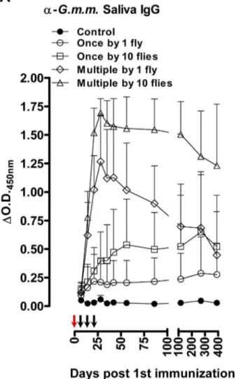
flies (1 versus 10 flies) as well as differences in frequency (single versus weekly exposure over 3 weeks) were detected by ELISA. Exposure to a single tsetse fly bite was sufficient to induce slightly elevated levels of antibodies against G. m. morsitans saliva and rTsal1 (Figure 1), although the recorded differences with control plasmas were not significant (p= 0.9843 and p= 0.6146 for the anti-rTsal1 and anti-G. m. morsitanssaliva tests respectively). Single exposure of mice to 10 flies resulted in significantly increased reactivity againstG. m. morsitanssaliva (p= 0.0160) but not against rTsal1 (p= 0.8618). With both antigens, statistically significant differences were recorded considering the entire sampling period between control mice and mice subjected to the repeated exposure to 1 and 10 flies (p,0.0001). Both the anti-rTsal1 and the anti-G. m. morsitanssaliva ELISA test were able to differentiate between these two repeated tsetse exposure schemes (p,0.0001 and p= 0.0012 respectively). Based on a standard curve generated using the plasma sample with the highest IgG titer (i.e. day 28, mouse exposed to the most intense biting regimen), antibody half-lives in five mice exposed multiple times to a single fly or 10 flies were determined by two-phase decay regressions (Figure 2). The average half-lives (T1/22) of the anti-saliva and anti-rTsal IgGs in a
second phase of decay after a first phase immediately after cessation of tsetse exposure were respectively 36 and 40 days (Figure 2). There was a significant individual variation within the different exposure groups with up to a 10-fold difference between the strongest and weakest responder. On average, a 3 to 4-fold difference in peak IgG titer was recorded between mice exposed to a low (multiple bites by a single fly) and high exposure regimen (multiple bites by 10 flies). The antibodies appeared relatively persistent over an evaluation period of more than 1 year. Following a long period of non-exposure, the bite of a single tsetse fly was sufficient to boost the anti-saliva and anti-rTsal1 IgG titers. This boosting appeared independent of the previous exposure intensity (Figure 3) as statistical analysis over the 6-week sampling period revealed the inability of the saliva and rTsal1-based tests to differentiate mice subjected to a low or high initial exposure regimen (p= 0.4141 and p= 0.9609 respectively). Anti-body titers against both antigens reached a plateau within 7 days after re-challenge, while in naive animals the responses were slightly lower (p= 0.0833 and p= 0.1925 for the anti-saliva and anti-rTsal1 IgG levels respectively) and only reached peak titers within 4 weeks after initial exposure (Figure 3).
Figure 1. Tsetse fly induced humoral responses in mice.Long-term follow-up of (A) the anti-G. m. morsitanssaliva IgG responses and (B) the anti-rTsal1 IgG responses in mice (n= 8/group) exposed to 4 different tsetse fly biting intensities (1 versus 10 flies) and frequencies (single versus weekly exposure for 3 weeks). Arrows indicate the tsetse exposure regimen for single exposed (red arrows) and multiple exposed mice (red and black arrows). Presented data are the meanDO.D.450 nmvalues and the 95% CI obtained with a 1:1600 dilution of the individual plasmas.
A strong correlation between anti-saliva and anti-rTsal1 ELISA results was recorded with a Spearman correlation coefficientrof 0.92. Intra-laboratory repeatability of the saliva and anti-rTsal1 detection assays was excellent (Spearman r= 0.98). A comparison of the diagnostic value of the anti-saliva and rTsal1 indirect ELISA was conducted using ROC curve analysis. Comparison of the area under the curve (AUC), based on the mouse plasmas from the experiment presented in Figure 1 and collected at least 3 weeks after the initial tsetse fly exposure, indicated that the anti-tsetse saliva ELISA has a better test performance than the rTsal1-based assay (AUC 0.94 versus 0.82, Figure S1).
Serological responses of mice exposed to various
Glossinaspecies
Mice were repeatedly exposed to the bites of variousGlossina species (G. m. morsitans,G. p. gambiensis,G. pallidipesandG. f. fuscipes).
It was noted that the feeding performance ofG. fuscipeson mice was inferior to those of the other species, resulting in a high mortality of these flies when maintained on mice.G. m. morsitans saliva and rTsal1 were next used as antigens in indirect ELISA to detect the elicited antibodies. A marked cross-reactivity of the anti-saliva IgGs elicited by threeGlossinaspecies (G. m. morsitans,G. p. gambiensisandG. pallidipes,p,0.0001 at the 1:100 plasma dilution) and a relatively weaker cross-reactivity ofG. f. fuscipes(p= 0.0053) was detected withG. m. morsitanssaliva as antigen (Figure 4A&B). Similarly, antibodies induced by exposure toG. m. morsitans,G. p. gambiensis and G. pallidipes significantly reacted with rTsal1 (respectivelyp,0.0001,p= 0.0157 and p= 0.0002), but no cross-reaction with rTsal1 was detected in mice exposed toG. f. fuscipes (p= 0.9883). This indicated that a number of antigens are sufficiently conserved in the saliva of several Glossina species to allow multi-species detection of exposure using G. m. morsitans saliva. The rTsal1 also enabled detection of exposure of mice to
Figure 2. Antibody kinetics in tsetse exposed mice.Evolution over time in the absence of tsetse fly challenge (from day 28 onwards) of the anti-G. m. morsitanssaliva IgG concentrations (A, B, C) and the anti-rTsal1 IgG concentrations (D, E, F) in mice (n= 5/group) exposed multiple times to a single fly (B, E) or 10 flies (A, D). Presented data are the IgG concentrations relative to the highest responder plasma and expressed as percent based on a standard dilution series. Box plots represent the means, with minimum and maximum and the 75-25% percentiles. The average antibody half-life (T1/22,C, F) over the 2 exposure groups (n= 10) was determined by a two-phase non-linear decay regression (dotted line) of the declining
antibody concentrations relative to the concentration recorded after cessation of the tsetse exposure (day 28 set as starting concentration of 100%). Scatter plots indicate the mean concentrations with the 95% CI.
various tested tsetse fly species, except for G. fuscipes (Palpalis subgenus).
Serological responses of mice exposed to various hematophagous insect species
Mice were experimentally immunized by 4 intradermal injections with the saliva of stable flies (Stomoxys calcitrans) or horse flies (Tabanus yao) and compared with control mice and mice that were immunized with tsetse fly saliva following the same immunization protocol. Immunization with Tabanus saliva did not result in IgGs that reacted with rTsal1 and totalG. morsitans saliva as coating antigens (p= 0.7882 andp= 0.7637 at the 1:100 plasma dilution, Figure 4D&E). However, a slight increase that did not reach statistical significance was observed in responsiveness of theStomoxysexposed plasmas in the rTsal1 and saliva-based assays (p= 0.0837 and p= 0.0639 respectively). At a standard 1:1600 dilution, no cross-reaction of the Stomoxys exposed plasmas with rTsal1 and saliva was detected (p= 0.9920 and p= 0.9915 respectively). Analysis of the published salivary transcriptome of Stomoxys calcitrans suggested the presence of a Tsal1 homologue from which a truncated sequence was published (GenBank Accession Nu: ACN69159, [26]). Within this region, only 36% identity in amino acid sequence was found, which could explain the slightly elevated anti-rTsal1 reactivity of Stomoxys exposed mice.
Serological responses in tsetse fly exposed pigs
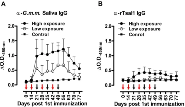
Pigs were experimentally exposed to a low or a high tsetse fly bite regimen, followed by assessment of the antibody production.
As compared to the mouse indirect ELISA, we have modified the porcine assay by including E. coli soluble extract in the sample diluent as described elsewhere [25] to reduce the background that was observed particularly onto the rTsal1 antigen which was produced in a bacterial expression system. Under the used assay conditions, anti-rTsal1 and anti-saliva IgGs were detected from day 21 after the first tsetse exposure onwards (Figure 5). The specific IgG titers in pigs correlated with the intensity of tsetse exposure. Anti-rTsal1 and anti-tsetse saliva IgGs were elevated in tsetse exposed groups as compared to the non-exposed control pig and pre-immune plasmas, with significant differences for the high exposure group (p= 0.0216 and p= 0.0030 respectively). The repeated exposure of pigs to 30 flies resulted in higher anti-rTsal1 and anti-tsetse saliva IgG titers as compared to the pigs exposed to a low tsetse challenge by 3 flies (p= 0.0014 and p= 0.0024 respectively). However, exposure to the low exposure scheme did not result in significantly elevated responses in both the rTsal1 and saliva-based ELISA (p= 0.8844 and p= 0.4261 respectively, Figure 5). Boosting of 2 pigs from the low exposure group after a 2-month non-exposure period by the bites of 10 flies resulted in elevated anti-saliva IgG titers but was only weakly detectable using the rTsal1-based ELISA and with higher individual variation (Figure 6). In general, a good correlation between anti-saliva and anti-rTsal1 ELISA results, obtained for the samples from the tsetse fly exposure experiment presented in Figure 5, was recorded with a Spearman correlation coefficient r of 0.79. Intra-laboratory repeatability of the anti-saliva and anti-rTsal1 detection assays was adequate (Spearmanr= 0.94). Comparison of the area under the curve (AUC) for the anti-tsetse saliva and anti-rTsal1 ELISA ROC curve indicated that the assay based on totalG. morsitanssaliva has
Figure 3. Effect of boosting in mice after a long period of non-exposure.Evolution of the anti-G. m. morsitanssaliva IgG responses (A) and the anti-rTsal1 IgG repsonses (B)was evaluated in mice (n= 6/group) originally exposed to the bites of a single or 10 flies and re-exposed to a single tsetse fly bite after a 1-year period of non-exposure (indicated by an arrow). Controls are the naive mice exposed to a single bite as depicted in Figure 1. The original control animals (n= 3) were not exposed and served as negative controls (dotted line). Presented data are meanDO.D.450 nm
a better average performance than the anti-rTsal1 IgG detection test (AUC 0.96 versus 0.83, Figure S2).
Discussion
Assessment of exposure of populations at risk to the bites of tsetse fly vectors could be an important step towards improved control of both HAT and AAT. Given that novel innovative vector control strategies are being developed (e.g. miniaturized insecti-cide-treated targets [27], the release of sterile male insects [8,9]) and deployed on increasingly large scales on the African continent, monitoring the impact of these new as well as conventional interventions on actual exposure to tsetse fly bites is a logical
follow-up. The tsetse fly species implicated in the transmission of trypanosomes are not uniform throughout the African continent as more than 30 tsetse species and subspecies exist that play differential roles in parasite transmission, have preferences for specific biotopes and display specific host feeding preferences. A number of species are strongly implicated in HAT transmission, such as flies of the Morsitans subgenus (e.g. used in this study:G. morsitans morsitans,G. pallidipes) and flies of the Palpalis group (e.g. used in this study:G. palpalis gambiensis,G. fuscipes fuscipes). In case of AAT, a large panel of tsetse species and other sympatric hematophagous arthropods are respectively involved in biological and mechanical transmission. Especially stable flies (Stomoxyssp.) and horse flies (Tabanussp.) play a role in mechanical transmission
Figure 4. Detection of humoral responses induced by various hematophagous insects.Serially diluted plasmas of control mice and mice repeatedly exposed to the bites ofG. morsitans morsitans,G. fuscipes fuscipes,G. palpalis gambiensisandG. pallidipes(n= 5/group) were tested in the
G. m. morsitans saliva-based (A) and rTsal1-based IgG detection ELISA (B). Serially diluted plasmas of control mice and mice experimentally immunized with harvested G. m. morsitans, Stomoxys calcitrans andTabanus yao saliva (n= 6/group) were analysed for responses against the respective saliva extracts (C) and were tested in theG. m. morsitanssaliva-based (D) and rTsal1-based IgG detection ELISA (E). Presented data are the meanDO.D.450 nmvalues with the 95% CI.
of animal trypanosomes such asT.vivax. However, these insects are not able to biologically transmit trypanosomes as the parasite cannot complete its life cycle in these insects.
Research on a number of hematophagous arthropod vectors has resulted in the concept of exploiting salivary components as specific biomarkers of exposure [15,28,29,30,31,32,33]. An advantage of this approach is that relatively simple serological tests could provide information on actual exposure to the bites of
disease vectors and provide a risk indicator of contracting a vector-transmitted disease without the need of strenuous entomological surveys. Whole salivary gland extracts of sand flies [33,34,35], triatomine bugs [30,31] and various mosquito species [36,37,38] can be used to assess biting exposure. It has been suggested that this type of serological approach would enable the detection of very low levels of exposure that could remain undetected by entomological trappings [39]. Also differences in exposure level
Figure 5. Tsetse fly induced humoral responses in pigs.Follow-up of the anti-G. m. morsitanssaliva IgG responses (A) and the anti-rTsal1 IgG responses (B) in pigs exposed to 2 different tsetse fly biting intensities (two-weekly exposure for 6 weeks to 3 flies (low exposure group, n = 4) or weekly exposure for 7 weeks to 30 flies (high exposure group, n = 5)). One non-exposed animal was included as a negative control. Arrows indicate the tsetse exposure regimen for the low exposure (red arrows) and high exposure group (red and black arrows). Presented data are the mean
DO.D.450 nmvalues and the 95% CI obtained with a 1:1600 dilution of the individual plasmas.
doi:10.1371/journal.pntd.0002911.g005
Figure 6. Effect of boosting in pigs after a period of non-exposure.Evolution of the anti-G. m. morsitanssaliva IgG responses and the anti-rTsal1 IgG repsonses was evaluated in pigs (n= 2) originally exposed two-weekly for 6 weeks to the bites of 3 flies ( = low exposure group) and re-exposed to the bites of 10 flies (indicated by an arrow) after a 2-month period of non-exposure. One non-re-exposed animal sampled during the priming experiment (Figure 5) served as a negative control. Presented data are meanDO.D.450 nmvalues with the 95% CI obtained with a 1:1600 dilution of
the individual plasmas.
due to vector control interventions (e.g. insecticide treated bednets) could be elucidated on the basis of salivary proteins as immunological probes [29,34,40,41]. While total salivary extracts can be used in various ELISA and immunoblot formats, some studies have moved towards the use of recombinant proteins or peptides which could lead to the development of more standardized immune assays [29,42,43,44,45,46,47,48,49]. Sever-al strategies could be envisaged either aiming at species-specific or pan-species exposure detection.
Studies using various tsetse fly species have shown that salivary components are immunogenic in mice, rabbits, cattle and humans [15,17,18,19,20,23]. Immunoblotting of salivary proteins separat-ed on 1D or 2D protein gels have highlightseparat-ed immunogenic proteins corresponding to several protein families including endonuclease (Tsal), adenosine deaminase (TSGF), 59nucleotidase (59Nuc) and Antigen 5 (Ag5) related proteins [19]. In addition, the immunogenic nature ofG. m. morsitans sgp1,sgp2andsgp3resulted in their identification on the basis of immune screening of a phage cDNA expression library [50]. We have previously observed by Western blot analysis that human plasma samples collected in Tororo (Uganda, [51]) whereG.fuscipes fuscipesis the predominant tsetse fly, commonly recognized the 43–45 kDa Tsal proteins inG. m. morsitans saliva [15]. This suggested thatG. m. morsitans Tsal-based immune screening of these Ugandan samples cross-detected exposure to G. fuscipes fuscipes, responsible for Trypanosoma brucei rhodesiense transmission in that area [52]. Plasma samples from tsetse fly exposed individuals in Guinean HAT foci also displayed strong reactivity against the highly abundant Tsal proteins inG. palpalis gambiensissaliva in immunoblots [19]. As the Tsal protein band is commonly recognized in samples from Glossina-exposed humans [15,19], Tsal proteins could have the potential of a pan-Glossinaspecies exposure marker. However, reactivity with Tsal1 and Tsal2 was also observed with a pool of human control plasmas from Bobo-Dioulasso (Burkina Faso) [19]. Based on our current study we anticipate that the inclusion criteria for these urban unexposed plasma donors (not having traveled outside of the city for at least three months, [21]) might have been insufficiently stringent to exclude circulating anti-Tsal antibodies. Indeed, tsetse flies are present at sites outside the city and given the high immunogenicity and anticipated long antibody persistence, this could explain the documented positive reactions in the immuno-blots.
Production of a recombinant version of the Tsal1 protein allowed evaluating its effectiveness for detecting tsetse fly exposure in an antibody-detection ELISA. By experimentally exposing mice and pigs to different tsetse fly biting regimens, the high immunogenicity of total G. m. morsitans saliva and Tsal1 as a major constituent was confirmed. In mice, a single bite was proven sufficient to induce a detectable immune response in naive animals and to boost antibody levels in previously exposed animals. Kinetics of the antibody clearance was assessed in mice over a.1 year period, which revealed a long persistence with an average half-life of 36–40 days. In pigs, the apparent antibody clearance rate was faster and although the time of sampling and the number of experimental animals was limited, the anti-saliva IgG half-life could be estimated to be 15 days (data not shown). This clearance rate was consistent with the fast decline of anti-G. m. submorsitans saliva IgG levels observed in cattle within 10 weeks after cessation of tsetse exposure [20]. Both in mice and pigs, boosting of prior induced anti-saliva immunity was observed within 7 days by the anti-G. m. morsitanssaliva and anti-rTsal1 ELISA. However, with the used sample sizes and experimental conditions, both the rTsal1 and saliva-based ELISA were unable to statistically differentiate the control pig from pigs exposed to a low tsetse fly challenge.
Interestingly, the boosting of immunity in mice seemed indepen-dent of the previous exposure intensity. As such, surveys based on rTsal1 or totalG. m. morsitanssaliva in tsetse fly control intervention zones would give information on the actual tsetse exposure intensity rather than history and could provide a tool to monitor re-invasion. Consistent with this, serological results for cattle in South-West Burkina Faso based on G. palpalis gambiensis whole saliva extract related to the seasonality which directly impacts the intensity of host/vector contact [20]. Also humans living in HAT endemic areas display significant differences in anti-G. p. gambiensis saliva IgGs depending on the study site [19]. Similar results were obtained with a peptide derived from the adenosine deaminase-related protein TSGF1, although obtained ELISA signals seemed very low [21]. In both the cattle and human ELISA tests, large inter-individual differences were observed [15,19,20,21]. It should be noted that in our study the individual variation in antibody titers in the different exposure groups also varied significantly. In controlled experimental conditions with outbred mice, we recorded up to 10-fold differences in anti-saliva and anti-rTsal1 antibody concentrations between the strongest and weakest responders. Consequently, using the tsetse salivary antigens as immunological probes has the limitation of inherent variability due to individual differences in immunological responsiveness. Never-theless, we observed a clear overall difference in anti-rTsal1 and anti-saliva IgG levels between the different exposure groups of mice and pigs, with an average 3–4 fold difference in specific antibody concentration between a low exposure (repeated exposure to 1 fly for mice or to 3 flies for pigs) and a high exposure regimen (repeated exposure to 10 flies for mice or to 30 flies for pigs). This is in line with observations made for cattle that were experimentally exposed to different biting intensities [20]. Beside population-level studies, individual serological follow-up of selected animals or sentinel animals using the recombinant Tsal1 or the totalG. morsitanssaliva could provide a measure for tsetse prevalence, provide evidence for the impact of an intervention strategy or could be a sensitive tool to detect re-invasion of previously cleared areas or the efficacy of a barrier protecting a cleared area. Here, rTsal1 and total G. morsitans saliva seem to provide an indication of exposure to a broad range of Glossina species. Only for G. f. fuscipes, detection of induced IgGs is hampered which could relate to the genetic distance of this species as member of the Palpalis group (althoughG. palpalis gambiensiswas efficiently cross-detected). Nevertheless, serological responses against rTsal1 were detected in humans from Uganda principally exposed toG. fuscipes, which suggests that sensitivity and specificity parameters related to a single antigen may vary considerably depending on the host species. It also remains to be evaluated whether the intensity and persistence of the anti-Tsal1 IgG responses would not result in a saturation in populations persistently exposed to tsetse bites. This could possibly limit application of the rTsal1-based assay to examining subjects in tsetse-free areas that are at risk of invasion or to monitoring sentinel animals in order to evaluate tsetse presence in endemic regions.
presence of a homologue (GenBank Accession Nu: ACN69159, [26]) with relatively limited degree of identity to Tsal1. Given the strong immunogenicity of the Tsal proteins and based on our ELISA analyses with the experimentally exposed pigs and mice we propose to use a 1:1600 plasma dilution in favor of an increased specificity and an overall reduced risk of detecting cross-reactive antibodies. For the porcine ELISA, E. coli soluble extract was added to the sample diluent to increase the specificity similar to what was proposed for other pig serological tests [25]. Under these stringent experimental conditions, the recombinant Tsal1 was able to detect the high levels of exposure to tsetse fly bites and with a good correlation with the data obtained for total G. m. morsitans saliva as an antigen.
Collectively and combined with our previous analyses using a large panel of East-African human plasmas [15], this study indicates that a recombinant version of theG. m. morsitans Tsal1 fulfills the criteria of a candidate exposure biomarker for a broad range of tsetse fly species. We believe that the high sensitivity of the rTsal antigen and the broad species recognition could be an added value to the immunoassays that are tested in the framework of other studies based on tsetse salivary peptides.
Supporting Information
Figure S1 Repeatability and specificity/sensitivity anal-ysis of the antibody detection test in mice. Scatter plot analysis of the anti-G. m. morsitanssaliva IgG responses (A) and the anti-rTsal1 IgG repsonses (B) (DO.D.450 nm) in two separate tests
performed on a panel of 1:1600 diluted mouse plasma samples (n= 352). Test repeatability was analyzed by the non-parametric Spearman correlation test. Sensitivity and specificity of the two
assays were assessed by receiver operating characteristic (ROC) curve analysis of theDO.D. values of exposed and non-exposed mice (C). The area under the ROC curve (AUC) is reported as a measure for the test performance.
(TIF)
Figure S2 Repeatability and specificity/sensitivity anal-ysis of the antibody detection test in pigs. Scatter plot analysis of the anti-G. m. morsitanssaliva IgG responses (A) and the anti-rTsal1 IgG repsonses (B) (DO.D.450 nm) in two separate tests
performed on a panel of 1:1600 diluted porcine plasma samples (n= 80). Test repeatability was analyzed by the non-parametric Spearman correlation test. Sensitivity and specificity of the two assays were assessed by receiver operating characteristic (ROC) curve analysis of theDO.D. values of exposed and non-exposed mice (C). The area under the ROC curve (AUC) is reported as a measure for the test performance.
(TIF)
Acknowledgments
We acknowledge Peter Van Den Bossche for his involvement in the initiation of the project and who tragically died in a car accident on 11/11/ 2010. We wish to acknowledge Pe´lagie Atrou, Serge Mpouam and Ke´ita Moulaye who were involved as master students in parts of this project.
Author Contributions
Conceived and designed the experiments: GC RDD LD FG JVDA. Performed the experiments: GC RD RDD SS JVDA. Analyzed the data: GC RD LD JVDA. Contributed reagents/materials/analysis tools: GC RDD LD FG JVDA. Wrote the paper: GC RD RDD SS LD JVDA.
References
1. Simarro PP, Cecchi G, Paone M, Franco JR, Diarra A, et al. (2010) The Atlas of human African trypanosomiasis: a contribution to global mapping of neglected tropical diseases. Int J Health Geogr 9: 57.
2. Aksoy S (2011) Sleeping sickness elimination in sight: time to celebrate and reflect, but not relax. PLoS Negl Trop Dis 5: e1008.
3. Simarro PP, Jannin J, Cattand P (2008) Eliminating human African trypanosomiasis: where do we stand and what comes next? PLoS Med 5: e55. 4. Cattand P, Food and Agriculture Organization, Programme Against African Trypanosomiasis (Paat) (2010) Linking sustainable human and animal African trypanosomiasis control with rural development strategies. PAAT technical and scientific series 10. Food and Agriculture Organization of the United Nations. 76 pp.
5. Vreysen MJ, Seck MT, Sall B, Bouyer J (2013) Tsetse flies: their biology and control using area-wide integrated pest management approaches. J Invertebr Pathol 112 Suppl: S15–25.
6. Schofield CJ, Kabayo JP (2008) Trypanosomiasis vector control in Africa and Latin America. Parasit Vectors 1: 24.
7. Torr SJ, Hargrove JW, Vale GA (2005) Towards a rational policy for dealing with tsetse. Trends Parasitol 21: 537–541.
8. Vreysen MJ, Saleh KM, Ali MY, Abdulla AM, Zhu ZR, et al. (2000)Glossina austeni(Diptera: Glossinidae) eradicated on the island of Unguja, Zanzibar, using the sterile insect technique. J Econ Entomol 93: 123–135.
9. Sow A, Sidibe I, Bengaly Z, Bance AZ, Sawadogo GJ, et al. (2012) Irradiated male tsetse from a 40-year-old colony are still competitive in a Riparian forest in Burkina Faso. PLoS One 7: e37124.
10. Fontaine A, Diouf I, Bakkali N, Misse D, Pages F, et al. (2011) Implication of haematophagous arthropod salivary proteins in host-vector interactions. Parasit Vectors 4: 187.
11. Alves-Silva J, Ribeiro JM, Van Den Abbeele J, Attardo G, Hao Z, et al. (2010) An insight into the sialome ofGlossina morsitans morsitans. BMC Genomics 11: 213.
12. Caljon G, De Ridder K, De Baetselier P, Coosemans M, Van Den Abbeele J (2010) Identification of a tsetse fly salivary protein with dual inhibitory action on human platelet aggregation. PLoS One 5: e9671.
13. Caljon G, Van Den Abbeele J, Stijlemans B, Coosemans M, De Baetselier P, et al. (2006) Tsetse fly saliva accelerates the onset ofTrypanosoma bruceiinfection in a mouse model associated with a reduced host inflammatory response. Infect Immun 74: 6324–6330.
14. Cappello M, Bergum PW, Vlasuk GP, Furmidge BA, Pritchard DI, et al. (1996) Isolation and characterization of the tsetse thrombin inhibitor: a potent
antithrombotic peptide from the saliva ofGlossina morsitans morsitans. Am J Trop Med Hyg 54: 475–480.
15. Caljon G, Van Den Abbeele J, Sternberg JM, Coosemans M, De Baetselier P, et al. (2006) Tsetse fly saliva biases the immune response to Th2 and induces anti-vector antibodies that are a useful tool for exposure assessment. Int J Parasitol 36: 1025–1035.
16. Caljon G, De Ridder K, Stijlemans B, Coosemans M, Magez S, et al. (2012) Tsetse salivary gland proteins 1 and 2 are high affinity nucleic acid binding proteins with residual nuclease activity. PLoS One 7: e47233.
17. Poinsignon A, Remoue F, Rossignol M, Cornelie S, Courtin D, et al. (2008) Human IgG antibody response toGlossinasaliva: an epidemiologic marker of exposure to Glossina bites. Am J Trop Med Hyg 78: 750–753.
18. Poinsignon A, Cornelie S, Remoue F, Grebaut P, Courtin D, et al. (2007) Human/vector relationships during human African trypanosomiasis: initial screening of immunogenic salivary proteins ofGlossinaspecies. Am J Trop Med Hyg 76: 327–333.
19. Dama E, Cornelie S, Bienvenu Somda M, Camara M, Kambire R, et al. (2013) Identification ofGlossina palpalis gambiensisspecific salivary antigens: towards the development of a serologic biomarker of human exposure to tsetse flies in West Africa. Microbes Infect 15: 416–427.
20. Somda MB, Bengaly Z, Dama E, Poinsignon A, Dayo GK, et al. (2013) First insights into the cattle serological response to tsetse salivary antigens: A promising direct biomarker of exposure to tsetse bites. Vet Parasitol 197: 332– 340.
21. Dama E, Cornelie S, Camara M, Somda MB, Poinsignon A, et al. (2013) Identification of a Candidate Synthetic Peptide (Tsgf1) to Monitor Human Exposure to Tsetse Flies in West Africa. PLoS Negl Trop Dis 7: e2455. 22. Caljon G, Broos K, De Goeyse I, De Ridder K, Sternberg JM, et al. (2009)
Identification of a functional Antigen5-related allergen in the saliva of a blood feeding insect, the tsetse fly. Insect Biochem Mol Biol 39: 332–341. 23. Ellis JA, Shapiro SZ, ole Moi-Yoi O, Moloo SK (1986) Lesions and
saliva-specific antibody responses in rabbits with immediate and delayed hypersensi-tivity reactions to the bites ofGlossina morsitans centralis. Vet Pathol 23: 661–667. 24. Stevens WJ, Van den Abbeele J, Bridts CH (1996) Anaphylactic reaction after bites by Glossina morsitans(tsetse fly) in a laboratory worker. J Allergy Clin Immunol 98: 700–701.
26. Wang X, Ribeiro JM, Broce AB, Wilkerson MJ, Kanost MR (2009) An insight into the transcriptome and proteome of the salivary gland of the stable fly, Stomoxys calcitrans. Insect Biochem Mol Biol 39: 607–614.
27. Lindh JM, Torr SJ, Vale GA, Lehane MJ (2009) Improving the cost-effectiveness of artificial visual baits for controlling the tsetse flyGlossina fuscipes fuscipes. PLoS Negl Trop Dis 3: e474.
28. Schwartz BS, Ribeiro JM, Goldstein MD (1990) Anti-tick antibodies: an epidemiologic tool in Lyme disease research. Am J Epidemiol 132: 58–66. 29. Drame PM, Poinsignon A, Besnard P, Cornelie S, Le Mire J, et al. (2010)
Human antibody responses to theAnophelessalivary gSG6-P1 peptide: a novel tool for evaluating the efficacy of ITNs in malaria vector control. PLoS One 5: e15596.
30. Schwarz A, Sternberg JM, Johnston V, Medrano-Mercado N, Anderson JM, et al. (2009) Antibody responses of domestic animals to salivary antigens ofTriatoma infestansas biomarkers for low-level infestation of triatomines. Int J Parasitol 39: 1021–1029.
31. Volf P, Grubhoffer L, Hosek P (1993) Characterisation of salivary gland antigens of Triatoma infestans and antigen-specific serum antibody response in mice exposed to bites ofT. infestans. Vet Parasitol 47: 327–337.
32. Remoue F, Cisse B, Ba F, Sokhna C, Herve JP, et al. (2006) Evaluation of the antibody response toAnophelessalivary antigens as a potential marker of risk of malaria. Trans R Soc Trop Med Hyg 100: 363–370.
33. Rohousova I, Ozensoy S, Ozbel Y, Volf P (2005) Detection of species-specific antibody response of humans and mice bitten by sand flies. Parasitology 130: 493–499.
34. Gidwani K, Picado A, Rijal S, Singh SP, Roy L, et al. (2011) Serological markers of sand fly exposure to evaluate insecticidal nets against visceral leishmaniasis in India and Nepal: a cluster-randomized trial. PLoS Negl Trop Dis 5: e1296. 35. Vinhas V, Andrade BB, Paes F, Bomura A, Clarencio J, et al. (2007) Human
anti-saliva immune response following experimental exposure to the visceral leishmaniasis vector,Lutzomyia longipalpis. Eur J Immunol 37: 3111–3121. 36. Orlandi-Pradines E, Almeras L, Denis de Senneville L, Barbe S, Remoue F, et
al. (2007) Antibody response against saliva antigens ofAnopheles gambiaeandAedes aegyptiin travellers in tropical Africa. Microbes Infect 9: 1454–1462. 37. Drame PM, Poinsignon A, Besnard P, Le Mire J, Dos-Santos MA, et al. (2010)
Human antibody response toAnopheles gambiaesaliva: an immuno-epidemiolog-ical biomarker to evaluate the efficacy of insecticide-treated nets in malaria vector control. Am J Trop Med Hyg 83: 115–121.
38. Doucoure S, Mouchet F, Cornelie S, DeHecq JS, Rutee AH, et al. (2012) Evaluation of the human IgG antibody response toAedes albopictussaliva as a new specific biomarker of exposure to vector bites. PLoS Negl Trop Dis 6: e1487. 39. Poinsignon A, Cornelie S, Ba F, Boulanger D, Sow C, et al. (2009) Human IgG
response to a salivary peptide, gSG6-P1, as a new immuno-epidemiological tool for evaluating low-level exposure toAnophelesbites. Malar J 8: 198.
40. Sagna AB, Sarr JB, Gaayeb L, Drame PM, Ndiath MO, et al. (2013) gSG6-P1 salivary biomarker discriminates micro-geographical heterogeneity of human exposure toAnophelesbites in low and seasonal malaria areas. Parasit Vectors 6: 68.
41. Schwarz A, Juarez JA, Richards J, Rath B, Machaca VQ, et al. (2011) Anti-triatomine saliva immunoassays for the evaluation of impregnated netting trials against Chagas disease transmission. Int J Parasitol 41: 591–594.
42. Poinsignon A, Cornelie S, Mestres-Simon M, Lanfrancotti A, Rossignol M, et al. (2008) Novel peptide marker corresponding to salivary protein gSG6 potentially identifies exposure toAnophelesbites. PLoS One 3: e2472.
43. Elanga Ndille E, Doucoure S, Damien G, Mouchet F, Drame PM, et al. (2012) First attempt to validate human IgG antibody response to Nterm-34 kDa salivary peptide as biomarker for evaluating exposure toAedes aegyptibites. PLoS Negl Trop Dis 6: e1905.
44. Ali ZM, Bakli M, Fontaine A, Bakkali N, Vu Hai V, et al. (2012) Assessment of Anophelessalivary antigens as individual exposure biomarkers to species-specific malaria vector bites. Malar J 11: 439.
45. King JG, Vernick KD, Hillyer JF (2011) Members of the salivary gland surface protein (SGS) family are major immunogenic components of mosquito saliva. J Biol Chem 286: 40824–40834.
46. Vlkova M, Rohousova I, Hostomska J, Pohankova L, Zidkova L, et al. (2012) Kinetics of antibody response in BALB/c and C57BL/6 mice bitten by Phlebotomus papatasi. PLoS Negl Trop Dis 6: e1719.
47. Teixeira C, Gomes R, Collin N, Reynoso D, Jochim R, et al. (2010) Discovery of markers of exposure specific to bites of Lutzomyia longipalpis, the vector of Leishmania infantumchagasi in Latin America. PLoS Negl Trop Dis 4: e638. 48. Souza AP, Andrade BB, Aquino D, Entringer P, Miranda JC, et al. (2010) Using
recombinant proteins fromLutzomyia longipalpissaliva to estimate human vector exposure in visceral Leishmaniasis endemic areas. PLoS Negl Trop Dis 4: e649. 49. Sanders ML, Jaworski DC, Sanchez JL, DeFraites RF, Glass GE, et al. (1998) Antibody to a cDNA-derived calreticulin protein fromAmblyomma americanumas a biomarker of tick exposure in humans. Am J Trop Med Hyg 59: 279–285. 50. Van Den Abbeele J, Caljon G, Dierick JF, Moens L, De Ridder K, et al. (2007)
TheGlossina morsitanstsetse fly saliva: general characteristics and identification of novel salivary proteins. Insect Biochem Mol Biol 37: 1075–1085.
51. Maclean L, Odiit M, Sternberg JM (2006) Intrathecal cytokine responses in Trypanosoma brucei rhodesiensesleeping sickness patients. Trans R Soc Trop Med Hyg 100: 270–275.