Opposing Roles for Interferon Regulatory Factor-3 (IRF-3)
and Type I Interferon Signaling during Plague
Ami A. Patel1,2, Hanni Lee-Lewis1,2, Jennifer Hughes-Hanks1., Craig A. Lewis3., Deborah M. Anderson1,2 *
1Department of Veterinary Pathobiology, University of Missouri, Columbia, Missouri, United States of America,2Laboratory for Infectious Disease Research, University of Missouri, Columbia, Missouri, United States of America,3Starling Enterprise, LLC, Columbia, Missouri, United States of America
Abstract
Type I interferons (IFN-I) broadly control innate immunity and are typically transcriptionally induced by Interferon Regulatory Factors (IRFs) following stimulation of pattern recognition receptors within the cytosol of host cells. For bacterial infection, IFN-I signaling can result in widely variant responses, in some cases contributing to the pathogenesis of disease while in others contributing to host defense. In this work, we addressed the role of type I IFN duringYersinia pestisinfection in a murine model of septicemic plague. Transcription of IFN-b was induced in vitro and in vivo and contributed to pathogenesis. Mice lacking the IFN-I receptor,Ifnar, were less sensitive to disease and harbored more neutrophils in the later stage of infection which correlated with protection from lethality. In contrast, IRF-3, a transcription factor commonly involved in inducing IFN-bfollowing bacterial infection, was not necessary for IFN production but instead contributed to host defense.In vitro, phagocytosis ofY. pestisby macrophages and neutrophils was more effective in the presence of IRF-3 and was not affected by IFN-bsignaling. This activity correlated with limited bacterial growthin vivoin the presence of IRF-3. Together the data demonstrate that IRF-3 is able to activate pathways of innate immunity against bacterial infection that extend beyond regulation of IFN-bproduction.
Citation:Patel AA, Lee-Lewis H, Hughes-Hanks J, Lewis CA, Anderson DM (2012) Opposing Roles for Interferon Regulatory Factor-3 (IRF-3) and Type I Interferon Signaling during Plague. PLoS Pathog 8(7): e1002817. doi:10.1371/journal.ppat.1002817
Editor:Ralph R. Isberg, Tufts University School of Medicine, United States of America
ReceivedOctober 20, 2011;AcceptedJune 10, 2012;PublishedJuly 26, 2012
Copyright:ß2012 Patel et al. This is an open-access article distributed under the terms of the Creative Commons Attribution License, which permits unrestricted use, distribution, and reproduction in any medium, provided the original author and source are credited.
Funding:This work was supported by the NIH/NIAID Midwest Regional Center of Excellence for Biodefense and Emerging Infectious Diseases (U54157160). The funders had no role in study design, data collection and analysis, decision to publish, or preparation of the manuscript.
Competing Interests:Starling Enterprises offers statistical analysis of biological data as well as consulting on study design and analysis. Dr. Lewis is the chief officer of this company and there are no associated commercial products. Dr. Lewis was provided with all raw data collected for survival, titer, cytokine and real time PCR experiments shown in the paper and he returned appropriate statistical analyses on these data. There was no competing financial interest with Starling Enterprise. This does not alter our adherence to all PLoS Pathogens policies on sharing data and materials.
* E-mail: [email protected]
.These authors contributed equally to this work.
Introduction
Type I interferons (IFN-I) are expressed by macrophages and epithelial cells as part of the first line of defense against infection, and the IFN-I receptor (IFNAR) is expressed by most cells [1]. IFN-I signaling following bacterial infection leads to the produc-tion of pro-inflammatory cytokines and chemokines and promotes apoptosis of infected cells [2]. In some cases, a pathologic role for IFN-I activation has also been described [3]. Interferon regulatory factor 3, IRF-3, is a major transcription factor that induces IFN-I following cytosolic detection of a pathogen [4]. Toll-like receptor (TLR) activation or other host pattern recognition receptors signal independent of the adaptor MyD88 to phosphorylate IRF-3 and activate IRF-3 dependent innate immune defenses [5]. Following phosphorylation, IRF-3P is found in the nucleus where it forms a complex with p300 which can act as a potent transcription factor, binding to interferon stimulated response elements (ISREs) on target genes, including Ifnb [6,7]. Secreted IFN-b binds IFNAR and signaling through STAT-1 and STAT-2 (signal transducers and activators of transcription) induces transcription of hundreds of ISRE-containing genes including many pro-inflammatory cytokines and chemokines. Further amplification of IFN-b
expression occurs through an autocrine loop that requires IFNAR, IRF-3 and a second transcription factor IRF-7 and all three proteins play key roles in the expression of IFN-I [8].
Intracellular pathogens, such as Listeria monocytogenes, trigger activation of IRF-3 and subsequent induction of IFN-bas a result of their escape from the phagolysosome and replication within the host cytosol [9]. Subsequent IFN-b signaling in leukocytes, not necessarily infected byL. monocytogenes, promotes apoptosis, which is thought to result in a reduction of the number of effector cells available to defend against the infection [10,11,12]. Further immune suppression is caused by an IRF-3-dependent down-regulation of the IFN-creceptor on macrophages, rendering them unresponsive to type II IFN [13]. Thus, forListeria, IFN-I signaling is exploited as a virulence mechanism, allowing the bacterium to disarm the host immune system. However, other intracellular bacterial pathogens, such asLegionella pneumophilaare successfully combated by the IFN-I response, and the replication of these bacteria appears directly affected [14,15,16].
of IFN signaling can be dependent on the mechanism of activation.
Some bacteria, such asYersinia pestis,are highly inflammatory even though they synthesize an altered lipopolysaccharide structure that poorly stimulates TLRs [24]. Deletion of Tlr2or
Tlr4does not increase sensitivity or resistance toY. pestisinfection in mouse models, indicating that these pathways are likely not activated during the infection [24,25,26]. In the lungs, Y. pestis
establishes an anti-inflammatory environment that is permissive to bacterial replication [27]. Following this initial anti-inflammatory state, robust neutrophil recruitment in response to the pathogen occurs but is ineffective [28]. Loss of CXC-chemokine signaling, a major pathway for neutrophil recruitment and activation, results in increased sensitivity to plague [29]. Though neutrophils are recruited early to infected lungs of Cxcr22/2 mice, they have
reduced capacity to limit bacterial growth. Together these data suggest that wild type Y. pestis induces multiple pathways of neutrophil chemotaxis and is at least partially resistant to neutrophil killing. In contrast, if Y. pestis lack the pigmentation locus (pgm2), a 102 kb chromosomal deletion that attenuates
virulence, CXCR2 is not required for host defense suggesting that the pgm locus may be involved in neutrophil resistance.
Infection by Y. pestis causes plague, a lethal disease that is characterized by rapid bacterial growth, massive pro-inflammatory responses and tissue necrosis which lead to the rapid demise of mammalian hosts including humans [28,30,31,32]. Late stage disease involves vascular dissemination, rapid replication of extracellular bacteria and the development of high titer septicemia. In contrast, early infection may involve an intracellular stage, as the bacteria survive well but grow slowly inside activated macrophages [28,33]. Perhaps due to this intracellular form,Y. pestisis thought to initially suppress inflammation causing an apparent biphasic inflammatory response to infection [28,32,34,35]. Production of IFN-c, normally an effective response to activate macrophages and other phagocytic cells to destroy extracellular pathogens, is initially prevented and when given as a therapeutic to mice during the first 24 hrs post-infection, the bacteria are cleared without development of disease [36,37,38].
Recently, a related pathogen,Y. pseudotuberculosis, was shown to induce MyD88-independent type I IFN and NF-kB activation following infection of macrophagesin vitro[39]. Infection resulted
to the pathogenesis of plague. Neutrophil depletion in the bone marrow became pronounced in wild type mice compared to
Ifnar2/2 even though both strains of mice initially developed a
similar systemic infection. In contrast, the IFN-b transcription factor IRF-3 was required for host defense in a manner that was not dependent on IFN. Bacterial growth and inflammation proceeded more rapidly in Irf32/2 mice and in vitro, Y. pestis
infection of bone marrow derived macrophages and neutrophils from the mutant mice resulted in decreased phagocytosis. Together, the data demonstrate the importance of IRF-3 to the basic process of phagocytosis, suggesting an interferon-indepen-dent role for the transcription factor during bacterial infection.
Results
Type I interferon is induced early following pulmonaryY. pestisinfection
Intranasal infection of non-pigmented (pgm2) strains ofY. pestis
leads to lethal septicemic plague, with little to no bacterial growth in the lungs, effectively slowing the progression of disease [40]. In this model, mice that were pre-treated with inorganic iron more uniformly progressed towards lethal septicemic plague compared to untreated mice over 5–9 days. In order to identify host genes that may be important to Y. pestisinfection, we challenged wild type C57BL/6 mice that had been pre-treated with iron with the pgm2strain KIM D27 by intranasal infection. On days 2, 4 and 7, mice were euthanized, lungs, liver and spleen homogenized in sterile PBS and used to measure bacterial load and host gene expression. We found that bacteria disseminated early and could be recovered from the liver and spleen at 2 days post-infection after which they typically either replicated and caused lethal disease (5 of 6 mice survived until day 7) or appeared to be slowly clearing the infection (Figure 1A). As early as 2 days post-infection, we observed a significant increase in transcription ofIfnb in the lungs which peaked on day 4 then declined (Figure 1B). Ip10
(Cxcl10), a pro-inflammatory cytokine activated by IFN-b, also appeared induced in the lungs on day 2. Other IFN-responsive genes such asMx1were also significantly increased on day 2 post-infection whereasIfnawas not induced byY. pestis(Table S1). In contrast, mRNA levels of cytokines Ifnc and Tnfa peaked late during infection on day 7, a time where animals showed signs of acute disease. Together, it appears thatYersinia pestisactivate type I IFN during pulmonary infection. Furthermore, similar to previous reports, we found the expression of type II IFN and the NF-k B-responsive Tnfa to be initially suppressed, only being activated after the onset of disease [28,32].
RAW 264.7 cells, a monocyte-derived macrophage cell line, were infected withY. pestisKIM D27 and expression of cytokines was measured by real time PCR. These results demonstrated a necessary for an early stage of phagocytosis, IFNAR
significant increase in expression ofIfnbandIp10mRNA at 4 hrs post-infection whereas Ifna4 was not induced (Figure 1C). Together the data suggest that macrophages produce IFN-b
following infection byY. pestisand may respond to it by activating expression of pro-inflammatory cytokines. We therefore sought to understand the role of IFN-bin the progression of plague.
IFN-I signaling contributes to the pathogenesis of plague
To understand the role of type I IFN signaling toYersinia pestis
infection, we studied susceptibility of mice lacking the IFN-I receptor, IFNAR, to pulmonary infection byY. pestis. Mice lacking
Ifnarwere not more sensitive to infection, and in fact, they were more resistant with a significant reduction in mortality and increase in time to disease (Figure 2A). Bacterial growth in the lungs, liver and spleen ofIfnar2/2mice appeared similar to wild
type early during infection and in each, growth by more than 3 orders of magnitude was seen between days 2 and 4 (Figure 2B). In contrast, wild type mice continued to progress and increased bacterial titers were recovered on day 7 whereas bacterial clearance inIfnar2/2 mice had occurred at this time point and
only one mouse had recoverable bacteria.
We also examined serum cytokines in these mice. Along with bacterial titer, pro-inflammatory cytokines in the serum of both strains of mice were similar early and no gross differences were apparent on days 2 and 4 post-infection for any cytokine (Figure 2C–H). By day 7, most pro-inflammatory cytokines were lower inIfnar2/2mice compared to wild type, consistent with their
reduction in bacterial load. Together these data suggest that IFN-I signaling becomes detrimental during the later stage of infection. Given that differences in host responses and bacterial clearance were evident between days 4 and 7 post-infection, we assayed the infection on day 5 in more detail to identify the host responses that differed between strains and determine how these influenced disease pathology. Similar to what we observed on days 2 and 4, bacterial load in the lung, liver and spleen on day 5 appeared reduced in Ifnar2/2 mice compared to wild type but these
differences were not statistically significant (Figure 3A). In addition, serum samples revealed similarities in the secretion of pro-inflammatory cytokines (Figure 3B shows a subset of cytokines measured). Fixed tissues from these mice were examined by histopathology and immunohistochemistry. Pathological analysis indicated similarities in overall severity of lesions and degree of inflammation between wild type and mutant, with correlation between increased bacterial titers and disease progression (Figure 3C). Upon further examination, we found a correlation between infiltration of polymorphonuclear cells with reduced bacterial titer in the liver and spleen ofIfnar2/2mice whereas for
wild type mice, polymorphonuclear cells appeared less common in these tissues. Immunohistochemistry suggested these PMNs were Gr-1+
and therefore were likely neutrophils and/or monocytes (Figure S1). Caspase-3 staining appeared similar in both the liver and spleen of wild type and Ifnar22/2 mice, with the most
pronounced staining in the necrotic lesions (data not shown). Together, the results suggest a systemic, IFN-b-dependent
Figure 1. Type I interferon is produced following infection of C57BL/6 mice.(A–B) Wild type female C57BL/6 mice were challenged by intranasal infection withY. pestisKIM D27. Groups of 3 mice were analyzed for bacterial load and gene expression on days 2, 4 and 7 post-infection. (A) Bacterial titer recovered in the lungs, liver and spleen. Bars depict the median titer (n = 6 total per group, 2 independent trials); *P,0.05 as determined by Kruskal Wallis rank sum test. (B) mRNA expression in the lungs. Gene expression values were normalized to the housekeeping geneywhazand are presented as a ratio compared to
age-matched female C57BL/6 mice that were not infected. Data shown are a subset of a total of 17 analyzed from two independent experiments (n = 6 mice total per time point); error bars depict the standard deviation from the mean. (C) RAW 264.7 cells were infected by
Y. pestis KIM D27, mRNA isolated and probed for expression of the indicated genes by real time PCR. Data were normalized to housekeeping gene ywhaz. Data shown is representative of two independent trials; *P,0.05 compared to not infected as determined by Wilcoxan matched pairs rank test.
reduction in neutrophil/monocyte populations in wild type mice in the late stage of infection.
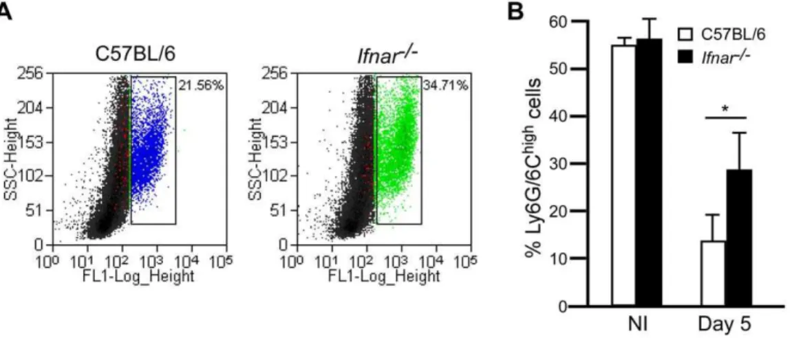
Since we observed an apparent systemic neutropenia that might be dependent on IFNAR, we asked whether neutrophils in the bone marrow were also reduced in wild type mice as disease progressed. Bone marrow was isolated from tibia and femurs, then fixed and stained with anti-Ly6G/6C and the cells examined by flow cytometry. Bone marrow from wild type andIfnar2/2mice
that were not infected contained similar numbers of neutrophils (Ly6Ghi, Figure 4). However, on day 5 post-infection, wild type mice harbored significantly fewer Ly6Ghicells in the bone marrow
compared toIfnar2/2mice. Thus, on day 5, neutropenia is more
pronounced in wild type mice than in Ifnar2/2 mice. This
observation may explain why wild type mice lose control over the infection, while Ifnar2/2 mice are able to contain it. Together
these data support a model whereby IFNAR signaling contributes to the depletion of immune cells duringY. pestisinfection.
Irf32/2mice develop septicemic plague at an accelerated
rate
We also studied the sensitivity of mice lacking the IFN-b
transcription factorsIrf3orIrf7. We challenged wild type,Irf32/2 Figure 2. Type I IFN receptor deficient mice are more resistant to infection byY. pestis.Groups of 5–10 wild type C57BL/6 andIfnar2/2mice
were challenged by intranasal infection ofY. pestisKIM D27. (A) Survival curve (collected from 3 independent trials, n = 30 for wild type mice, n = 20 for
Ifnar2/2mice); P = 0.0378 compared to wild type mice, analyzed by Gehan Wilcoxan test (B) Bacterial load from lungs, liver and spleen on days 2, 4 and 7;
P.0.05, analyzed by Kruskal Wallis rank sum test, for all tissues between strains (data shown are representative of two independent trials, n = 6 mice per group); (C–H) Serum from these mice was analyzed for 19 cytokines and chemokines; a subset of 6 are shown for a representative trial (two independent trials, n = 6 mice per group): (C) IL-6, (D) TNF-a, (E) IFN-c, (F) CCL-5, (G) IL-1b, (H) IP-10. Open symbols represent wild type, closed symbols represent
Ifnar2/2tissues; *P,0.1 wild type compared withIfnar2/2mice; significance was determined by Kruskal Wallis rank sum test.
andIrf72/2mice by intranasal infection ofY. pestisKIM D27 and
followed development of acute disease over a 14 day period. At this challenge dose, 30% of wild type mice developed lethal disease
on days 5–9 post-infection (Figure 5A). Similarly, 50% ofIrf72/2
mice developed plague with a similar time to lethal disease. However,Irf32/2mice were significantly more sensitive and 90%
developed lethal disease in only 4 days. Pre-treatment of mice with iron did not substantially impact development of disease inIrf32/2
mice as those that did not receive iron also developed an increased rate of mortality compared to wild type (Figure S2).
To determine whether IRF-3 was responsible for synthesis of type I IFN during Y. pestis infection, we challenged wild type,
Irf32/2andIrf72/2mice withY. pestisKIM D27 and measured gene expression in the lungs on days 2 and 4 post-infection. While it appeared that Ifnb expression was decreased in Irf32/2 and Irf72/2 mice compared to wild type on day 4 post-infection,
expression of this cytokine was not fully dependent on either transcription factor (Figure 5B). Similarly,Ip10expression was not dependent on IRF-3 or IRF-7, and in fact, increased expression of
Ip10 was observed in both strains of mutant mice. Thus, Ifnb
expression is not likely to depend on IRF-3 or IRF-7 followingY. pestisinfection of the lung.
IRF-3 contributes to the control of bacterial growthin vivo
Bacterial load was examined in the lungs, liver, and spleen of wild type and mutant mice on days 2, 3 and 4 post-infection. Results showed similar bacterial titers in all three tissues on day 2 post-infection, and in several animals in all groups the bacteria were undetectable (Figure 6A, Figure S3). In striking contrast, rapid bacterial growth occurred over the next 24 hrs inIrf32/2
mice but not in wild type orIrf72/2mice.
We also examined disease pathology by staining formalin fixed lungs, liver and spleen with hematoxylin and eosin (H&E). Total pathological severity scoring of lungs, liver and spleen indicated a disease inIrf32/2 mice consistent with bacterial sepsis on day 3 post-infection, with a large degree of necrosis in infected tissues and increased inflammation, while wild type mice appeared to have less necrosis and inflammation (Figure 6B). All mice harbored mild lesions on day 2 post-infection, and in the liver of Irf32/2
mice there were increased inflammatory foci, many of which contained dying cells while in wild type mice small neutrophilic foci formed that typically contained intact cells (Figure 6C). Disease in Irf32/2 mice progressed rapidly and on day 3,
degenerating neutrophilic inflammatory foci were still present in the liver, but multiple large necrotic lesions were also observed in both the liver and spleen (Figure 6D–E). In contrast, neutrophilic foci in the liver of wild type mice remained comprised primarily of intact cells and there was minimal damage to the spleen. Bacterial colonies could be seen in some areas of necrosis in the liver of mutant mice and these appeared larger and more numerous as disease progressed suggesting that rapid bacterial growth caused accelerated plague in theIrf32/2 mice (Figure 6F). Noticeably,
intact neutrophils were generally absent in areas of bacterial colonies which were instead surrounded by necrotic tissue. Likewise, spleens were necrotic in moribund mice (Figure 6G), and, overall, pathology in moribund mice appeared similar in both groups. Lungs of wild type mice had neither inflammation nor disease while Irf32/2 mice had developed mild inflammation in the lungs on day 3 (Figure S4). Together the data suggest that
Irf32/2 and wild type mice succumbed to septicemic plague
though the mutant mice developed severe disease more rapidly. In contrast toIrf32/2mice, all tissues ofIrf72/2mice examined had
fewer lesions, mild to moderate inflammatory foci, and were indistinguishable from wild type (Figure S5).
We also analyzed serum cytokine production in wild type and
Irf32/2mice on days 2 and 3 post-infection. Both strains harbored
Figure 3. IFNAR dependent depletion of neutrophils correlates with disease.Wild type C57BL/6 andIfnar2/2mice were challenged
by intranasal infection ofY. pestisKIM D27. On day 5 post-infection, 6 mice per group were euthanized and tissues processed for (A) bacterial load, (B) serum cytokines and (C) histology. Samples for all assays were obtained for each mouse; open shapes are wild type, closed shaped are
Ifnar2/2 mice; representative data are shown; n = 12 mice per strain
assayed in 2 independent trials. All values for titers and cytokines were not statistically significant (P.0.9) between wild type andIfnar2/2mice
as determined by Kruskal Wallis rank sum test. (C) H&E stains ofIfnar2/2
liver and spleen (top panels, left to right) and wild type liver and spleen (bottom panels, left to right). Images shown are representative of the mice with positive bacterial titers. Scale bar indicates 100mm.
relatively low levels of pro-inflammatory cytokines in the serum on day 2 with no statistical significance between wild type andIrf32/2
mice for all cytokines analyzed (Figure 7A–H). In striking contrast, however,Irf32/2mice produced significantly higher levels of
pro-inflammatory cytokines in the serum on day 3 post-infection compared to wild type, whereas IL-17, IL-5 and MIP-1awere not detectably different between the strains (data not shown). Together, the data suggest a burst in the production of inflammatory cytokines occurred between days 2 and 3 post-infection forIrf32/2mice. Since this is the same time point where
both bacterial growth and tissue necrosis became substantially greater inIrf32/2mice compared to wild type, these data suggest
that bacterial growth or the subsequent tissue injury may have triggered a massive pro-inflammatory response.
Immunohistochemistry confirmed the identity of macrophages, monocytes and neutrophils and revealed the presence of apoptosis in the red pulp of the spleen (Figure S6). Inflammatory foci of the liver appeared to contain both macrophages and neutrophils and there was no caspase-3 staining in the degenerated foci. In the spleen, red pulp necrosis that stained positive for cleaved caspase-3
Figure 4. IFNAR-dependent depletion of Ly6G+cells in the bone marrow.
Bone marrow cells of wild type C57BL/6 andIfnar2/2mice that
were not infected (NI) (n = 3 mice per group assayed in 2 independent trials) or on day 5 post-challenge by intranasal infection withY. pestisKIM D27 (n = 13 mice per group assayed in 3 independent trials) and stained with anti-Ly6G/6C-FITC followed by flow cytometry analysis. Data was gated on high stained cells as indicated in the scatter plot (A) and are shown for mice with the greatest percentage neutrophils in each group from a single trial. (B) Mean percent neutrophils from representative trial, error bars indicate the standard deviation from the mean. White bars indicate wild type mice, black bars indicateIfnar2/2mice; *P,0.05 between wild type andIfnar2/2mice determined by two-tailed Kruskal Wallis test.
doi:10.1371/journal.ppat.1002817.g004
Figure 5.Irf32/2mice are more sensitive to pulmonary infection byY. pestis.(A) Groups of 5–7 male and female wild type C57BL/6,Irf32/2,
andIrf72/2 mice were challenged by intranasal infection ofY. pestisKIM D27 and monitored for 14 days; *P,0.05 compared to wild type as
determined by Gehan-Wilcoxan test; data shown are combined from three independent experiments for each strain (n = 20 for wild type, n = 13 for
Irf32/2mice, n = 11 forIrf72/2mice). (B) Groups of 3 wild type C57BL/6,Irf32/2andIrf72/2mice were challenged by intranasal infection withY. pestis
KIM D27. On days 2 and 4 post-infection, mice were euthanized by CO2asphyxiation, lungs harvested and processed for RNA isolation and real time PCR. mRNA expression values were normalized to housekeeping geneywhaz[19]; y-axis represents fold change compared to mice that were not infected. Error bars depict the standard deviation from the mean. Data shown were collected in a single trial (n = 3 for all day 2 samples; only 2Irf32/2
andIrf72/2mice survived until day 4, n = 3 for wild type C57BL/6 on day 4). *P,0.05, ***P,0.005 as determined by one way ANOVA followed by
appeared to involve both macrophages and neutrophils. Together the phenotypic data suggest that Irf32/2 mice experience more
extensive and accelerated tissue damage that correlates with an increase in bacterial growth.
IRF-3 limits intracellular bacterial growth in bone marrow derived macrophages
Previous work has shown that IFN-I helps limit intracellular growth ofL. pneumophilain alveolar epithelial cells while, duringL. monocytogenes infection of macrophages, IFN-I signaling reduces expression of IFN-c receptor thereby preventing activation of macrophages by IFN-c[13,14]. We therefore sought to determine whether IRF-3 and IFN-I had an impact on bacterial uptake and
survival in macrophages. Towards this end, we isolated bone marrow derived macrophages (BMDMs) from wild type,Irf32/2
andIfnar2/2mice and performed a gentamicin protection assay to
enumerate intracellular bacteria following infection. BMDMs were pre-treated with PBS, anti-IFN-b or IFN-c for 4 hrs prior to infection withY. pestisKIM D27. Bacteria (16107CFU) grown at 37uC were added to the macrophages at a multiplicity of infection of 10 (time 0) and incubated for 30 min before adding gentamicin to kill extracellular bacteria. After 90 min incubation in gentami-cin (time 2 hr), macrophages were lysed and intracellular bacteria enumerated by plating on agar medium. The results showed that 5–6% of Y. pestis was intracellular in wild type and Ifnar2/2
BMDMs while only 2–3% ofY. pestiswas intracellular inIrf32/2
Figure 6.Irf32/2mice develop septicemic plague at an accelerated rate.Groups of 3 wild type C57BL/6 andIrf32/2mice were challenged by
intranasal infection ofY. pestisKIM D27. (A) Bacterial load for lungs, liver and spleen of wild type andIrf32/2mice on days 2 and 3 post-infection;
open symbols indicate wild type, closed symbols indicateIrf32/2; *P,0.05 for all three tissues compared to wild type as determined by Kruskal Wallis
rank sum test. (B) Mean severity scores for histopathology of lungs, liver and spleen on day 3; error bars indicate the standard deviation from the mean scores. (C–G) Wild type tissues are the left panels,Irf32/2tissues are the right panels; Day 2 (C), 3 (D–E), and moribund (F–G) hematoxylin and
eosin (H&E) stain for liver (C–D,F) or spleen (E, G). Scale bar indicates 50mm. Data shown are representative from 2 independent experiments (n = 6
mice per strain).
BMDMs, a difference that was reproducible and statistically significant (Figure 8A). This happened in the presence and absence of anti-IFN-b showing that IFN-b signaling does not appear to affect phagocytosis. Further, BMDMs fromIfnar2/2mice had no defect in phagocytosis compared to wild type. Treatment with IFN-c yielded results similar to untreated in all mouse strains. BMDMs from wild type and mutant mice were able to kill intracellular Y. pestis similarly as approximately 0.5–1% of the bacteria found at 2 hrs were still present at 6 hrs post-infection. IFN-ctreatment had little effect in all three mouse strains, with each showing 0.5–1% recovery of bacterial titer between 2 and 6 hrs post-infection. These data suggest Irf32/2 macrophages
have a defect in an early stage of phagocytosis. We also measured cell death in these samples by determining LDH release caused by the infection compared with detergent-lysis of BMDMs. These results showed no detectable differences in cytotoxicity between wild type,Ifnar2/2andIrf32/2mice suggesting that macrophage
viability following infection is not dependent on IRF-3 (Figure 8B). Neutrophils play a key role in restriction ofY. pestisgrowthin vivo
[41]. Furthermore, neutrophilic function is modified following injection of YopH by the Yersinia type III secretion system, a virulence mechanism that blocks intracellular calcium signaling and contributes to resistance of extracellular bacteria to neutro-philic killing [42]. We therefore wondered if the increase in susceptibility ofIrf32/2mice to plague might be due to a defect in
bacterial phagocytosis by neutrophils. To address this, we measured phagocytosis of Y. pestis by bone marrow derived neutrophils (BMNs) from wild type andIrf32/2mice. Similar to
macrophages, approximately 10% of the bacteria were taken up by neutrophils from wild type mice whereas only 1% of the infecting dose was taken up byIrf32/2neutrophils suggesting IRF-3 is necessary for phagocytosis (Figure 8C). However, once internalized, neutrophils fromIrf32/2bone marrow were capable
of killing Y. pestis to a similar degree as wild type neutrophils. When examined for cell death by LDH release, BMNs showed no detectable differences between wild type orIrf32/2 cells during
this time period (Figure 8D). Further, no significant differences were detected in anti-IFN-btreated compared to untreated BMNs from either wild type orIrf32/2 mice over the 6 hr time period
examined. Together these data suggest that IRF-3 may be required for efficient phagocytosis ofY. pestis. Further, since anti-IFN-bdid not affect phagocytosis, it appears the effect of IRF-3 on this pathway is not mediated by IFN-b. Since neutrophils are known to be important to host defense againstY. pestis, these data suggest that a defect in phagocytosis may be responsible for the increase in susceptibility ofIrf32/2mice [43].
IRF-3-mediated protection is lost in the presence of theY. pestispigmentation locus
Our septicemic plague model involves intranasal delivery of the non-pigmented laboratory strainY. pestis KIM D27 which lacks 102 kb of chromosomal DNA including a high pathogenicity island [44]. We therefore wanted to understand the relevance of IRF-3 following challenge with fully virulentY. pestis. Intranasal infection of the wild typeY. pestis strain KIM5 leads to primary pneumonic plague over a period of 3–4 days, andIrf32/2 mice succumbed to disease with an indistinguishable time course and mortality rate (Figure 9). Similar results were obtained following challenge withY. pestisCO92, another fully virulent strain from the
Orientalis, rather thanMediavalis, biovar (data not shown). Together the data suggest that the wild type bacteria bypass IRF-3 either because its contribution to host defense is less pronounced against the fully virulent bacteria or wild type Y. pestis carry virulence factors in the pgm locus that silence IRF-3’s role in host defense. We also tested an isogenic non-pigmented mutant strain of KIM5 isolated by plating on congo red agar to verify that the role of IRF-3 is not limited to a lab-adaptedY. pestisstrain isolated in 1965 [45]. This strain (KIM52) was then used to challenge wild
type andIrf32/2mice by intranasal inoculation. Similar to D27, challenge withY. pestisKIM52led to an apparent acceleration of
disease in Irf32/2 mice compared to wild type, though for the
single trial we performed, these differences were not significant
Figure 7.Irf32/2mice express pro-inflammatory responses that correlate with disease progression.Wild type C57BL/6 andIrf32/2mice
were challenged by intranasal infection ofY. pestisKIM D27. On days 2 and 3 post-infection, serum was collected by cardiac puncture and analyzed by cytokine bead array. A subset of 8 of the 19 cytokines measured are shown, open bars are from wild type mice; closed bars are fromIrf32/2mice: (A)
IL-6, (B) TNF-a, (c) IFN-c, (D) KC, (E) CCL-5, (F) IL-1b, (G) IP-10, (H) MCP-1. *P,0.05 as determined by Kruskal Wallis rank sum test; data shown are representative from two independent experiments, concurrent with the samples shown in figure 3 (n = 6 mice per strain).
(p = 0.28, Figure S7). These data suggest that the role of IRF-3 in host defense is not specific to bacterial strain isolate and support a role for IRF-3 in preventing septicemic plague.
Discussion
Yersinia pestisrequires high blood titer to enable its transmission to fleas and persistence in the environment [46]. Its virulence,
therefore, has evolved to promote replication to high titers in the blood, an environment that becomes overwhelmed by anti-bacterial defense mechanisms that attempt to prevent this massive growth. Multiple interactions between virtually all arms of the innate immune system and Yersinia determine the outcome of infection, with each having downstream consequences that may contribute further to disease. In this work, we addressed the role of type I interferon (IFN-I) activation in mice following Y. pestis Figure 8. Decreased phagocytosis ofY. pestisin bone marrow derived macrophages (BMMs) and neutrophils (BMNs) fromIrf32/2
mice.BMMs (A–B) or BMNs (C–D) from wild type and mutant mice were pre-treated with PBS, anti-IFN-bor IFN-cbefore infecting with 16107CFUY.
pestisKIM D27 at MOI of 10. At 30 min post-infection, gentamicin was added to the medium and the infection continued for a total of 6 hrs. Bacteria were enumerated by plating in triplicate (A, C) following detergent lysis of macrophages at t = 2 and 6 hr post-infection. Cell death was also assessed by collecting the culture supernatant at the same time points prior to detergent lysis and measuring the percent of LDH released compared to lysed control cells that were not infected (B, D). Data shown indicate the mean values collected in 2 independent trials; each sample was run in triplicate in each trial; error bars indicate the standard deviation from the mean. **P,0.005, significance was determined by one way ANOVA followed by Bonferroni’s multiple comparison test (BMMs) or two-tailed Wilcoxan rank sum test (BMNs).
infection. We identified production of IFN-b in the lungs early following pulmonary infection and found this was not dependent on the transcription factors IRF-3 or IRF-7. Production of type I IFN led to increased susceptibility to plague in a manner that correlated with systemic neutrophil depletion. IRF-3, however, was necessary for host defense and its expression led to decreased susceptibility to plague in a manner that correlated with decreased bacterial growth. Together these data raise new insight into the innate immune response.
Wild typeY. pestis can survive and even replicate in macro-phages yet cause disease due to exponential growth of extracellular bacteria that secrete cytotoxins (Yops) and other virulence factors into host cells [34,47,48,49]. Thus, the bacteria interface with the host in multiple environments where they may be recognized by surface-located and intracellular pattern recognition receptors or may perturb intracellular signaling through direct interactions involving the Yops of the type III secretion system (Figure 10). Bacterial pathogen recognition by one or more of these pathways, leads to phosphorylation of IRF-3 and perhaps also activation other interferon regulatory factors. Phosphorylation of IRF-3 causes its migration to the nucleus where it can form an active DNA binding complex, likely with p300, but possible alternative DNA binding complexes cannot be ruled out. In this manner, IRF-3 not only activates transcription of IFN-b but may also stimulate up-regulation of key mediators of phagocytosis. Addi-tional work is needed to identify genes that are affected by IRF-3, whether the same IRF-3 has alternative phosphorylation or interactions in the nucleus that affect its activity or if other interferon regulatory factors are responsible for IFN-bproduction. IRF-3 phosphorylation allows its nuclear migration and multiple phosphorylation sites of IRF-3 have been demonstrated [6]. TRIF activation by other bacterial pathogens typically leads to the production of IFN-b which can limit the infection by stimulating the expression of pro-inflammatory cytokines and chemokines such as IP-10 or RANTES, thereby inducing neutrophil recruitment [20,50]. Here we found evidence that phagocytosis may require IRF-3 activation. Though we do not yet know if this requirement is caused by production of a secreted factor such as a cytokine or other inflammatory mediator, expression of proteins involved in bacterial uptake, or even if additional neutrophil or macrophage functions are dependent on
pestisinfection will be important to identify the proteins that are necessary for the antibacterial effects of IRF-3.
Wild typeY. pestisare known to survive better than pgm2strains inside activated macrophages and, in our analyses, we showed that the absence of IRF-3 had minimal effect on protection against infection by wild type bacteria. It is conceivable that the wild type pulmonary infection, which results in death due to acute bronchopneumonia, progresses so rapidly that IRF-3 protection is overwhelmed by the developing lung injury or that resident phagocytic cells of the lung are unable to activate the anti-bacterial defense mechanism mediated by IRF-3. Alternatively, IRF-3-dependent phagocytosis may be neutralized by one or more gene products encoded by the pgm locus. Pgm-encoded ripA is a virulence factor necessary for intracellular bacterial growth, but not phagocytosis, in IFN-c activated bone marrow derived macrophages [56]. At least two additional virulence factors are believed to be encoded in the pgm locus, only one of which is known: the siderophore, yersiniabactin, whose deletion severely attenuates virulence [57]. Future studies combining mouse and bacterial genetics will facilitate an understanding of the mecha-nism whereby wild typeY. pestisinduce or evade IRF-3.
Our results suggest that it is likely that multiple transcription factors activate IFN-I duringY. pestisinfection, as the absence of IRF-3 or IRF-7 did not result in a complete loss of Ifnb
production. Recently, expression of IRF-1 was shown to be induced followingY. pseudotuberculosisinfection though the role of this transcription factor during Yersinia infection has not been investigated [39]. Thus, it is conceivable that IRF-1 or even another IRF is responsible for IFN-bproduction. Alternatively, it may be that small amounts of IFN-I that remain inIrf32/2and Irf72/2 mice are sufficient to cause immune cell depletion such that deletion of multiple IRFs are required to achieve the phenotype caused by deletion of IFNAR.
DuringListeriainfection, surface expression of IFN-creceptor is down-regulated on macrophages making them less able to kill intracellular bacteria. We therefore analyzed phagocytosis and bacterial killing in macrophages lacking IFN-I signaling in the presence and absence of IFN-cactivation. In these experiments, we did not detect differences between wild type andIfnarmutant macrophages in their ability to be activated by IFN-c. Further, we did not observe a decrease in the production of pro-inflammatory cytokines such as KC or MCP-1 that are normally IFN-c -dependent in theIfnar2/2mice. Together these data suggest that
the IFN-creceptor is not down-regulated by IFN-I duringY. pestis
infection. It is therefore likely that the downstream effects of IFN-I are somewhat pathogen-specific.
Our data found a link between IFN-I and neutrophil depletion and we propose that either neutrophils or another population responsible for their maturation and/or recruitment undergoes
the absence of theY. pestispigmentation locus.Groups of five male and female wild type C57BL/6 andIrf32/2mice were challenged
by intranasal infection of 16104CFUY. pestisKIM5+
and monitored over 14 days for development of disease. Data shown were collected in 3 independent experiments, n = 5 mice per group in each experiment; 15 mice total per strain. P = 0.7295 between wild type andIrf32/2mice as
cell death in response to IFN-I signaling duringY. pestisinfection (Figure 10). Depletion of neutrophils or other immune cells that is associated with type I IFN has been reported for several pathogens, includingFrancisella,Listeriaand influenza virus. Over 40 genes known to be involved in regulating apoptosis are ISGs, thus a role for IFN-induced immune cell death in host susceptibility to plague will require the study of potentially many genes and their effects onY. pestispathogenesis.
Y. pestis joins a growing list of pathogens, such as Listeria monocytogenes, Mycobacterium tuberculosis, Staphylococcus aureus and
Francisella tularensis that enhance virulence through IFNAR [10,11,12,58,59,60,61,62]. Consistent in all of these models, the detrimental effect of IFN-I appears more pronounced under conditions of high pathogen burden. Yet, the molecular mecha-nism remains elusive. Because of the clear benefits of IFN-I on viral and cancer defense, use of this cytokine in humans is currently under investigation. Thus it is imperative that its potential side effects against bacterial infection be recognized and understood so they can be avoided while gaining the full therapeutic benefits of type I IFN.
Materials and Methods
Bacterial strains
Fully virulentY. pestisKIM5+
was grown fresh from frozen stock by streaking for isolation onto heart infusion agar (HIA) plates supplemented with 0.005% Congo Red and 0.2% galactose to screen bacteria that retain the pigmentation locus [63]. For pneumonic plague challenge studies, a single pigmented colony was used to inoculate heart infusion broth (HIB) supplemented
with 2.5 mM CaCl2and grown 18–24 hrs at 37uC, 120 rpm. All handling of samples containing liveY. pestisKIM5+was performed in a select agent authorized BSL3 facility under protocols approved by the University of Missouri Institutional Biosafety Committee. Non-pigmented Y. pestis strain KIM D27 was routinely grown fresh from frozen stock on HIA, followed by aerobic growth at 27uC in HIB overnight prior to use in experiments.
Isolation ofY. pestisnon-pigmented mutant (KIM52)
A naturally occurring non-pigmented variant of the fully virulentY. pestisKIM5+
strain was isolated following plating on Congo Red agar [63,64]. Deletion of the pigmentation locus on the chromosome was verified by PCR as previously described prior to use in experiments [40].
Animals
All animal procedures were in strict accordance with the Office of Laboratory Animal Welfare and the National Institutes of Health Guide for the Care and Use of Laboratory Animals and were approved by the University of Missouri Animal Care and Use Committee. Wild type C57BL/6 mice were commercially obtained from Charles River Laboratories (MA, USA). C57BL/6 mice were the inbred strain background of theIrf32/2,Irf72/2,
andIfnar2/2mice which were kind gifts of Drs Michael Diamond
and Herbert Virgin [65,66]. Mice were bred and raised at the University of Missouri barrier housing facilities. Male and female wild type and mutant mice, ranging from 15–30 g were used for challenge experiments. During challenge with fully virulentY. pestis
and the isogenic non-pigmented mutant, mice were maintained in
Figure 10. Model forYersinia pestisactivation of IRF-3 and type I interferon.Extracellular bacteria inject Yops via the type III secretion system into the host cell where they may activate signaling pathways that terminate in IRF-3 phosphorylation. Bacteria may also be taken up by phagocytosis where they are able to survive inYersiniacontaining vacuoles or within the cytoplasm where they may be detected by pattern recognition receptors leading to IRF-3 phosphorylation. Following phosphorylation, IRF-3P migrates to the nucleus where it may bind p300 (a) or may form an alternative DNA binding complex (b) and activate transcription of IFN-band other, currently unknown genes (X). IFN-bexpression may also be dependent on other interferon response factors (IRFs) (c) and IFN-bsignaling leads to neutropenia perhaps by activation of programmed cell death. Phagocytosis is enhanced by the product of gene X.
intranasal infections involving pgm2 Y. pestis strains, mice were
given 50mg FeCl2 by intraperitoneal injection just prior to
challenge. For challenge with fully virulentY. pestis, bacteria were diluted to 26103CFU/0.02 ml sterile PBS. All animals were lightly anesthetized by isoflurane inhalation just prior to infection.
Quantification of bacterial load in tissues
Immediately after euthanasia, blood was collected directly from the heart by cardiac puncture. Lungs, spleens and livers were removed, and half of each tissue was processed for bacterial load by homogenizing in 1 ml sterile PBS. Serial dilutions of homogenized tissues were then plated in triplicate onto HIA plates for quantification of bacterial titer (CFU/organ). Serum was collected from the blood following centrifugation and stored at
280uC until analyzed.
Histopathologic evaluation of tissues
Approximately half of each tissue was placed in 10% formalin for 96 hrs. Lungs were first perfused with sterile PBS, removed and sectioned, then perfused with 10% formalin for histological analysis. Fixed tissues were embedded in paraffin, trimmed and stained with hematoxylin and eosin (H&E). For histologic scoring of tissues, slides were evaluated in a single blind fashion by a veterinarian with expertise in pathology. Lesions observed as well as the severity scores (0 to 3) were documented for lungs, liver and spleen. For enumeration of pyogranulomatous inflammatory foci in the liver, 10 fields per tissue were counted on each slide.
Immunohistochemistry
Tissues that had been fixed in 10% formalin and paraffin-embedded as described above were sectioned for immunohisto-chemical analysis. Sections were stained with anti-rat F4/80 (AbD Serotec, Oxford, UK), monoclonal antibody NIMP-R14 (Santa Cruz Biotechnology, CA, USA) [67] or anti-rat Caspase-3 (Trevigen, MD, USA) and detection was achieved by secondary staining with biotinylated rabbit anti-rat IgG and HRP-streptavi-din (DAKO, CA, USA). Staining and detection were carried out according to the manufacturer’s guidelines. For scoring, ten fields were counted for positive caspase-3 staining on each slide and scored from 0–3 (0 = no 3 staining, 1 = infrequent caspase-3 staining, 2 = moderately frequent caspase-caspase-3 staining, caspase-3 = positive caspase-3 staining in majority of tissue).
RNA isolation and Real Time PCR
Approximately half of the lung tissue was homogenized in RNAlater (Qiagen, CA, USA). RNA isolation was performed using RNeasy Mini Kit according to manufacturer’s instructions (Qiagen, CA, USA). Total RNA was treated with Turbo DNase
instructions and analyzed by Illuminex using IS 100 software (Qiagen, CA, USA). IL-2, IL-4 and IL-9 were undetectable in all samples and therefore they were removed from further analysis.
Bone marrow-derived macrophage (BMDM) isolation
BMDMs were isolated from C57BL/6, Irf32/2 and Ifnar2/2
mice essentially as previously described by culturing for 6 days in Dulbecco’s modified Eagle’s medium (DMEM) containing 20 ng/ ml M-CSF (eBiosciences, CA, USA) in place of L cell media [68]. Twenty-four hours prior to infection, 16106cells were seeded into 12-well plates with DMEM containing 20 ng/ml M-CSF, 10% fetal bovine serum (FBS). Where indicated, macrophages were pre-treated with 1mg/ml anti-IFN-b or 500mg/ml IFN-c
(Abcam, Cambridge, MA, USA) per well for 4 hrs prior to infection.
Bone marrow neutrophil isolation
Primary bone marrow-derived neutrophils were isolated from femurs of wild type C57BL/6 orIrf32/2mice. Neutrophils were
enriched by separation on a three-layer Percoll (Sigma-Aldrich, St. Louis, MO) gradient. Purity of the isolated cells was assessed by microscopy at.95% neutrophils. Neutrophils were used in assays within 1 h of purification. Where indicated, neutrophils were pre-treated with PBS or anti-IFN-b10 min prior to infection.
Intracellular survival assay
Overnight cultures of Y. pestis KIM D27 were diluted 1:20, incubated at 27uC for 3 hrs then shifted to 37uC for 1 hr. Bacteria were infected at a multiplicity of infection (MOI) of 10 and the plates were centrifuged at 866g for 5 min before incubating at
37uC, 5%CO2. Following 30 min, cells were washed with sterile PBS and incubated in cell culture medium containing 40mg/ml gentamicin and the infection continued for an additional 5.5 hrs. For bacterial titer, media was aspirated and wells were washed once with sterile PBS. Infected macrophages and neutrophils were lysed with 300ml of 0.1% Triton X-100 in PBS, cells were then washed once with PBS, serially diluted and plated in triplicate on HIA for enumeration of colony forming units. To ensure that only intracellular bacteria had been enumerated, 10ml of aspirated
media was plated on HIA for each well and no bacterial growth was recovered in these samples (data not shown).
Cytotoxicity assay
comparing the amount of LDH in the supernatant to that recovered from control BMDMs that were not infected following lysis by 0.1% triton X-100.
Flow cytometry
Bone marrow cells from C57BL/6 and Ifnar2/2 mice were
isolated just prior to infection or on day 5 post-infection (dpi) from tibia and femurs using cold PBS. Cells were incubated with Fc receptor blocking solution (BioLegend, CA, USA) to eliminate non-specific binding for 10 min prior to fixing in 4% paraformal-dehyde for 20 min. Cells were then washed 26with cold PBS at 12006g for 8 min and stained with FITC-conjugated anti-mouse
Ly6G/6C antibody (BD Biosciences, CA, USA) for 30 min. Cells were washed 36with cold PBS and analyzed on MoFlo XDP using Summit software (Beckman Coulter, UT, USA).
Statistical analysis
Data from all trials were analyzed for statistical significance. Statistical analyses were performed using R (Gehan Wilcoxan, Kruskal Wallis) or GraphPad prism (Wilcoxan match paired, Student’s t test, ANOVA, Mantel Cox) software [69].
Supporting Information
Figure S1 Increased GR-1+
F4/80+
inflammatory foci in Ifnar2/2mice correlates with bacterial clearance. Wild
type C57BL/6 and Ifnar2/2 mice were challenged by intranasal
infection of 16106CFUY. pestisKIM D27 after pre-treatment with 50mg Fe+2
. Formalin fixed livers (left panels) and spleens (right panels) were prepared from wild type C57BL/6 (bottom) orIfnar2/2 (top)
mice on day 5 post-infection, followed by sectioning and immuno-histochemistry staining with anti-F4/80 (A) or anti-NIMP (B). Images shown are representative of two independent experiments, n = 6 per group, and were taken from mice from which bacteria could be recovered from both tissues. Scale bar indicates 50mm.
(TIF)
Figure S2 Increase in sensitivity ofIrf32/2mice occurs in the absence of pre-treatment with iron.Groups of five wild type C57BL/6,Irf32/2, andIrf72/2mice were challenged by
intranasal infection of 16106CFU Y. pestis KIM D27 and monitored for survival over 14 days (n = 5 mice per group, single trial).
(TIF)
Figure S3 IRF-7 is dispensable for host defense against Y. pestis.Wild type C57BL/6 andIrf72/2mice were challenged
by intranasal infection ofY. pestisKIM D27. On days 2, 3 and 4 post-infection, lungs (circle), liver (triangle), and spleen (diamond) were harvested, homogenized in sterile PBS and plated to enumerate bacterial load per tissue. Open shapes are wild type and black shapes are Irf72/2; data were collected in two
independent experiments, each with 3 mice per group. (TIF)
Figure S4 Increased inflammation develops in the lungs ofIrf32/2mice.Groups of three wild type C57BL/6 andIrf32/ 2 mice were challenged by intranasal infection ofY. pestisKIM
D27. On day 3 post-infection, lungs were harvested, fixed in 10% formalin and analyzed by histochemistry; wild type (left),Irf32/ .(-right). Scale bar indicates 100mm. Images are representative of 6 mice.
(TIF)
Figure S5 Irf72/2mice develop inflammatory foci in the
liver with little tissue necrosis on day 3 following pulmonary infection. Groups of three Irf72/2 mice were challenged by intranasal infection of 16106CFU Y. pestis KIM
D27. On day 3 post-infection, animals were euthanized, tissues collected and fixed in 10% formalin. Hematoxylin and eosin (H&E) staining for lungs (top panels), liver (bottom left) and spleen (bottom right) ofIrf72/2mice. Histology for wild type mice on day
3 post-infection was shown in Figure 3. Scale bar indicates 100mm. Images are representative of 6 mice.
(TIF)
Figure S6 Increased apoptosis in phagocytic cells of Irf32/2 mice on day 3 correlates with accelerated disease progression.Formalin fixed livers (A–F) and spleens (G–I) tissues collected from wild type C57BL/6 andIrf32/2mice
on days 2 (A, C) and 3 (B, D, F–I) post-infection (see figure 3) were analyzed by immunohistochemistry with anti-F4/80 (A–B, G), NIMP R14 (C–D, H), and anti-caspase-3 (F, I). For each panel of stains, wild type tissues are on the left,Irf32/2tissues are on the right. Scale bar indicates 50mm. Images are representative of 6
mice per group, collected in two independent experiments. (E) Day 3 foci of inflammation containing intact neutrophils in the liver were quantified by counting in 10 non-overlapping fields and the mean number counted from all fields was determined (n = 6 mice per group). (J) Mean severity scoring (0–3, with 3 indicating positive stain on the majority of the tissue) for caspase-3+
staining in lungs, liver and spleen on day 3 (n = 6 mice per group); *P,0.05 between wild type and Irf32/2 tissue, analyzed by unpaired
Student’s t-test. (TIF)
Figure S7 Increase in sensitivity of Irf32/2 mice is
dependent on the absence of theY. pestispigmentation locus.Male and female wild type C57BL/6 (n = 5) andIrf32/2
mice (n = 6) were challenged by intranasal infection of 16106CFUY. pestisKIM52and monitored for 14 days. (TIF)
Table S1 Expression of inflammatory molecules follow-ing pulmonary infection byY. pestisKIM D27.
(DOCX)
Table S2 List of primers used for quantitative RT-PCR in this study.
(DOCX)
Acknowledgments
We are indebted to Drs. Michael Diamond and Herbert Virgin IV for their generosity in providing mice and helpful discussions. We also wish to thank Robert Perry for the kind gift of theY. pestisKIM5+
strain; Susan Gater for assistance with flow cytometry; and Susan Cushing for use of the Illuminex equipment. We are also grateful to members of our laboratory for assistance with the BSL3 experiments and helpful discussions and to Dr. Kristen Peters for critical comments on the manuscript. Histology services were provided by the Research Animal Diagnostic Laboratory (RADIL) at the University of Missouri; flow cytometry was performed in the MRCE Innate Immunity Core at the University of Missouri Laboratory for Infectious Disease Research.
Author Contributions
8. Sato M, Hata N, Asagiri M, Nakaya T, Taniguchi T, et al. (1998) Positive feedback regulation of type I IFN genes by the IFN-inducible transcription factor IRF-7. FEBS Lett 441: 106–110.
9. O’Riordan M, Yi C, Gonzales R, Lee K, Portnoy D (2002) Innate recognition of bacteria by a macrophage cytosolic surveillance pathway. Proc Natl Acad Sci USA 99: 13861–13866.
10. Auerbach V, Brockstedt D, Meyer-Morse N, O’Riordan M, Portnoy D (2004) Mice lacking the type I interferon receptor are resistant toListeria monocytogenes. J Exp Med 200: 527–533.
11. Carrero J, Calderon B, Unanue E (2004) Type I interferon sensitizes lymphocytes to apoptosis and reduces resistance toListeria infection. J Exp Med 200: 535–540.
12. O’Connell R, Saha S, Vaidya S, Bruhn K, Miranda G, et al. (2004) Type I interferon production enhances susceptibility toListeria monocytogenesinfection. J Exp Med 200: 437–445.
13. Rayamajhi M, Humann J, Penheiter K, Andreasen K, Lenz L (2010) Induction of IFN-abenablesListeria monocytogenesto suppress macrophage activation by IFN-c. J Exp Med 207: 327–337.
14. Opitz B, Vinzing M, van Laak V, Schmeck B, Heine G, et al. (2006)Legionella pneumophilainduces IFNbin lung epithelial cells via IPS-1 and IRF3, which also control bacterial replication. J Biol Chem 281: 36173–36179.
15. Monroe K, McWhirter S, Vance R (2009) Identification of host cytosolic sensors and bacterila factors regulating the type I interferon response to Legionella pneumophila. PLoS Path 5: e1000665.
16. Plumlee C, Lee C, Berg A, Decker T, Shuman H, et al. (2009) Interferons direct an effective innate response toLegionella pneumophilainfection. J Biol Chem 284: 30058–30066.
17. Parker D, Martin F, Soong G, Harfenist B, Aguilar J, et al. (2011)Streptococcus pneumoniaeDNA initiates type I interferon signaling in the respiratory tract. MBio 2: e00016–00011.
18. Charrel-Dennis M, Latz E, Halmen K, Trieu-Duot P, Fitzgerald K, et al. (2008) TLR-independent type I interferon induction in response to an extracellular bacterial pathogen via intracellular recognition of its DNA. Cell Host Microbe 4: 543–554.
19. Joyce E, Popper S, Falkow S (2009) Streptococcus pneumoniae nasopharyngeal colonization induces type I interferons and interferon-induced gene expression. BMC Genomics 10: 404–420.
20. Carrigan S, Junkins R, Yang Y, MacNeil A, Richardson C, et al. (2010) IFN regulatory factor 3 contributes to the host response duringPseudomonas aeruginosa lung infection in mice. J Immunol 185: 3602–3609.
21. Opitz B, van Laak V, Eitel J, Suttorp N (2010) Innate immune recognition in infectious and noninfectious diseases of the lung. Am J Respir Crit Care Med 181: 1294–1309.
22. Jones J, Kayagaki N, Broz P, Henry T, Newton K, et al. (2010) Absent in melanoma 2 is required for innate immune recognition ofFrancisella tularensis. Proc Natl Acad Sci USA 107: 9771–9776.
23. Guarda G, Braun M, Staehi F, Tardivel A, Mattmann C, et al. (2011) Type I interferon inhibits interleukin-1 production and inflammasome activation. Immunity 34: 213–223.
24. Montminy S, Khan N, McGrath S, Walkowicz M, Sharp F, et al. (2006) Virulence factors ofYersinia pestisare overcome by a strong lipopolysaccharide response. Nat Immunol 7: 1066–1073.
25. Pouliot K, Pan N, Wang S, Lu S, Lien E, et al. (2007) Evaluation of the role of LcrV-Toll like receptor 2-mediated immunomodulation in the virulence of Yersinia pestis. Infect Immun 75: 3571–3580.
26. DePaolo R, Tang F, Kim I, Han M, Levin N, et al. (2008) TLR6 drives differentiation of tolerogenic DC and contributes to LcrV-mediated plague pathogenesis. Cell Host Microbe 4: 350–361.
27. Price P, Jin J, Goldman W (2012) Pulmonary infection byYersinia pestisrapidly establishes a permissive environment for microbial proliferation. Proc Natl Acad Sci USA 109: 3083–3088.
28. Lathem W, Crosby S, Miller V, Goldman W (2005) Progression of primary pneumonic plague: A mouse model of infection, pathology, and bacterial transcriptional activity. Proc Natl Acad Sci 102: 17786–17791.
36. Nakajima R, Brubaker R (1993) Association between virulence ofYersinia pestis and suppression of Gamma Interferon and Tumor Necrosis Factor Alpha. Infect Immun 61: 23–31.
37. Parent M, Wilhelm L, Kummer L, Szaba F, Mullarky I, et al. (2006) Gamma Interferon, Tumor Necrosis Factor Alpha, and Nitric Oxid Synthase 2, key elements of cellular immunity, perform critical protective functions during humoral defense against lethal pulmonaryYersinia pestisinfection. Infect Immun 74: 3381–3386.
38. Kummer L, Szaba F, Parent M, Adamovicz J, Hill J, et al. (2008) Antibodies and cytokines independently protect against pneumonic plague. Vaccine 26: 6901– 6907.
39. Auerbuch V, Golenbock D, Isberg R (2009) Innate Immune Recognition of Yersinia pseudotuberculosisType III Secretion. PLoS Path 5: 1000686.
40. Lee-Lewis H, Anderson D (2010) Absence of inflammation and pneumonia during infection with non-pigmentedYersinia pestisreveals new role for the pgm locus in pathogenesis. Infect Immun 78: 220–230.
41. Laws T, Davey M, Titball R, Lukaszewski R (2010) Neutrophils are important in early control of lung infection byYersinia pestis. Microbes Infect 12: 331–335. 42. Andersson K, Magnusson K, Majeed M, Stendahl O, Fallman M (1999)Yersinia
pseudotuberculosis-induced calcium signaling in neutrophils is blocked by the virulence effector YopH. Infect Immun 67: 2567–2574.
43. Spinner JL, Cundiff JA, Kobayashi SD (2008)Yersinia pestistype III secretion system-dependent inhibition of human polymorphonuclear leukocyte function. Infect Immun 76: 3754–3760.
44. Buchrieser C, Prentice M, Carniel E (1998) The 102-kilobase unstable region of Yersinia pestis comprises a high-pathogenicity island linked to a pigmentation segment which undergoes internal rearrangement. J Bacteriol 180: 2321–2329. 45. Brubaker R, Beesley E, Surgalla M (1965)Pasteurella pestis: Role of Pesticin I and
iron in experimental plague. Science 149: 422–424.
46. Lorange E, Race B, Sebbane F, Hinnebusch B (2005) Poor vector competence of fleas and the evolution of hypervirulence inYersinia pestis. J Infect Dis 191: 1907–1912. 47. Straley S, Harmon P (1984) Growth in mouse peritoneal macrophages ofYersinia pestislacking established virulence determinants. Infect Immun 45: 649–654. 48. Grabenstein J, Marceau M, Pujol C, Simonet M, Bliska J (2004) The response
regulator PhoP of Yersinia pseudotubeculosis is important for replication in macrophages and for virulence. Infect Immun 72: 4973–4984.
49. Noel B, Lilo S, Capurso D, Hill J, Bliska J (2009)Yersinia pestiscan bypass protective antibodies to LcrV and activation with gamma interferon to survive and induce apoptosis in murine macrophages. Clin Vacc Immunol 16: 1457–1466. 50. Cai S, Batra S, Shen L, Wakamatsu N, Jeyaseelan S (2009) Both TRIF- and
MyD88-dependent signaling contribute to host defense against pulmonary Klebsiellainfection. J Immunol 183: 6629–6638.
51. Grandvaux N, Servant M, tenOever B, Sen G, Balachandran S, et al. (2002) Transcriptional Profiling of Interferon Regulatory Factor 3 Target Genes: Direct Involvement in the Regulation of Interferon-Stimulated Genes. J Virol 76: 5532–5539.
52. Elco C, Guenther J, Williams B, Sen G (2005) Analysis of genes induced by Sendai virus infection of mutant cell lines reveals essential roles of Interferon Regulatory Factor 3, NF-kB, and Interferon but not Toll-like Receptor 3. J Virol 79: 3920–3929.
53. Chattopadhyay S, Yamashita M, Zhang Y, Sen G (2011) The IRF-3/Bax-mediated apoptotic pathway, activated by viral cytoplasmic RNA and DNA, inhibits virus replication. J Virol 85: 3708–3716.
54. Fensterl V, Sen G (2011) The ISG56/IFIT1 gene family. J Interfer Cyto Res 31: 71–78.
55. Terenzi F, White C, Pal S, Williams B, Sen G (2007) Tissue-specific and inducer-specific differential induction of ISG56 and ISG54 in mice. J Virol 81: 8656–8665.
56. Pujol C, Grabenstein J, Perry R, Bliska J (2005) Replication ofYersinia pestisin interferon g-activated macrophages requires ripA, a gene encoded in the pigmentation locus. Proc Natl Acad Sci 102: 12909–12914.
58. Martin F, Gomez M, Wetzel D, Memmi G, O’Seaghdha M, et al. (2009) Staphyloccus aureusactivates type I IFN signaling in mice and humans through the Xr repeated sequences of protein A. J Clin Invest 119: 1931–1939. 59. Shahangian A, Chow E, Tian X, Kang J, Ghaffari A, et al. (2009) Type I IFNs
mediate development of posinfluenza bacterial pneumonia in mice. J Clin Invest 119: 1910–1920.
60. Henry T, Kirimanjeswara G, Ruby T, Jones J, Peng K, et al. (2010) Type I IFN signaling constrains IL-17A/F secretion by gamma-delta T cells during bacterial infections. J Immunol 184: 3755–3767.
61. Chessler A, Caradonna K, Da’dara A, Burleigh B (2011) Type I interferons increase host susceptibility toTrypanosoma cruzi infection. Infect Immun 79: 2112–2119.
62. Manca C, Tsenova L, Freeman S, Barczak A, Tovey M, et al. (2005) Hypervirulent M. tubercuolosisw/Beijing strains upregulate type I IFNs and increase expression of negative regulators of the Jak-Stat pathway. J Interfer Cyto Res 25: 694–701.
63. Surgalla M, Beesley E (1969) Congo red-agar plating medium for detecting pigmentation inPasteurella pestis. App Microbiol 18: 834–837.
64. Hare J, McDonough K (1999) Highfrequency RecAdependent and -independent mechanisms of Congo Red binding mutations inYersinia pestis. J Bacteriol 181: 4896–4904.
65. Sato M, Suemori H, Hata N, Asagiri M, Ogasawara K, et al. (2000) Distinct and essential roles of transcription factors IRF-3 and IRF-7 in response to viruses for IFN-a/bgene induction. Immunity 13: 539–548.
66. Purtha W, Chachu K, Virgin HI, Diamond M (2008) Early B-cell activation after West Nile virus infection requires alpha/beta interferon but not antigen receptor signaling. J Virol 82: 10964–10974.
67. Tacchini-Cottier F, Zweifel C, Belkaid Y, Mukankundiye C, Vasei M, et al. (2000) An immunomodulatory function for neutrophils during the induction of a CD4+
Th2 response in BALB/c mice infected withLeishmainia major. J Immunol 165: 2628–2636.
68. Celada A, Gray P, Rinderknecht E, Schreiber R (1984) Evidence for a gamma-interferon receptor that regulates macrophage tumoricidal activity. J Exp Med 160: 55–74.