Financial support: FAPEMIG, CNPq, UFMG.
(1)Departamento de Parasitologia, Instituto de Ciências Biológicas, UFMG, Belo Horizonte, MG, Brasil.
(2)Departamento de Bioquímica e Imunologia, Instituto de Ciências Biológicas, UFMG, Belo Horizonte, MG, Brasil.
Correspondence to: Antonio C.R. Leite. Cx. P. 486, 31270-901 Belo Horizonte, MG, Brasil. Phone: +55.31.34092837. Fax: +55.31.34092970.E-mail: rios@icb.ufmg.br
SPLEEN CELL PROLIFERATION DURING AND AFTER SKIN MYIASIS BY
HUMAN BOT FLY
Dermatobia hominis
Jomara Mendes GONÇALVES(1), Maria Fernanda Alves do NASCIMENTO(1), Natália Martins BREYNER(2), Viviane Cristina FERNANDES(2), Alfredo Miranda de GÓES(2) & Antonio César Rios LEITE(1)
SUMMARY
Spleen cells from mice were examined at 5, 10, 15, 20 and 25 days post-infection (dpi) with Dermatobia hominis larva and at 5, 10, 15, 30 and 60 days post-larval emergence (dple). Cell proliferation in vitro assays were carried out with RPMI-1640 medium and larval secretory product (LSP) of D. hominis at 5, 10, 15, 20 and 25 days. When each group of mice was tested against each medium, significance was only seen for 25 dpi, with increasing order: LSP-10 d, -25 d, -5 d, -20 d, -15 d and RPMI. Significant results were also observed when each medium was tested against mice at each dpi or dple. Each dple group vs. each medium produced significant results only for 10 dple, with increasing order: LSP-5 d, -20 d, -25 d, -10 d, -15 d and RPMI. Comparative tests were also carried out between groups to refine certain observations. The LSPs were also analyzed using SDS-PAGE. The results prove that myiasis caused depletion of spleen cells, particularly under the effect of the LSP-10 and -15, but the cells tended to increase up to 60 dple. This in vitro assay may represent the real systemic immune response in the relationship LSP-D. hominis-host.
KEYWORDS: Dermatobia hominis; Mice; Skin myiasis; Spleen cell; Blastogenesis.
INTRODUCTION
Cutaneous obligatory myiasis by the human bot fly Dermatobia hominis (L. Jr.) larva is a Neotropical zoonotic disease that produces severe economic losses to livestock production and also has public health importance11. The developmental cycle of D. hominis is unusual
in that adults are aphagous, mated females capturing and depositing their eggs on several species of flies, including Musca domestica and Stomoxys calcitrans. After about a week, when the phoretic host feeds or lands on a mammalian host, the bot fly larvae hatch and immediately penetrate the animal’s skin. Although the pathological and immunological aspects of human bot fly myiasis have been little studied under field conditions6,26, mice have proved to be a suitable laboratory model for
the relationship between D. hominis and its hosts5,13,18,21. The response
of mouse spleen cells during and after infection by larvae of D. hominis is described here.
MATERIAL AND METHODS
Animal infection: Each animal from 10 groups of six male Swiss mice (20-25g) was submitted to skin infection with one newly hatched larvaof D. hominis reared in our laboratory9. The mice were sacrificed
and spleens obtained from groups 1-5 at 5, 10, 15, 20 and 25 days-post-infection (dpi), respectively. Spleens from groups 6-10 were collected at 5, 10, 15, 30 and 60 days post-larval-emergence (dple) respectively.
Spleens from corresponding negative control 11th group (without infection
= 0 dpi) were also examined.
Larval secretory product (LSP): Five groups with five male adult Wistar rats (190-220g) were infested with four newly hatched larvae of D. hominis and sacrificed at 5, 10, 15, 20 and 25 dpi to collect the larvae from the skin tissues9. Larvae at each infested date were washed three
times with RPMI-1640 medium (Sigma) and conditioned in Falcon tubes with 2 mL of RPMI for incubation at 37 ºC in a 5% CO2 atmosphere for 24 h. Crude D. hominis LSP was obtained at 5 d (LSP-5d) from 30 first larvae (L1); at 10 d (LSP-10 d) or 15 d (LSP-15 d) from 15 second larvae (L2) and at 20 d (LSP-20 d) or 25 d (LSP-25 d) from 10 third larvae (L3). The LSP of each different days or larval instars was stored in liquid nitrogen until ready for use.
Concentration and electrophoreses of LSP: Protein concentration of the LSP was determined using the Bradford (1976) technique and subjected to continuous electrophoresis using 10% SDS polyacrylamide gels, under reducing conditions14. The protein concentration used to
electrophoresis was of 50 µg/LPG µL. The separated proteins were stained with Coomassie blue.
Erythrocytes were lysed by re-suspending the pellet in 300 µL Tris-ammonium chlorite plus 30 mL MEM (minimum essential medium eagle, Sigma) and washing with the same solution. The spleen cells were suspended in 20 mL of RPMI, diluted in 10 mL Tweck solution and counted under a Neubauer camera. Cell proliferation assays (in triplicate) were done in response to RPMI and LSP at 5 d, 10 d, 15 d, 20 d and 25 d. Spleen cells (1x10⁶) were cultured in 200 µL culture medium
in 96-well flat-bottom plates. Except for the control group, 25 µg/mL of LSP was introduced in the culture medium in each plate and incubated at 37 ºC in a CO2 incubator for 5 d. At 18 h before the final incubation, 0.5 µCi of titrated thymidine (specific activity, 37 Ci/mL; New England Nuclear, Boston, MA, USA) was added to each well. Cells were harvested onto glass fiber paper and the incorporated radioactivity was measured by a liquid scintillation β-counter. The results were expressed as mean ± S.E. of c.p.m. values.
Statistical analyses: The results of the blastogenesis were analyzed by non-parametric methods (GraphicPad Prism 4.03 software) by comparison among all groups (Kruskal-Wallis test) and between pairs of groups (Mann-Whitney U test) using independent variables. The data are represented in box and whisker plots, the middle line corresponding to the median and the lower and upper line representing the 25 and 75 percentiles, respectively (50% of the data fall within the box, and 25% each above the below). The whiskers above and below the box plot indicate 95% and 5% percentiles, respectively. Values were considered significant at p < 0.05.
RESULTS
Animal infection: The L1 of D. hominis changed to L2 at six dpi, the L3 moulted at 16 dpi and the mature warbles escaped from the host around 30 dpi. A scar formed on the skin of each mouse 7-10 days later. In the laboratory L3 left the experimentally infested mice and pupated, resulting in fertile adult D. hominis.
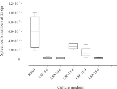
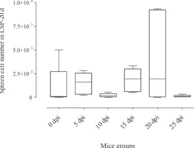
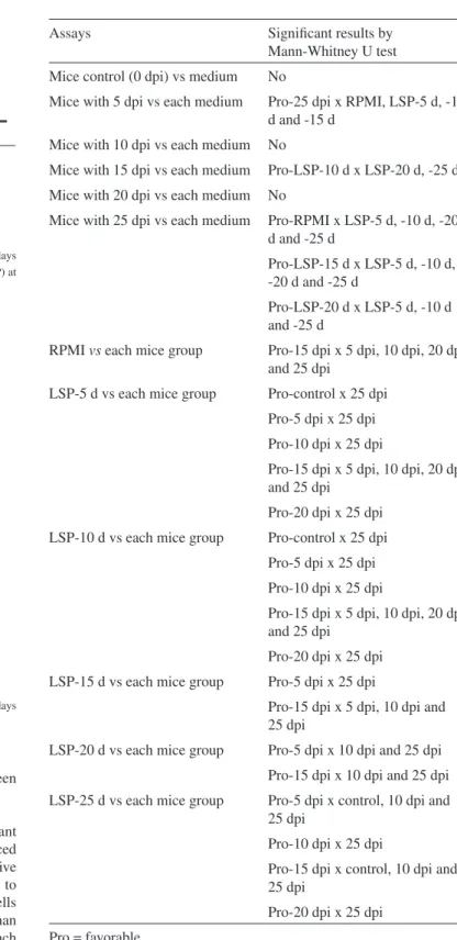
In vitro spleen cell proliferation from infested mice: When the response of each group (1-5) with each substrate was analyzed, significance was only seen at 25 days of infection (group 5) increasing as follows: LSP-10 d, -25 d, -5 d, -20 d, -15 d and RPMI (Fig. 1). In the reciprocal analysis, i.e., of each substrate with each mouse group, significance was observed in all assays, highest values being seen for RPMI (Fig. 2): 25, 5, 20, 10, 15 dpi and control; LSP-5 d (Fig. 3): 25, 5, 10, 20, 15 dpi and control; LSP-10 d (Fig. 4): 25, 10, 5, 20, 15 dpi and control; LSP-15 d (Fig. 5): 25, 5, 10, 20, 15 dpi and control; LSP-20 d (Fig. 6): 25, 10, 5, 15 dpi, control and 20 dpi; LSP-25 d (Fig. 7): 25, 10 dpi, control, 5, 20 and 15 dpi. The results of between-group analyses (Mann-Whitney U test) are summarized in Table 1.
In vitro spleen cell proliferation from mice at days-post-larval-emergence: Analyses of each mouse group vs each medium were only significant for 10 dple, values increasing in the following order: LSP-5 d, -20 d, -25 d, -10 d, -15 d and RPMI. Significant results were only obtained for three assays, when each medium was analyzed with each mouse group, values increasing as follows: to RPMI: 30, 10, 15, 60, 5 dple and control; 10 d: 30, 10, 15, 5, 60 dple and control and LSP-15 d: 30, 10, LSP-15 dple, control, 5 and 60 dple. Additional test results are also shown in Table 2.
Concentration and electrophoreses of LSP: Separation of LSP molecules by SDS-PAGE revealed multiple protein bands (Fig. 8), their molecular weights ranging from 12-50 kDa. Major stained protein bands of 12-25 kDa were seen in all larval stages.
DISCUSSION
The metamorphosis of D. hominis inside mammalian skin occurs below the point of L1 penetration, in deep dermis21. This contrasts with
other oestrid species, e.g. rodent bot fly (Cuterebra) and cattle bot fly (Hypoderma) where the L1 migrate within the host skin before the second and third instars fix in the cutaneous tissues to form the mature warble16,24,25. The multi-host parasitism exhibited by D. hominis also
differs from the host specificity seen in other bot flies. There is usually a balance between the host and the three larval stages6. Preliminary studies
Fig. 1 - Box and whisker plots representing the numbers of spleen cells from mice at 25 days post-infection (dpi) by Dermatobia hominis submitted to RPMI and larval-secretory-product (LSP).
Fig. 3 - Box and whisker plots representing the numbers of spleen cells from mice control and at days post-infection (dpi) by Dermatobia hominis submitted to larval-secretory-product (LSP) at five days.
Fig. 4 - Box and whisker plots representing the numbers of spleen cells from mice control and at days post-infection (dpi) by Dermatobia hominis submitted to larval-secretory-product (LSP) at 10 days.
Fig. 5 - Box and whisker plots representing the numbers of spleen cells from mice at days post-infection (dpi) by Dermatobia hominis submitted to larval-secretory-product (LSP) at 15 days (by Kruskal-Wallis test).
Fig. 6 - Box and whisker plots representing the numbers of spleen cells from mice at days post-infection (dpi) by Dermatobia hominis submitted to larval-secretory-product (LSP) at 20 days.
have demonstrated that bot burden, instar type, infection or re-infection can regulate the host’s inflammatory process as well as its humoral and cellular immune responses4,6,7,17,20.
As soon as D. hominis L1 invades the host’s skin, cellular and molecular changes occur in response to the mechanical presence of the larval stage as well as its secretory products. A chronological recruitment of the cells to skin lesions has been studied during and after infection in rats21,22. Cellular or humoral responses against human bot fly myiasis
using rabbits, cattle and mice were recently re-examined17. However,
cytological tissue changes (or damage) in warble-infested skin has been poorly studied under natural or controlled conditions. Circulating leucocytes in hosts with D. hominis myiasis are reported in cattle1,8 and
rats10. Except for observations of alterations in response to migration of
the L1 of Cuterebra and Hypoderma, there have been few studies of the host cellular pathways in skin myiasis6,26.
Oestrid LSP has been most studied in H. lineaum L1 midgut, from which enzymes (hypodermins) with immune regulation properties were isolated. Both hypodermin (trypsins) A and B15,28 deplete
complement cytolytic activity2,3, B also reducing leukocyte and
lymphocyte blastogenesis, as well as the production of interleukin-2 and prostaglandin19. Although molecules from D. hominis LSP have not
serine-Table 1
Analyses of spleen cells from mice control and days-post-infestation (dpi) by
Dermatobia hominis larvae in response to media (RPMI and larval secretory
product = LSP) of warbles at 5, 10, 15, 20 and 25 days and each medium vs each group
Assays Significant results by Mann-Whitney U test Mice control (0 dpi) vs medium No
Mice with 5 dpi vs each medium Pro-25 dpi x RPMI, LSP-5 d, -10 d and -15 d
Mice with 10 dpi vs each medium No
Mice with 15 dpi vs each medium Pro-LSP-10 d x LSP-20 d, -25 d Mice with 20 dpi vs each medium No
Mice with 25 dpi vs each medium Pro-RPMI x LSP-5 d, -10 d, -20 d and -25 d
Pro-LSP-15 d x LSP-5 d, -10 d, -20 d and -25 d
Pro-LSP-20 d x LSP-5 d, -10 d and -25 d
RPMI vs each mice group Pro-15 dpi x 5 dpi, 10 dpi, 20 dpi and 25 dpi
LSP-5 d vs each mice group Pro-control x 25 dpi Pro-5 dpi x 25 dpi Pro-10 dpi x 25 dpi
Pro-15 dpi x 5 dpi, 10 dpi, 20 dpi and 25 dpi
Pro-20 dpi x 25 dpi LSP-10 d vs each mice group Pro-control x 25 dpi
Pro-5 dpi x 25 dpi Pro-10 dpi x 25 dpi
Pro-15 dpi x 5 dpi, 10 dpi, 20 dpi and 25 dpi
Pro-20 dpi x 25 dpi LSP-15 d vs each mice group Pro-5 dpi x 25 dpi
Pro-15 dpi x 5 dpi, 10 dpi and 25 dpi
LSP-20 d vs each mice group Pro-5 dpi x 10 dpi and 25 dpi Pro-15 dpi x 10 dpi and 25 dpi LSP-25 d vs each mice group Pro-5 dpi x control, 10 dpi and
25 dpi
Pro-10 dpi x 25 dpi
Pro-15 dpi x control, 10 dpi and 25 dpi
Pro-20 dpi x 25 dpi Pro = favorable
Fig. 7 - Box and whisker plots representing the numbers of spleen cells from mice at days post-infection (dpi) by Dermatobia hominis submitted to larval-secretory-product (LSP) at 25 days.
Fig. 8 - SDS-PAGE (10 %) of larval-secretory-product from Dermatobia hominis: at five days (first instar), 10 or 15 days (second instar) and 20 or 25 days (third instar).
proteases occur in such larvae23. Our data reveal a reduction in spleen
cell numbers during D. hominis myiasis.
medium vs. each dpi, spleen cell suppression was also seen at the end of the infection. In this case more cells also found at -15 dpi, indicating similar significance with RPMI, LSP-5d and -10 d (Table 2).
Among control and dple mice vs. each medium the unique significant result at 10 dple indicates that RPMI and LSP-15 d have similar effects on spleen cell numbers, producing more cells than all the other assays. Otherwise, irrespective of the medium used vs. dple the lowest cell numbers in mice occurred at 30 dple. However, at 60 dple mice usually have more cells than in other assays. If 60 dple mice are compared with control group (uninfested mice) there is equalization between them.
Table 2
Analyses of spleen cells from mice control and days-post-larval-emergence
(dple) of Dermatobia hominis in response to media (RPMI and larval secretory
product = LSP) of warbles at 5, 10, 15, 20 and 25 days, and each medium versus each group
Assays Significant results by Mann-Whitney U test Mice with 5 dple vs each medium No
Mice with 10 dple vs each medium Pro-RPMI x 5 d, 10 d, 20 d and 25 d
Pro-10d x 20 d
Pro-15d x 5 d, 10 d, 20 d and 25 d
Mice with 15 dple vs each medium No Mice with 30 dple vs each medium No Mice with 60 dple vs each medium No
RPMI vs each mice group Pro-5 dple x 30 dple Pro-10 dple x 30 dple Pro-15 dple x 30 dple Pro-60 dple x 10 dple, 15 dple and 30 dple
LSP-5 d vs each mice group Pro-60 dple x 30 dple LSP-10 d vs each mice group Pro-control
Pro-10 dple x 30 dple Pro-60 dple x 10 dple, 15 dple and 30 dple
LSP-15 d vs each mice group Pro-10 dple x 30 dple
Pro-60 dple x control, 5 dple, 15 dple, 30 dple
LSP-20 d vs each mice group Pro-15 dple x 10 dple, 30 dple Pro-60 dple x 10 dple, 30 dple LSP-25 d vs each mice group Pro-10 dple x 30 dple
Pro-60 dple x 10 dple, 15 dple, 30 dple
Pro = favorable
Based on SDS-PAGE, two marked bands of small peptides expressed in D. hominis LSP revealed the presence of homologous molecules in the three parasite instars. In contrast with L1 and L2, large amounts of peptides were observed in L3 at 20d after infection. It is possible that at this point the L3 displays more LSP due to voracious parasitism. This is borne out by the results of histopathological studies21. The LSP of D.
hominis may have similar peptides (23 and 18 kDa) to those described in O. ovis: L1, L2 and L3 (larval extract), LSP and salivary gland of L312. The
warble fly H. lineatum (De Villers) has protease (trypsin-like) molecules which can break down bovine C33. The peptide of approximately 38
kDa present in L2 and L3 O. ovis27 may also be present in D. hominis. A spectrum of molecules from 60-97 kDa was detected in crude extract of D. hominis L2 and L38. Proteinases, probably serine-proteases of m.w.
13 and 22 kDa, were identified in crude extracts of D. hominis L2 and L3, and an unidentified molecule of 50 kDa also seen in both instars23.
Further studies of D. hominis LSP are in progress in our laboratory, which should provide new information on human bot fly myiasis.
RESUMO
Proliferação de células do baço durante e após miíase por Dermatobia hominis
Células do baço de camundongos foram examinadas aos 5, 10, 20 e 25 dias pós-infecção (dpi) com Dermatobia hominis e examinadas aos 5, 10, 15, 30 e 60 dias pós-emergência da larva (dpel). As células foram cultivadas em meio RPMI-1640 contendo, ou não (controle), produtos de secreção das larvas (PSL) de D. hominis com idade de 5, 10, 15, 20 e 25 dias. Em cada grupo com cinco camundongos testados nos meios de cultura, o número de células foi significativo para 25 dpi, com crescente aumento na seguinte ordem: PSL-10 d, -25 d, -5 d, -20 d, -15 d e RPMI. Resultados significantes foram também observados nos testes entre cada meio contendo células tanto de camundongos dpi ou dpel. Em cada dpel grupo versus meio significância foi somente verificada para 10 dpel, na ordem crescente: PSL-5 d, -20 d, -25 d, -10 d, -15 d e RPMI. Testes comparativos foram também realizados entre grupos. O PSL foi analisado sob SDS-PAGE. Os resultados provam que a miíase causou depleção de células do baço, particularmente sob efeito do PSL-10 e -15, mas ocorreu normalidade do número de células aos 60 dpel. Este ensaio in vitro pode representar uma resposta imune sistêmica na relação PSL-D. hominis-hospedeiro.
ACKNOWLEDGMENTS
Thanks to Dr Bruce Alexander for revision of this manuscript. REFERENCES
1. BARBOSA, C.G.; SANAVRIA, A. & BARBOSA, D.P.R.C. - Alterações hematológicas em bovinos infestados experimentalmente com larvas de Dermatobia hominis (Diptera: Cuterebridae). Rev. bras. Parasit. vet., 12: 61-67, 2003.
2. BOULARD, C. & BENCHARIF, F. - Changes in the haemolytic activity of bovine serum complement by Hypoderma lineatum (insect oestridae) larval proteinases in naïve and immune cattle. Paras. Immunol., 6: 459-467, 1984.
3. BOULARD, C. - Degradation of bovine C3 by serine proteases from parasites
4. BOULARD, C. - Larval-host parasite relationships. Part A: Hypodermatinae host-parasite interactions. In: COLWELL, D.D.; HALL, M.J.R. & SCHOLL, P.J.The Oestrid flies: biology, host-parasite relationships, impact and management. Oxfordshire, CAB International, 2006. p. 167-179.
5. CHAIA, G.; BORJA, G.E.; CHIARI, L.; SANTOS, C.N. & ABREU, T.L. - Experimental chemotherapy of dermatobiosis in laboratory animals. Rev. Inst. Med. trop. S. Paulo, 17: 298-306, 1975.
6. COLWELL, D.D.; HALL, M.J.R. & SHOLL, P.J. - The Oestrid flies: biology, host-parasite relationships, impact and management. Oxfordshire, CAB International, 2006.
7. DORCHIES, P.; TABOURET, G.; HOSTE, H. & JACQUIET, P. - Larval-host parasite relationships. Part D: Oestrinae host-parasite interactions. In: COLWELL, D.D.; HALL, M.J.R. & SCHOLL, P.J.The Oestrid flies: biology, host-parasite relationships, impact and management. Oxfordshire, CAB International, 2006. p. 191-200.
8. FERNANDES, N.L.M.; SOCOL, V.T.; PINTO, S.B.; MINOZZO, J.C & OLIVEIRA, C.A.L. - Resposta imuno-humoral e celular em bovinos da raça nelore imunizados com extratos de larvas (L2 e L3) de Dermatobia hominis (Linnaeus Jr., 1781). Cienc.
rural, 37: 789-795, 2007.
9. FILIPPIS, T. & LEITE, A.C.R. - Morphology of the second- and third-instar larvae of Dermatobia hominis by scanning electro microscopy. Med. vet. Entomol., 12:
160-168, 1998.
10. GONÇALVES, J.M.; PEREIRA, M.C.T.; EVANGELISTA, L.G. & LEITE, A.C.R. - Expression of circulating leucocytes before, during and after myiasis by Dermatobia hominis in experimental infested rat. Rev. Inst. Med. trop. S. Paulo, 49: 289-292, 2007.
11. GUIMARÃES, J.H. & PAPAVERO, N. - Myiasis caused by obligatory parasites. VI. Dermatobia hominis (Linnaeus Jr.) (Cuterebridae). In: GUIMARÃES, J.H. & PAPAVERO, N. Myiasis in man and animals in the Neotropical region. São Paulo, Plêide, 1999. p. 257-302.
12. INNOCENTI, L.; MASETTI, M.; MACCHIONI, G. & GIORGI, F. - Larval salivary gland proteins of the sheep nasal bot fly, (Oestrus ovis L.), are major immunogens in infested sheep. Vet. Parasit., 60: 273-282, 1995.
13. JOBSEN, J.A. & MOURIER, H. - The morphology of the larval instars and pupa of Dermatobia hominis (Diptera: Cuterebridae). Entomol. Berich, 32: 218-224,
1972.
14. LAEMMLI, U.K. - Cleavage of structural proteins during the assembly of the head of bacteriophage T4. Nature, 227: 680-685, 1970.
15. LECROISEY, A.; TONG, N.T. & KEIL, B. -Hypodermin B, a trypsin-related enzyme from the insect Hypoderma lineatum. Comparison with Hypodermin A and
Hypoderma collagenase, two serine proteinases from the same source. Europ. J.
Biochem., 134: 261-267, 1983.
16. LEITE A.C.R. & WILLIAMS, P. - Experimental infection of rodents with larvae of
Metacuterebra apicalis, a neotropical cuterebrid. Rev. bras. Parasit. vet., 8: 35-39,
1999.
17. LELLO, E. - Larval-host parasite relationships. Part B: Cuterebrinae host-parasite interactions. In: COLWELL, D.D.; HALL, M.J.R. & SCHOLL, P.J. The Oestrid
flies: biology, host-parasite relationships, impact and management. Oxfordshire, CAB International, 2006. p. 179-189.
18. LELLO, E. & ROSIS, A.M.B. - Inflammatory reaction to the human bot-fly,
Dermatobia hominis, in infested and reinfested mice. Med. vet. Entomol., 17: 55-60, 2003.
19. NICOLAS-GAULARD, I.; MOIRE, N. & BOULARD, C. - Effect of the parasite enzyme, hypodermin A, on bovine lymphocyte proliferation and interleukin-2 production via the prostaglandin pathway. Immunology, 85: 160-165, 1995.
20. OTRANTO, D. - The immunology of myiasis: parasite survival and host defense strategies. Trends Parasit., 17: 176-182, 2001.
21. PEREIRA, M.C.T.; LEITE, V.H.R. & LEITE, A.C.R. - Experimental skin lesions from larvae of the bot fly Dermatobia hominis. Med. vet. Entomol., 15: 22-27, 2001.
22. PEREIRA, M.C.T. & LEITE, A.C.R. - Eosinophil and mast cell expression in host skin during larval development of the human bot fly Dermatobia hominis. Parasite, 9: 333-339, 2002.
23. PIRES, F.A.; MOYA-BORJA, G.E.; BARREIRA, J.D.; PINHO, R.T. & ALVES, C.R. - The main proteinases in Dermatobia hominis second and third instar larvae are serine-proteases. Vet. Parasit., 145: 326-331, 2007.
24. SABROSKY, C.W. - North American species of Cuterebra, the rabbit and rodent bot flies (Diptera: Cuterebridae). Thomas Say Foundation, 11: 1-240, 1986. 25. SCHOLL, P.J. - Biology and control of cattle grubs. Ann. Rev. Entomol., 38: 53-70,
1993.
26. SLANKY, F. - Insect/man associations: effects of Cuterebrid bot fly parasites on their hosts. Ann. Rev. Entomol., 52: 17-36, 2007.
27. TABOURET, G.; PREVOT, F.; BERGEAUD, J.P.; DORCHIES, P.H. & JACQUIET, P. - Oestrus ovis (Diptera: Oestridae): sheep humoral immune response to purified excreted/secreted salivary gland 28 KDa antigen complex from second and third instar larvae. Vet. Parasit., 101: 53-66, 2001.
28. TONG, N.T.; IMHOFF, J.M.; LECROISEY, A. & KEIL, B. - Hypodermin A, a trypsin-like neutral proteinase from the insect Hypoderma lineatum. Biochim. Biophys. Acta, 658: 209-219, 1981.