Loop-Mediated Isothermal Amplification for
Laboratory Confirmation of Buruli Ulcer
Disease
—
Towards a Point-of-Care Test
Marcus Beissner1*, Richard Odame Phillips2, Florian Battke3, Malkin Bauer1, Kossi Badziklou4, Fred Stephen Sarfo2, Issaka Maman4, Agata Rhomberg1, Ebekalisai Piten5, Michael Frimpong6, Kristina Lydia Huber1, Dominik Symank1, Moritz Jansson1, Franz Xaver Wiedemann7, Abiba Banla Kere4, Karl-Heinz Herbinger1, Thomas Löscher1, Gisela Bretzel1
1Department of Infectious Diseases and Tropical Medicine (DITM), University Hospital, Ludwig-Maximilians-University, Munich, Germany,2Komfo Anokye Teaching Hospital (KATH), Kwame Nkrumah University of Science and Technology (KNUST), Kumasi, Ghana,3Battke Scientia GmbH, Taufkirchen, Germany,4Institut National d’Hygiène (INH), Ministère de la Santé, Lomé, Togo,5Centre Hospitalier Régional Maritime (CHR-Maritime), Tsévié, Togo,6Kumasi Centre for Collaborative Research in Tropical Medicine (KCCR), Komfo Anokye Teaching Hospital (KATH), Kwame Nkrumah University of Science and Technology (KNUST), Kumasi, Ghana,7German Leprosy and Tuberculosis Relief Association, Togo office (DAHW-T), Lomé, Togo
*beissner@lrz.uni-muenchen.de
Abstract
Background
As the major burden of Buruli ulcer disease (BUD) occurs in remote rural areas, develop-ment of point-of-care (POC) tests is considered a research priority to bring diagnostic ser-vices closer to the patients. Loop-mediated isothermal amplification (LAMP), a simple, robust and cost-effective technology, has been selected as a promising POC test candi-date. Three BUD-specific LAMP assays are available to date, but various technical chal-lenges still hamper decentralized application. To overcome the requirement of cold-chains for transport and storage of reagents, the aim of this study was to establish a dry-reagent-based LAMP assay (DRB-LAMP) employing lyophilized reagents.
Methodology/Principal Findings
Following the design of an IS2404based conventional LAMP (cLAMP) assay suitable to apply lyophilized reagents, a lyophylization protocol for the DRB-LAMP format was devel-oped. Clinical performance of cLAMP was validated through testing of 140 clinical samples from 91 suspected BUD cases by routine assays, i.e. IS2404dry-reagent-based (DRB) PCR, conventional IS2404PCR (cPCR), IS2404qPCR, compared to cLAMP. Whereas qPCR ren-dered an additional 10% of confirmed cases and samples respectively, case confirmation and positivity rates of DRB-PCR or cPCR (64.84% and 56.43%; 100% concordant results in both assays) and cLAMP (62.64% and 52.86%) were comparable and there was no signifi-cant difference between the sensitivity of the assays (DRB PCR and cPCR, 86.76%; cLAMP,
a11111
OPEN ACCESS
Citation:Beissner M, Phillips RO, Battke F, Bauer M, Badziklou K, Sarfo FS, et al. (2015) Loop-Mediated Isothermal Amplification for Laboratory Confirmation of Buruli Ulcer Disease—Towards a Point-of-Care Test. PLoS Negl Trop Dis 9(11): e0004219. doi:10.1371/journal.pntd.0004219
Editor:Pamela L. C. Small, University of Tennessee, UNITED STATES
Received:June 22, 2015
Accepted:October 17, 2015
Published:November 13, 2015
Copyright:© 2015 Beissner et al. This is an open access article distributed under the terms of the
Creative Commons Attribution License, which permits unrestricted use, distribution, and reproduction in any medium, provided the original author and source are credited.
Data Availability Statement:All relevant data are within the paper and its Supporting Information files.
Funding:This work was supported by the European Community's Seventh Framework Programme (FP7/ 2007-2013) under grant agreement N° 241500 (BuruliVac) (ROP, FXW, GB). The funders had no role in study design, data collection and analysis, decision to publish, or preparation of the manuscript.
83.82%). Likewise, sensitivity of cLAMP (95.83%) and DRB-LAMP (91.67%) were compara-ble as determined on a set of 24 samples tested positive in all routine assays.
Conclusions/Significance
Both LAMP formats constitute equivalent alternatives to conventional PCR techniques. Pro-vided the envisaged availability of field friendly DNA extraction formats, both assays are suitable for decentralized laboratory confirmation of BUD, whereby DRB-LAMP scores with the additional advantage of not requiring cold-chains. As validation of the assays was con-ducted in a third-level laboratory environment, field based evaluation trials are necessary to determine the clinical performance at peripheral health care level.
Author Summary
Buruli ulcer disease (BUD) mainly occurs in remote rural areas of Sub-Saharan Africa, affects skin and soft tissue, and may lead to severe disabilities. Therefore, early diagnosis and treat-ment with antimycobacterial therapy are essential whereby the WHO recommends labora-tory confirmation of 70% of the cases. As the current diagnostic gold standard (polymerase chain reaction [PCR]) is restricted to third-level laboratories, development of confirmatory point-of-care (POC) tests for BUD applicable at primary health care level has become a research priority to bring diagnosis closer to where the patients are. Loop-mediated isother-mal amplification (LAMP) has been selected by the WHO as one of the promising candidate technologies for POC tests. The aim of this study was to establish and validate a LAMP assay applying lyophilized reagents which are stable at ambient temperature, thus avoiding the need for cold-chains. The results from this study suggest that the assay provides a valuable alternative to other PCR tests as currently used for laboratory confirmation of BUD.
Introduction
Buruli ulcer disease (BUD), caused byMycobacterium ulcerans, is an infectious disease affect-ing skin, soft tissues and sometimes the bones. The major endemic foci occur in rural areas of Sub-Saharan Africa where BUD mainly affects children below the age of 15 years.
Antimycobacterial therapy can cure up to 80% of patients diagnosed in early stages of the disease. If treated in advanced stages or left untreated, extensive destruction of tissue followed by fibrous scarring and contractures may lead to severe sequelae such as functional limitation of affected joints, which occur in up to 25% of cases. In the absence of proven preventive strategies, early diagnosis and treatment are therefore crucial to avoid disease related disabilities [1–2].
The WHO recommends laboratory confirmation of at least 70% of clinically suspected BUD cases per country [3]. Application of the 100%M.ulceransspecific diagnostic reference standard for clinical samples, i.e. amplification of the multicopy insertion sequence (IS)2404
12], molecular IS2404detection formats applicable as point-of-care (POC) tests are urgently needed to bring diagnosis closer to where the patients live [13].
Behind this background, an expert group convened by the Foundation for New Innovative Diagnostics (FIND) and the WHO in November 2013 selected loop-mediated isothermal amplification (LAMP) as promising nucleic acid based candidate POC technology applicable for decentralized diagnosis at primary health care level [14].
The salient features of LAMP technology are attributable to theBacillus stearothermophilus -derivedBstpolymerase, which is characterized by strand-displacement activity (without 5’-3’ exo-nuclease activity), enzyme activity at constant temperature (~ 65 +/- 3°C) without the need of steps for denaturation of double-stranded DNA or primer annealing at different temperatures, high amplification efficiency (up to 1010copies in 60 minutes) and low susceptibility to classical PCR inhibitors (e.g. melanin, collagen, humic acids). Furthermore, the ability to specifically amplify target sequences by the use of four distinct primers recognizing 6 distinct regions in a sin-gle step without the need for sophisticated laboratory equipment made this nucleic acid detection method promising as POC test. LAMP applications were thus established and validated for the diagnosis of various human pathogens such as (protozoan) parasites (e.g.Plasmodium falciparum,
Leishmania spp.,Trypanosoma brucei,Giardia duodenalis,Schistosoma mansoni/haematobium,
Taenia solium), bacteria (e.g.Listeria monocytogenes,Staphylococcus aureus,Mycobacterium tuberculosis) as well as viruses and fungi in settings with limited resources [15–25].
To date, three different LAMP assays for laboratory confirmation of BUD were published. The assay described by de Souza et al. targets the enoyl reductase gene of theM.ulcerans viru-lence plasmid, technical validation of the assay however was conducted only on a limited num-ber of samples [26]. Njiru et al. and Ablordey et al. reported two LAMP assays amplifying different regions of the IS2404ofM.ulcerans. Both assays underwent validation on various clinical and environmental samples of BUD patients and infected animals from Ghana and Australia, were 100%M.ulceransspecific (without any false positive result) and revealed ana-lytical sensitivities of 20 [27] as well as 30–300 [28] copies of the respective IS2404target sequence, which equals 0.1 to 1.5 genome equivalents ofM.ulcerans, respectively. These ana-lytical sensitivities approach that of cPCR [6,27–28], but not that of qPCR [7,29]. However, both assays were evaluated under optimal laboratory conditions applying high-standard DNA extraction and purification procedures in third level laboratories or national research centers, which may not be practicable at primary health care level. To simulate technical feasibility under field conditions, crude (i.e. boiled) DNA extracts were used without further purification for LAMP testing of clinical samples and led to a significant decrease in sensitivity [28]. More-over, all LAMP assays described so far require unlimited cold-chains as well as shipment of reagents on dry-ice, which is a major cost factor for endemic settings and not always feasible at decentralised facilities. Therefore, technical advancement of LAMP technology and DNA extraction into utterly field friendly formats is unanimously recommended [27–28].
Against this background, the aim of this study was to establish an IS2404detection based LAMP assay employing lyophilized reagents (dry-reagent-based [DRB] LAMP) which provides significant benefit for application under tropical climate conditions, to validate the assay on clinical samples including fine needle aspirates (FNA) which were largely omitted in previous studies, and to provide a prototype assay for future large-scale field testing.
Materials and Methods
Ethical statement
diagnostic purposes. Written informed consent was obtained from all study participants and/ or their legal representative, if aged below 18 years.
Study participants, clinical samples and data collection
Clinically suspected BUD patients were recruited from two study sites in Ghana (Agogo Pres-byterian Hospital, Asante Akim North District, n = 12; Tepa Government Hospital, Ahafo Ano North District, n = 20) and one study site in Togo (“Centre Hospitalier Régional de Tsévié”, region“Maritime”, n = 59) and 140 diagnostic samples (FNA, n = 66; swab samples, n = 32; punch biopsy samples, n = 42) were collected according to standardized procedures. Briefly, swabs were taken by circling the undermined edges of ulcerative lesions, and FNA or 3mm punch biopsies were obtained from the center of non-ulcerative lesions. Samples were trans-ported to the Kumasi Center for Collaborative Research in Tropical Medicine (KCCR, Kumasi, Ghana) or the“Institut National d’Hygiène”(INH, Lomé, Togo) in 2 ml screw cap tubes con-taining 700μl (swab and punch biopsy samples) or 300μl (FNA samples) cell lysis solution
(CLS; Qiagen, Hilden, Germany) within one day at ambient temperature [10,30–33].
Clinical, epidemiological and routine laboratory data were collected by means of WHO BU 01.N forms [34] and standardized project specific laboratory data entry forms, and were entered in a web-based database as previously described [10].
Laboratory confirmation by PCR
Whole genome DNA was extracted from clinical samples in CLS at KCCR or INH using the Gentra Puregene DNA extraction kit (Qiagen) with minor modifications of the manufacturer’s instructions as described inS1 Protocol[6]. DNA extracts were stored at 4–8°C (up to one week) or -18°C (long-term storage).
For routine on-site laboratory confirmation DNA extracts were subjected to IS2404DRB PCR at KCCR and INH as previously described [6,10,35]. For comparative testing in the con-text of external quality assurance programs with conventional, gel-based IS2404PCR (cPCR) [4–5,33,35,36] and a recently described modified IS2404qPCR based on the assay published by Fyfe et al. [7,29] aliquots of DNA extracts were shipped to the Department of Infectious Diseases and Tropical Medicine (DITM), Munich, Germany by courier service at ambient tem-perature [10].
Development of an IS
2404
detection-based LAMP assay
The development and validation of the LAMP assay was conducted in the laboratories of DITM.
DNA extracts. For establishment and technical validation of an IS2404LAMP assay, sequencing confirmed DNA extracts of fiveM.ulceransstrains from cultures (i.e.“must detect”
samples), twelve mycobacterial species (i.e.“must not detect”samples:M.avium,M.chelonae,
M.fortuitum,M.gordonae,M.intracellulare,M.kansasii,M.marinum,M.smegmatis,M. szul-gai,M.tuberculosis,M.xenopiandM.lentiflavum) were available at DITM [29,37–38].
IS2404plasmid standard. To generate a plasmid standard applicable as positive control
and calibration template with known copy numbers for regions amplified by gel-based IS2404
(DRB) PCR, IS2404qPCR as well as the novel IS2404LAMP assays, respectively, the complete IS2404sequence was amplified by conventional PCR from a sequencing confirmedM.ulcerans
culture extract. The primers were IS2404-fwd (5`-3`: ATG GCT TTG TTG GCG ATC GC) and IS2404-rev (5`-3`: TTA GCA GGC TTG TGA GCT GG). The reaction mixture contained 13.5μl molecular grade H2O (Carl Roth, Karlsruhe, Germany), 2.5μl 10-fold PCR buffer for
2.0μl MgSO4, 2.5μl dNTP mix (2 mM each), 10 pmol of each primer, 2μl DNA template and
0.5μl KOD Hot-Start Polymerase (Merck). The amplification was performed at 95°C for 2
min., followed by 35 cycles of 95°C for 20 sec., 57°C for 15 sec., 70°C for 20 sec. and a final incubation at 70°C for 2 min. The PCR product was purified from a 1.2% agarose TAE gel by means of the Double Pure Kit (Bio&SELL, Feucht, Germany) according to the manufacturer’s instructions. Then, a 3’A-overhang was added to the purified PCR product by incubating the following reaction mixture for 20 minutes at 72°C: 38μl purified PCR product, 5μl
PCR-reac-tion buffer (10-fold), 5μl MgCl2(25 mM), 1μl dATP (10 mM), 1μl Taq polymerase
(Bio&-SELL, each). The‘A’-tailed PCR product was again purified with the Double Pure Kit and then ligated into a pGEM-T-vector (Promega, Mannheim, Germany) using the following reaction mixture incubated at 4°C overnight: 5μl 2-fold Rapid Ligation Buffer, 3μl DNA (with 3’
A-overhangs), 1μl pGEM-T-vector and 1μl T4 DNA Ligase. For cloning the plasmid to bacteria,
2μl of the ligation reaction were mixed with 50μlE.coliJM109 z-competent cells (Zymo,
Frei-burg, Germany), inoculated onto LB agar plates with 100 mg/L ampicillin (Carl Roth) and incubated at 37°C overnight. The selectedE.coliclone was cultivated in 5 ml LB medium with 100 mg/L ampicillin (Carl Roth), incubated at 37°C overnight and then subjected to plasmid preparation using the matrix-based HiYield Plasmid Mini kit (Süd-Laborbedarf, Gauting, Ger-many) according to the manufacturer’s specification. The cloned plasmid sequence was con-firmed by direct DNA sequencing as previously described [37]. Purity of extracted IS2404
plasmids was assessed by photometry on a BioPhotometer plus (Eppendorf, Wesseling-Berz-dorf, Germany) and agarose gel-electrophoresis on a 1% TAE gel. Quantification of plasmid DNA extracts was done using the fluorescence quantification kit“Quant-It dsDNA Broad Range”on a Qubit (Life Technologies, Karlsruhe, Germany) according to the manufacturer’s instruction and plasmid standards were adjusted to 106copies perμl.
Analytical sensitivity was determined as lower limit of detection (LOD), i.e. lowest template concentration rendering positive amplification of 95% of samples. The LOD of each assay (including DRB PCR, cPCR and qPCR) was determined at DITM using the plasmid standard in 10-fold serial dilutions [39].
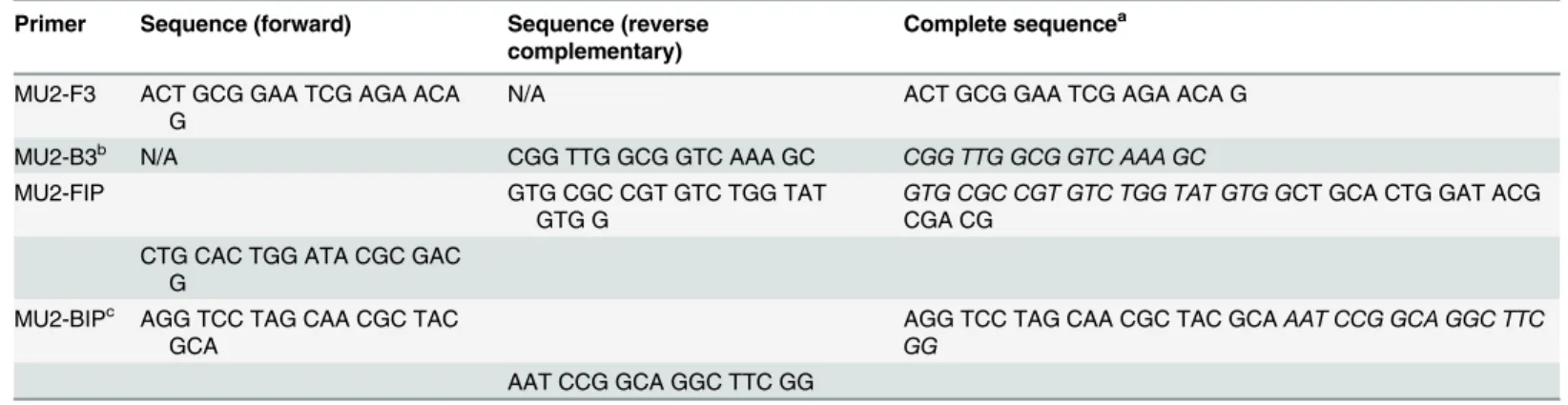
IS2404LAMP primers. A set of four primers was designed for amplification of theM. ulceransspecific IS2404by manually analyzing the target sequence and designing the primers according to the needs for LAMP amplification. Specificity of the primers forM.ulceranswas confirmed in silico by means of the basic local alignment search tool (BLAST, GenBank, NCBI) [40]. The primer set consisted of two smaller oligonucleotides with forward (MU2-F3) and reverse complementary (MU2-B3) sequences and two larger oligonucleotides (MU2-FIP and MU2-BIP) with a complex sequence construction. Primer sequences are provided in
Table 1and binding sites within the IS2404are displayed inFig 1. Primers MU2-B3 and MU2-BIP were first published by Ablordey et al. [28].
Conventional IS2404LAMP (cLAMP) protocol. Each cLAMP reaction mix consisted of
1μlBstDNA polymerase (large fragment, 8 U/μl; New England Biolabs [NEB], Frankfurt am
Main, Germany), 1.0μl dNTP mix (2 mM each, Merck), 1.0μl of primers MU2-F3 and
MU2-B3 (5 pmol/μl) and 2.0μl of primers MU2-FIP and MU2-BIP (10 pmol/μl), respectively
(TibMolBiol, Berlin, Germany), 2.0μl betaine (5 M; Sigma-Aldrich, Taufkirchen, Germany),
2.5μl 10-fold Thermopol buffer forBstDNA polymerase (NEB) and 11.5μl molecular grade
H2O (Carl Roth). Following the addition of 1μl DNA extract (template), cLAMP reactions
(final volume: 25μl) were carried out in 1.5 ml SafeSeal reaction tubes (Sarstedt, Nümbrecht,
Germany) at 65°C for 60 minutes in a conventional thermoblock (HLC Thermomixer MKR 13, HLC BioTech, Bovenden, Germany) and a final step at 80°C for 10 minutes terminated the amplification.
Table 1. Primer sequences of the IS2404LAMP assay.
Primer Sequence (forward) Sequence (reverse complementary)
Complete sequencea
MU2-F3 ACT GCG GAA TCG AGA ACA G
N/A ACT GCG GAA TCG AGA ACA G
MU2-B3b N/A CGG TTG GCG GTC AAA GC CGG TTG GCG GTC AAA GC
MU2-FIP GTG CGC CGT GTC TGG TAT
GTG G
GTG CGC CGT GTC TGG TAT GTG GCT GCA CTG GAT ACG CGA CG
CTG CAC TGG ATA CGC GAC G
MU2-BIPc AGG TCC TAG CAA CGC TAC GCA
AGG TCC TAG CAA CGC TAC GCAAAT CCG GCA GGC TTC GG
AAT CCG GCA GGC TTC GG
Table 1shows the sequences of a set of fourM.ulceransspecific LAMP primers targeting the IS2404. N/A, not applicable.
a
The reverse complementary sequence of nucleotides is displayed in italics.
bMU2-B3, the same primer sequence was originally published by Ablordey et al. as primer
“Buruli-B3”[28].
cMU2-BIP, the same primer sequence was originally published by Ablordey et al. as primer
“Buruli-BIP”[28].
doi:10.1371/journal.pntd.0004219.t001
Fig 1. Binding sites of LAMP primers within the IS2404.Fig 1shows binding sites of LAMP primers within the IS2404ofM.ulceransstrain Agy 99 (Genbank accession number: CP000325.1). Primer MU2-FIP consists of a first reverse complementary region“F1”and a second forward region“F2”. Primer MU2-BIP (first described by Ablordey et al. [28]) consists of a first forward region“B1”and a second reverse complementary region“B2”. Primers and corresponding regions are highlighted in colors; red: primer MU2-F3 (region“F3”); yellow: primer MU2-FIP (region“F2”); light blue: primer MU2-FIP (region “F1”); dark blue: primer MU2-BIP (region“B1”); green: primer MU2-BIP (region“B2”) and pink: primer MU2-B3 (region“B3”; first described by Ablordey et al. [28]).
IS2404dry-reagent-based LAMP (DRB LAMP) protocol. The DRB LAMP reaction mix
contained the same concentrations of reagents and primers as described for the conventional LAMP assay. However, as the wildtypeBstpolymerase large fragment contained glycerol it was not possible to lyophilize. Therefore, a customizedBstpolymerase dissolved in H2O and
reac-tion buffer (NEB) were applied for the DRB LAMP assay. The DRB LAMP reacreac-tion mix was prepared for each reaction in 1.5 ml reaction tubes and subjected to lyophilization by means of a RVC 2–25 CD plus Vacuum Concentrator (Christ, Osterode, Germany) at 1.0 mbar and a safety pressure of 1.000 mbar according to the manufacturer’s specifications. During the pro-cess of validation, DRB LAMP reaction tubes were stored at ambient temperature in the dark and reactions were carried out within one week after lyophilization as described for cLAMP following the addition of 1μl template DNA and adjustment with 24μl molecular grade H2O
(Carl Roth) to a final reaction volume of 25μl.
LAMP product visualization. LAMP products were detected by two different methods: i) gel-electrophoresis on a 0.5x TBE gel containing 0.01% GelRed (Biotium, Cologne, Germany) and ii) SYBR Green I (Life Technologies) staining (0.5μl of 1:5 diluted SYBR Green I staining
solution and 12.5μl of LAMP product) followed by UV-transillumination.
Comparative testing of clinical samples
Confirmation rates of routine assays (PCR [DRB PCR, cPCR] and qPCR) compared with cLAMP. The confirmation rate was defined as the number of patients with a positive PCR, qPCR or cLAMP test result divided by the number of all suspected BUD cases.
Sensitivity rates of PCR (DRB PCR, cPCR) and cLAMP among confirmed BUD patients. The sensitivity rate was defined as the number of patients with a positive PCR or cLAMP test result divided by the number of all qPCR confirmed patients.
Positivity rates of routine assays (PCR [DRB PCR, cPCR] and qPCR) and cLAMP among clinical samples from suspected BUD cases. The positivity rate was defined as the number of clinical samples from suspected BUD cases with a positive PCR, qPCR or cLAMP test result divided by the number of all samples tested.
Sensitivity rates of routine assays (PCR [DRB PCR, cPCR]) compared with cLAMP among clinical samples from confirmed BUD cases. The sensitivity rate was defined as the number of clinical samples from confirmed BUD patients with a positive PCR or cLAMP test result divided by the number of all samples with a positive qPCR result.
Positivity rates of DRB LAMP compared with routine assays (PCR [DRB PCR, cPCR] and qPCR) and cLAMP among clinical samples from suspected BUD cases. The positivity rate was defined as the number of clinical samples with a positive PCR, qPCR or cLAMP test result divided by the number of all samples tested.
Sensitivity rates of cLAMP and DRB LAMP among clinical samples from confirmed BUD cases. The sensitivity rate was defined as the number of clinical samples from confirmed BUD patients with a positive cLAMP or DRB LAMP test result divided by the number of all samples with a positive result in DRB PCR, cPCR and qPCR each.
Statistical analysis
Results
Performance characteristics of routine assays (DRB PCR, cPCR and
qPCR) compared with cLAMP
In silico analysis of the novel IS2404LAMP primers and testing of 17 DNA extracts from mycobacterial cultures (M.ulcerans, n = 5; other mycobacteria, n = 12) by cLAMP and DRB LAMP revealed 100% specificity of both assays forM.ulcerans. The LODs were 50 (DRB PCR and cPCR), 3 (qPCR) and 100 (cLAMP) copies of the target sequence IS2404, corresponding to 0.2, 0.01, and 0.5M.ulceransgenome equivalents, respectively.
Laboratory confirmation of suspected BUD cases
Out of the 91 patients with suspected BUD, 68 were laboratory confirmed as BUD patients (74.73%) by routine methods. Among 68 confirmed BUD patients, 40 patients (58.82%) were in age group 5–14 years (age range 5–56 years, mean 14 years, median 11 years), 33 patients (48.53%) were male, and 36 patients (52.94%) presented with non-ulcerative lesions.
Confirmation rates of routine assays (DRB PCR, cPCR and qPCR)
compared with cLAMP among suspected BUD cases
DRB PCR (on-site at KCCR or INH) and cPCR (DITM, 100% concordance between DRB and cPCR results) confirmed 59/91 (64.84%; 95%-CI: 55.02%-74.65%) of the suspected BUD cases, the qPCR confirmed 68/91 (74.73%; 95%-CI: 65.80%-83.65%), thus added an additional diag-nostic yield of 9.89%. The confirmation rate for cLAMP was 62.64% (95%-CI: 52.70%-72.58%; n = 57). Neither DRB PCR nor cPCR or cLAMP had false positive results compared with qPCR, and confirmation rates were not significantly different. According to McNemar test, there was no significant difference between DRB PCR or cPCR (100% concordant results) compared with qPCR (ORcrude= 1.60; 95%-CI: 0.81–3.20;P-value = 0.15), between cLAMP
compared with qPCR (ORcrude= 1.76; 95%-CI: 0.89–3.50;P-value = 0.08) and between DRB
PCR or cPCR compared with cLAMP (ORcrude= 1.10; 95%-CI: 0.57–2.11;P-value = 0.76).
Sensitivity rates of DRB PCR, cPCR and cLAMP among confirmed BUD
cases
Among the 68 BUD cases confirmed by qPCR, the sensitivity was 86.76% (95%-CI: 78.71%-94.82%; n = 59) for DRB PCR and cPCR and 83.82% (95%-CI: 75.07%-92.58%; n = 57) for cLAMP. According to McNemar test, there was no significant difference between DRB PCR and cPCR compared with cLAMP (ORcrude= 1.27; 95%-CI: 0.44–3.63;P-value = 0.63).
Positivity rates of routine assays (DRB PCR, cPCR and qPCR) and
cLAMP among clinical samples from suspected BUD cases
Among the 140 samples from 91 clinically suspected BUD cases, the positivity rate was 56.43% (95%-CI: 48.21%-64.64%; n = 79) for DRB PCR and cPCR, 67.14% (95%-CI: 59.36%-74.92%; n = 94) for qPCR, and 52.86% (95%-CI: 44.59%-61.13%; n = 74) for cLAMP. Neither DRB PCR nor cPCR or cLAMP revealed false positive results compared with qPCR. According to McNemar test, there was no significant difference between DRB PCR or cPCR compared with qPCR (ORcrude= 1.58; 95%-CI: 0.94–2.64;P-value = 0.07) and between DRB PCR or cPCR
compared with cLAMP (ORcrude= 1.16; 95%-CI: 0.70–1.90;P-value = 0.55), whereas the
differ-ence between cLAMP compared with qPCR was significant (ORcrude= 1.82; 95%-CI: 1.09–
Stratification into sample types did not reveal significant differences in positivity rates of DRB PCR, cPCR, qPCR and cLAMP among FNA, swab or punch biopsy samples.
Sensitivity rates of routine assays (DRB PCR and cPCR) compared with
cLAMP among clinical samples from confirmed BUD cases
Among the 94 samples from 68 BUD cases confirmed by qPCR, the sensitivity was 84.04% (95%-CI: 76.64%-91.45%; n = 79) for DRB PCR and cPCR, and 78.72% (95%-CI: 44.59%-61.13%; n = 74) for cLAMP. According to McNemar test, there was no significant difference between DRB PCR or cPCR compared with cLAMP (ORcrude= 1.42; 95%-CI: 0.64–3.18);P
-value = 0.35).
Performance characteristics of DRB LAMP
DRB LAMP revealed the same performance characteristics as determined for cLAMP (i.e. 100%M.ulceransspecificity and a LOD of 0.5M.ulceransgenome equivalents).
Positivity rates of DRB LAMP compared with routine assays (DRB PCR,
cPCR and qPCR) and cLAMP among clinical samples from suspected
BUD cases
To compare DRB LAMP with DRB PCR, cPCR, qPCR and cLAMP, 32 samples (FNA and swab samples, n = 16, respectively) from 32 suspected BUD cases were subjected to the assays. The positivity rate was 75.0% (95%-CI: 62.41%-87.59%; n = 24) for DRB PCR, cPCR and qPCR, 71.88% (95%-CI: 87.84%-100%; n = 23) for cLAMP, and 68.75% (95%-CI: 80.61%-100%; n = 22) for DRB LAMP. Neither cLAMP nor DRB LAMP revealed false positive results compared with DRB PCR, cPCR and qPCR.
According to McNemar test, there was no significant difference neither between DRB PCR, cPCR or qPCR compared with cLAMP (ORcrude= 1.17; 95%-CI: 0.34–4.10;P-value = 0.78),
nor between DRB PCR, cPCR or qPCR compared with DRB LAMP (ORcrude= 1.36; 95%-CI:
0.40–4.49;P-value = 0.58), nor between cLAMP compared with DRB LAMP (ORcrude= 0.86;
95%-CI: 0.26–2.87;P-value = 0.79).
Sensitivity rates of cLAMP and DRB LAMP among clinical samples from
confirmed BUD cases
Among the 24 samples from 24 BUD patients confirmed by DRB PCR, cPCR and qPCR, the sensitivity was 95.83% (95%-CI: 87.84%-100%; n = 23) for cLAMP and 91.67% (95%-CI: 80.61%-100%; n = 22) for DRB LAMP. According to McNemar test, there was no significant difference between cLAMP compared with DRB LAMP (ORcrude= 0.48; 95%-CI: 0.02–7.54);
P-value = 0.56).
IS
2404
LAMP detection methods
Out of 74 amplicons derived from cLAMP reactions, 74/74 (100%) were judged positive by gel-electrophoresis and 73/74 (98.65%) by SYBR Green I staining. All products derived from DRB LAMP were likewise analyzed and the concordance rate was 22/22 (100%) between both detec-tion methods.
Discussion
BUD belongs to the currently five neglected diseases in line for the IDM (innovative and inten-sified disease management) approach demanding a major scaling up of active detection, treat-ment, monitoring and surveillance. Development of diagnostic tests that bring health services closer to where NTDs are is considered a research priority. LAMP, a technology that features cost effectiveness, robustness and modest needs in terms of equipment, has recently been selected by the WHO as one of the promising tools for decentralized diagnostics [13–14,41].
Several investigators recently succeeded in developingM.ulceransspecific LAMP assays which showed performance characteristics comparable to conventional PCR formats [26–28]. Based on longstanding experience with a DRB PCR format for laboratory confirmation of BUD in Ghana and Togo [6,10,30,33,36,42], the development of a DRB LAMP assay
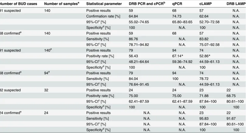
Table 2. Confirmation rates, sensitivity, specificity and significance of the applied molecular tests.
Number of BUD cases Number of samplesa Statistical parameter DRB PCR and cPCRb qPCR cLAMP DRB LAMP
91 suspected 140 Positive results 59 68 57 N.A.
Confirmation rate [%] 64.84 74.73 62.64 N.A.
95%-CIc[%] 55.02
–74.65 65.80–83.65 52.70–72.58 N.A.
Specificityd[%] 100 N.A. 100 N.A.
68 confirmede 140 Positive results 59 68 57 N.A.
Sensitivity [%] 86.76 N.A. 83.82 N.A.
95%-CIc[%] 78.71
–94.82 N.A. 75.07–92.58 N.A.
91 suspected 140# Positive results 79 94 74 N.A.
Positivity rate [%] 56.43 67.14* 52.86* N.A.
95%-CIc[%] 48.21
–64.64 59.36–74.92 44.59–61.13 N.A.
Specificityd[%] 100 N.A. 100 N.A.
68 confirmede 94# Positive results 79 94 74 N.A.
Sensitivity [%] 84.04 100 78.72 N.A.
95%-CIc[%] 76.64
–91.45 N.A. 44.59–61.13 N.A.
32 suspected 32 Positive results 24 24 23 22
Positivity rate [%] 75.00 75.00 71.88 68.75
95%-CIc[%] 62.41–87.59 62.41–87.59 87.84–100 80.61–100
Specificityd[%] 100 N.A. 100 100
24 confirmede 24 Positive results N.A. N.A. 23 22
Sensitivity [%] N.A. N.A. 95.83 91.67
95%-CIc[%] N.A. N.A. 87.84–100 80.61–100
Specificityd[%] N.A. N.A. 100 100
Table 2shows the results of DRB PCR, cPCR, qPCR, cLAMP, and DRB LAMP from clinical samples of clinically suspected and confirmed BUD cases recruited at Agogo Presbyterian Hospital, Ghana, Tepa Government Hospital, Ghana, and at Centre Hospitalier Régional de Tsévié, Togo.
N.A., not applicable.
aNumber of clinical samples tested.
#indicates if the presented results refer to the number of samples
–all other results refer to the number of patients or¶the number of patients and samples
was equal.
bResults of the DRB PCR and cPCR were 100% concordant for all samples tested. c95 percent con
fidence interval.
dSpeci
ficity was calculated as proportion of truly positive test results out of all positive results of the same test, based on the results of qPCR as reference test.
eLaboratory con
firmation was defined as positive IS2404qPCR test result of any sample tested per patient.
*Significantly different proportions of positive results among all clinical samples tested by two different tests, calculated by McNemar test.
applicable under tropical climate conditions at primary health care level was envisaged in this study. Thermodynamic reasons (i.e. leaving out an initial denaturation step for annealing of primers) required the design of modified primers, therefore as a first step a new cLAMP assay was established that constituted the basis for the DRB format. During development of the DRB assay lyophilization of the reaction mix initially constituted a major challenge. Due to the glyc-erol content ofBstpolymerase and reaction buffer as employed in previous cLAMP formats (including our own), customized glycerol-free reagents had to be obtained and adequate lyoph-ilization protocols had to be established.
The comparable performance of cLAMP, DRB LAMP and DRB PCR as well as cPCR sug-gests that both LAMP formats constitute a reliable alternative to conventional routine assays. Our data also show that the LAMP assays and the DRB PCR as well as cPCR have equal sensi-tivity for FNA samples. Both LAMP formats are applicable at primary health care level, the DRB format however provides significant advantages such as a simplified test layout and the possibility of storage of reagents at ambient temperature. Decentralized utilization of LAMP technology furthermore would lead to cost saving due to reduced expenditures for transporta-tion of samples to a reference center as well as reduced test costs, i.e. US$ 1–2 per LAMP reac-tion as compared to US$ 8–10 per DRB PCR or cPCR reaction.
In this study it was not possible to assess long-term storage of DRB-LAMP reaction tubes under tropical conditions. Long-term storability of DRB PCR reaction tubes was however pre-viously proven [6,36] which allows the conclusion that maximum storage periods of up to 12 months also apply for LAMP reagents.
Although in our study routine PCR and LAMP assays for the most part did not perform sig-nificantly different from qPCR, it must be assumed that qPCR renders an additional diagnostic yield of approximately 10% [10]. Therefore, regardless of the method used, confirmation of negative samples by qPCR e.g. through the global network of laboratories for confirmingM.
ulceransinfection [43] should be attempted. Likewise, participation of laboratories in external quality assurance programs as implemented by Eddyani et al. in collaboration with the WHO is strongly recommended [44].
While the amplification procedure of LAMP technology especially in the DRB format can be considered field friendly without restriction, current DNA extraction procedures are not yet entirely appropriate for POC testing and need optimization. As shown by Ablordey et al. the use of boiled crude DNA extracts led to a significant decrease in sensitivity [28]. Other options such as one-tube silica-membrane based extraction protocols [45] or one-tube enzyme-based lyophilized reactions are yet to be evaluated. A field friendly approach to storage of DNA extracts for purposes of quality assurance could be the filter paper technology as successfully applied for TBC [46].
In conclusion, the cLAMP and DRB LAMP formats evaluated in this study are equivalent alternatives to conventional PCR techniques and, provided the availability of field friendly DNA extraction formats, constitute valuable tools for decentralized laboratory confirmation of BUD. As in the case of other investigators who previously developed BUD specific LAMP assays, the validation of the LAMP assays presented in this study was conducted in a third-level laboratory environment, therefore field based evaluation trials are necessary to determine the clinical performance at peripheral health care level.
Supporting Information
S1 Protocol. Extraction of mycobacterial DNA from clinical specimens.
Acknowledgments
The authors thank Kerstin Helfrich and Carolin Mengele (DITM) for laboratory assistance, as well as Dr. Basile Kobara (“Programme National de Lutte contre l’Ulcère de Buruli, Lèpre et Pian”, Lomé, Togo) for excellent collaboration. This manuscript contains parts of the doctoral thesis of Dominik Symank and Moritz Jansson.
Author Contributions
Conceived and designed the experiments: MBe ROP FB GB. Performed the experiments: MBe MBa MF IM AR KLH DS MJ. Analyzed the data: MBe ROP FB MBa KB FSS FXW ABK KHH TL GB. Contributed reagents/materials/analysis tools: ROP FB KB FXW ABK KHH TL GB. Wrote the paper: MBe ROP FB MBa TL GB. Patient management: ROP FSS EP. Study logistics: FXW.
References
1. WHO. Treatment ofMycobacterium ulceransdisease (Buruli ulcer): guidance for health workers. Geneva, Switzerland: World Health Organization. 2012.
2. WHO. Buruli Ulcer. Fact sheet 199. Geneva, Switzerland: World Health Organization. 2007 updated 2015.
3. WHO. Recommendations for control of buruli ulcer. Geneva, Switzerland: World health organisation. 2013.
4. Ross BC, Johnson PDR, Oppedisano F, Marino L, Sievers A, Stinear T, et al. Detection of Mycobacte-rium ulceransin Environmental Samples during an Outbreak of Ulcerative Disease. Applied and Envi-ronmental Microbiology. 1997; 63(10):4135–38. PMID:9327583
5. Stinear T, Ross BC, Davies JK, Marino L, Robins-Browne RM, Oppedisano F, et al. Identification and Characterization of IS2404and IS2606: Two Distinct Repeated Sequences for Detection of Mycobac-terium ulceransby PCR. Journal of Clinical Microbiology. 1999; 37(4):1018–23. PMID:10074520 6. Siegmund V, Adjei O, Racz P, Berberich C, Klutse E, van Vloten F, et al. Dry-reagent-based PCR as a
novel tool for laboratory confirmation of clinically diagnosedMycobacterium ulcerans-associated dis-ease in areas in the tropics whereM.ulceransis endemic. J Clin Microbiol. 2005; 43(1):271–6. PMID: 15634982
7. Fyfe JAM, Lavender CJ, Johnson PDR, Globan M, Sievers A, Azuoloas J, et al. Development and Application of Two Multiplex Real-Time PCR Assays for the Detection ofMycobacterium ulceransin Clinical and Environmental Samples. Applied and Environmental Microbiology. 2007; 73(15):4733–40. PMID:17526786
8. Beissner M, Herbinger KH, Bretzel G. Laboratory diagnosis of buruli ulcer disease. Future Microbiol. 2010; 5(3):363–70. doi:10.2217/fmb.10.3PMID:20210548
9. WHO. Ed Portaels F. Laboratory diagnosis of buruli ulcer: a manual for health care providers. Geneva, Switzerland: World Health Organization; 2014.
10. Beissner M, Huber KL, Badziklou K, Halatoko WA, Maman I, Vogel F, et al. Implementation of a National Reference Laboratory for Buruli Ulcer Disease in Togo. PLoS Negl Trop Dis. 2013; 7(1): e2011. doi:10.1371/journal.pntd.0002011PMID:23359828
11. Vincent QB, Ardant MF, Adeye A, Goundote A, Saint-André JP, Cottin J, et al. Clinical epidemiology of laboratory-confirmed Buruli ulcer in Benin: a cohort study. Lancet Glob Health. 2014; 2(7):e422–30. doi:10.1016/S2214-109X(14)70223-2PMID:25103396
12. Philips RO, Sarfo FS, Abass MK, Abotsi J, Wilson T, Forson M, et al. Clinical and Bacteriological Effi-cacy of Rifampin-Streptomycin Combination for Two Weeks followed by Rifampin and Clarithromycin for Six Weeks for Treatment ofMycobacterium ulceransDisease. Antimicrob. Agents Chemother. 2014; 58(2):1161–66. doi:10.1128/AAC.02165-13PMID:24323473
13. WHO. Investing to overcome the global impact of neglected tropical diseases: Third WHO report on neglected tropical diseases. Geneva, Switzerland: World Health Organization; 2015.
14. WHO. Report of a WHO-FIND consulatative meeting on diagnostics for Buruli ulcer. Geneva, Switzer-land: World Health Organization; 2013.
16. Paris DH, Imwong M, Faiz AM, Hasan M, Yunus EB, Silamut K, et al. Loop-Mediated Isothermal PCR (LAMP) for the Diagnosis of Falciparum Malaria. Am J Trop Med Hyg. 2007; 77(5):972–76. PMID: 17984362
17. Pandey BD, Poudel A, Yoda T, Tamaru A, Oda N, Fukushima Y, et al. Development of an in-house loop-mediated isothermal amplification (LAMP) assay for detection ofMycobacterium tuberculosisand evaluation in sputum samples of Nepalese patients. J Med Microbiol. 2008; 57(4):439–43.
18. Abbasi I, King CH, Muchiri EM, Hamburger J. Detection ofSchistosoma mansoniandSchistosoma haematobiumDNA by Loop-Mediated Isothermal Amplification: Identification of Infected Snails from Early Prepatency. Am J Trop Med Hyg. 2010; 83(2):427–32. doi:10.4269/ajtmh.2010.09-0764PMID: 20682894
19. Nkouawa A, Sako Y, Li T, Chen X, Wandra T, Swastika IK, et al. Evaluation of a loop-mediated isother-mal amplification method using fecal specimens for differential detection of Taenia species from humans. J Clin Microbiol. 2010; 48(9):3350–52. doi:10.1128/JCM.00697-10PMID:20631114 20. Tang MJ, Zhou S, Zhang XY, Ou XY, Ge QL, Tang XJ, et al. Rapid and sensitive detection ofListeria
monocytogenesby loop-mediated isothermal amplification. Curr Microbiol. 2011; 63(6):511–16. doi: 10.1007/s00284-011-0013-3PMID:21935669
21. Neonakis IK, Spandidos DA, Petinaki E. Use of loop-mediated isothermal amplification of DNA for the rapid detection ofMycobacterium tuberculosisin clinical specimens. Eur J Clin Microbiol Infect Dis. 2011; 30:937–42. doi:10.1007/s10096-011-1195-0PMID:21331481
22. Khan MG, Bhaskar KR, Salam MA, Akther T, Pluschke G, Mondal D. Diagnostic accuracy of loop-medi-ated isothermal amplification (LAMP) for detection of Leishmania DNA in buffy coat from visceral leish-maniasis patients. Parasit Vectors. 2012; 5:280. doi:10.1186/1756-3305-5-280PMID:23206441 23. Poole CB, Tanner NA, Zhang Y, Evans TC Jr, Carlow CK. Diagnosis of brugian filariasis by
loop-medi-ated isothermal amplification. PLoS Negl Trop Dis. 2012; 6(12):e1948. doi:10.1371/journal.pntd. 0001948PMID:23272258
24. Lim KT, The CS, Thong KL. Loop-mediated isothermal amplification assay for the rapid detection of Staphylococcusaureus. Biomed Res Int. 2013; 2013:895816. doi:10.1155/2013/895816PMID: 23509796
25. Dhama K, Karthik K, Chakraborty S, Tiwari R, Kapoor S, Kumar A, et al. Loop-mediated isothermal amplification of DNA (LAMP): a new diagnostic tool lights the world of diagnosis of animal and human pathogens: a review. Pak J Biol Sci. 2014; 17(2):151–66. PMID:24783797
26. de Souza DK, Quaye C, Mosi L, Addo P, Boakye DA. A quick and cost effective method for the diagno-sis ofMycobacterium ulceransinfection. BMC Infect Dis. 2012; 12:8. doi:10.1186/1471-2334-12-8 PMID:22257432
27. Njiru ZK, Yeboah-Manu D, Stinear TP, Fyfe JAM. Rapid and sensitive detection ofMycobacterium ulceransusing a Loop-Mediated Isothermal Amplification test. J Clin Microbiol. 2012; 50(5):1737–41. doi:10.1128/JCM.06460-11PMID:22357495
28. Ablordey A, Amissah DA, Aboagye IF, Hatano B, Yamazaki T, Sata T, et al. Detection of Mycobacte-rium ulceransby the Loop Mediated Isothermal Amplification Method. PLoS Neglected Tropical Dis-eases. 2012; 6(4):e1590. doi:10.1371/journal.pntd.0001590PMID:22509415
29. Beissner M, Symank D, Phillips RO, Amoako YA, Awua-Boateng NY, Sarfo FS, et al. Detection of Via-bleMycobacterium ulceransin Clinical Samples by a Novel Combined 16S rRNA Reverse Transcrip-tase/IS2404 Real- Time qPCR Assay. PLoS Negl Trop Dis. 2012; 6(8):e1756. doi:10.1371/journal. pntd.0001756PMID:22953006
30. Herbinger KH, Adjei O, Awu-Boateng NY, Nienhuis WA, Kunaa L, Siegmund V, et al. Comparative study of the sensitivity of different diagnostic methods for the laboratory diagnosis ofBuruli ulcer dis-ease. Clin Infect Dis. 2009; 48(8):1055–64. doi:10.1086/597398PMID:19275499
31. Phillips RO, Sarfo FS, Osei-Sarpong F, Boateng A, Tetteh I, Lartey A, et al. Sensitivity of PCR Targeting Mycobacterium ulceransby Use of Fine-Needle Aspirates for Diagnosis of Buruli Ulcer. Journal of Clini-cal Microbiology. 2009; 47(4):924–26. doi:10.1128/JCM.01842-08PMID:19204098
32. Herbinger KH, Beissner M, Huber K, Awua-Boateng NY, Nitschke J, Thompson W, et al. Efficiency of Fine-Needle Aspiration Compared with Other Sampling Techniques for Laboratory Diagnosis of Buruli Ulcer Disease. Journal Of Clinical Microbiology. 2010; 48(10):3732–34. doi:10.1128/JCM.01549-10 PMID:20739480
33. Bretzel G, Huber KL, Kobara B, Beissner M, Piten E, Herbinger KH, Wiedemann FX, et al. Laboratory Confirmation of Buruli Ulcer Disease in Togo, 2007–2010. PLoS Negl Trop Dis. 2011; 5(7):e1228. doi: 10.1371/journal.pntd.0001228PMID:21811641
35. Bretzel G, Siegmund V, Nitschke J, Herbinger KH, Thompsopn W, Klutse E, et al. A stepwise approach to the laboratory diagnosis of Buruli ulcer disease. Trop Med Int Health. 2007; 12(1):89–96. PMID: 17207152
36. Siegmund V, Adjei O, Nitschke J, Thompson W, Klutse E, Herbinger KH, et al. Dry reagent-based poly-merase chain reaction compared with other laboratory methods available for the diagnosis of Buruli ulcer disease. Clin Infect Dis. 2007; 45(1):68–75. PMID:17554703
37. Beissner M, Awua-Boateng NY, Thompson W, Nienhuis WA, Klutse E, Agbenorku P, et al. A Genotypic Approach for Detection, Identification, and Characterization of Drug Resistance inMycobacterium ulceransin Clinical Samples and Isolates from Ghana. Am J Trop Med Hyg. 2010; 83(5):1059–65. doi: 10.4269/ajtmh.2010.10-0263PMID:21036838
38. Jansson M, Beissner M, Phillips RO, Badziklou K, Piten E, Maman I, et al. Comparison of Two Assays for Molecular Determination of Rifampin Resistence in Clinical Samples from Patients with Buruli Ulcer Disease. J. Clin. Microbiol. 2014; 52(4):1246. doi:10.1128/JCM.03119-13PMID:24478404
39. Taylor S, Wakem M, Dijkman G, Alsarraj M, Nguyen M. A practical approach to RT-qPCR—Publishing data that conform to the MIQE guidelines. Methods. 2010; 50(4):1–5.
40. Benson DA, Karsch-Mizrachi I, Lipman DJ, Ostell J, Wheeler DL. GenBank. Nucleic Acids Res. 2008; 36(D25-30).
41. Parida M, Sannarangaiah S, Dash PK, Rao PVL, Morita K. Loop mediated isothermal amplification (LAMP): a new generation of innovative gene amplification technique; perspectives in clinical diagnosis of infectious diseases. Rev Med Virol. 2008; 18:407–21. doi:10.1002/rmv.593PMID:18716992 42. Phillips RO, Phanzu DM, Beissner M, Badziklou K, Luzolo EK, Sarfo FS, et al. Effectiveness of routine
BCG vaccination on buruli ulcer disease: a case-control study in the Democratic Republic of Congo, Ghana and Togo. PLoS Negl Trop Dis. 2015; 9(1):e3457. doi:10.1371/journal.pntd.0003457PMID: 25569674
43. World Health Organization, Laboratory Support Network. Global network of laboratories for confirming Mycobacterium ulceransinfection (Buruli ulcer). Geneva, Switzerland: World Health Organization. Available:http://www.who.int/buruli/Global_network_laboratories_PCR.pdf. Accessed 28 Mai 2015. 44. Eddyani M, Lavender C, de Rijk WB, Bomans P, Fyfe J, de Jong B, et al. Multicenter External Quality Assessment Program for PCR Detection ofMycobacterium ulceransin Clinical and Environmental Specimens. PLoS One. 2014; 9(2):e89407. doi:10.1371/journal.pone.0089407PMID:24586755 45. McKenna. Refining Sample Prep for Molecular Dx. Genetic Engineering & Biotechnology News. 2013;
33(17):26–7.