online | memorias.ioc.fiocruz.br
Comparison of LAMP and PCR for molecular mass screening
of sand flies for
Leishmania martiniquensis
infection
Saruda Tiwananthagorn1/+, Hirotomo Kato2, Ranchana Yeewa1, Amontip Muengpan1, Raxsina Polseela3, Saovanee Leelayoova4
1Chiang Mai University, Faculty of Veterinary Medicine, Department of Veterinary Biosciences and Veterinary Public Health, Muang, Chiang Mai, Thailand 2Jichi Medical University, Department of Infection and Immunity, Division of Medical Zoology, Tochigi, Japan 3Naresuan University, Faculty of Medical Science, Department of Microbiology and Parasitology, Phitsanulok, Thailand
4Phramongkutklao College of Medicine, Department of Parasitology, Bangkok, Thailand
BACKGROUND Leishmaniasis caused by Leishmania martiniquensis infection has been reported in human and domestic animals of Martinique Island, Germany, Switzerland, USA, Myanmar and Thailand. The peculiar clinical features of disseminated cutaneous and visceral forms co-existence render the urgent need of specific diagnostic tool to identify the natural sand fly vectors for effective prevention and control strategies. Loop-mediated isothermal amplification (LAMP) of 18S rRNA gene as well as polymerase chain reaction (PCR) of minicircle kinetoplast DNA gene (PCR-mkDNA) have never been applied to detect
L. martiniquensis and L. siamensis in sand fly vectors.
OBJECTIVE The present study was aimed to validate malachite green-LAMP (MG-LAMP) and PCR-mkDNA techniques to detect L. martiniquensis in sand fly vectors, compared with the conventional PCR of internal transcribed spacer 1 (PCR-ITS1).
METHODS We compared the validity of LAMP of 18S rRNA gene and PCR-mkDNA, to PCR-ITS1 in simulation model of
L. martiniquensis infection in Sergentomyia gemmea sand flies. Attributable to the sensitivity and specificity, PCR-mkDNA was consecutively applied to detect L. martiniquensis in 380 female sand fly individuals captured in the newly identified affected region of Lamphun Province, Thailand.
FINDINGS AND MAIN CONCLUSIONS Results showed that PCR-mkDNA could detect at least one promastigote per sand fly, which was 10-time superior to LAMP and PCR-ITS1. In addition, PCR-mkDNA was more specific, able to differentiate L. martiniquensis from other viscerotropic Leishmania species, such as L. siamensis, L. (L.) donovani, and L. (L.) infantum.
Consecutively, mass screening of L. martiniquensis in 380 female sand fly individuals by PCR-mkDNA was implemented in a new affected area of Thailand where a patient with leishmaniasis/HIV co-infection resides; however Leishmania DNA was undetected. In conclusion, PCR-mkDNA is a promising tool for molecular mass screening of L. martiniquensis infection in outbreak areas where several species of Leishmania and sand flies co-exist.
Key words: Leishmania martiniquensis - PCR - minicircle kinetoplast DNA - Loop-mediated isothermal amplification - molecular screening - individual sand fly
doi: 10.1590/0074-02760160254
Financial support: Thailand Research Fund and Chiang Mai University (grant no. TRG5780028), CMU Short Term Research Fellowships in Overseas, and the MEXT of Japan (grant no. 25257501).
+ Corresponding author: stiwananthagorn@gmail.com Received 5 June 2016
Accepted 25 October 2016
Leishmaniasis is a vector-borne protozoan disease caused by several species of the genus Leishmania. Main clinical manifestations include cutaneous leishmaniasis (CL), mucocutaneous leishmaniasis (MCL), and visceral leishmaniasis (VL), generally associated with the Leish-mania species. Leishmania promastigote develop in the gut of female sand flies, and differentiate into intracellu-lar amastigote forms in vertebrate hosts after transmis-sion. The spread of disease depends on the distribution of the vectors and reservoir animal hosts. Autochtho-nous cutaneous and VL is now considered an emerging disease in Thailand (Leelayoova et al. 2013, Chiewchan-vit et al. 2015). Characterisation of Leishmania isolates
in Thailand is based on sequence analysis of the internal transcribed spacer 1 (ITS1) and the large subunit of RNA polymerase II genes suggesting that two distinct spe-cies belonging to L. enriettii complex are the causative agents; L. siamensis and L. martiniquensis (Pothirat et al. 2014, Chiewchanvit et al. 2015). In contrast to L. sia-mensis that was isolated only in one patient from Trang Province (Leelayoova et al. 2013), L. martiniquensis is more dominant and has a wider geographical distribu-tion, including France, Germany, Switzerland, USA, Myanmar and Thailand (Chiewchanvit et al. 2015).
im-munodeficiency virus co-infection. Regarding to animal reservoirs and vectors, Leishmania DNA was identified in black rats (Rattus rattus) and in two species of sand flies; Sergentomyia (Neophlebotomus) gemmea and S. barraudi in southern Thailand (Kanjanopas et al. 2013, Chusri et al. 2014). Between 2011 and 2014, at least five cases of L. martiniquensis infection have occurred in northern Thailand including one case in Chiang Rai province (Phumee et al. 2013); one case in Chiang Mai province (Chiewchanvit et al. 2015) and three cases in Lamphun province (BVBD/MoPH 2013, Pothirat et al. 2014, Chiewchanvit et al. 2015). Due to the continually increasing number of cases of L. martiniquensis in Thai-land, the development of a specific diagnostic tool to identify Leishmania infection in circulating sand flies in the affected areas is urgently needed.
Detecting and identifying Leishmania species in sand flies and animal reservoirs are important to predict the risk and transmission of the disease in outbreak and sur-rounding areas (Kato et al. 2007, 2010). Molecular tech-niques, such as polymerase chain reaction (PCR) and PCR-restriction fragment length polymorphism (PCR-RFLP) have been applied to detect and identify Leishma-nia species in reservoir hosts and sand fly vectors with high sensitivity and specificity (Kato et al. 2007, 2010). Due to various limitations in the microscopic detection of Leishmania in sand flies, a molecular mass screening method for Leishmania infection of sand fly individuals has been established (Kato et al. 2007, 2010). This method is a powerful tool for research confirmed on prevalent sand fly species and vector-host-parasite inter-relation-ships (Kato et al. 2007, 2010, Tiwananthagorn et al. 2012). PCR targeting various genes, such as ITS1, small sub-unit 18S ribosomal RNA (18S rRNA), minicircle kineto-plast DNA (mkDNA), mitochondrial cytochrome b(cyt b), have been used to identify Leishmania infection in sand flies (Kato et al. 2010, Kanjanopas et al. 2013, Chusri et al. 2014), human patients and animal reservoirs (Leelay-oova et al. 2013, Chusri et al. 2014, Hitakarun et al. 2014, Chiewchanvit et al. 2015). PCR targeting the mkDNA gene (PCR-mkDNA) has high sensitivity even when only one
Leishmania parasite exists in a sample (Kato et al. 2007). PCR targeting the ITS1 gene (PCR-ITS1) showed high sensitivity to detect L. siamensis as low as 0.05 promasti-gotes/µL (Hitakarun et al. 2014) and is the classical tech-nique to detect L. siamensis and L. martiniquensis in sand fly vectors (Kanjanopas et al. 2013, Chusri et al. 2014).
A colorimetric malachite green based Loop-mediated isothermal amplification (MG-LAMP) assay targeting the
18S rRNA gene has been developed for the robustness and superior sensitivity for mass screening of L. mexicana and
L. major infection in sand flies, with a detection sensitivity of 0.01 parasite (Nzelu et al. 2014). Recently, the LAMP as-say has been developed for simple detection of L. siamen-sis in clinical samples with the low detection limit as 103 parasites/mL whole blood or 2.5 parasites/tube (Sriworarat et al. 2015). However, LAMP as well as PCR-mkDNA have never been applied to detect L. martiniquensis and
L. siamensis in sand fly vectors. The present study, there-fore, was aimed to validate MG-LAMP and PCR-mkDNA techniques to detect L. martiniquensis in sand fly vectors,
compared with the conventional PCR-ITS1. Attributable to the sensitivity and specificity, PCR-mkDNA was consecu-tively applied to detect L. martiniquensis in 380 female sand fly individuals captured in the newly identified af-fected region of Lamphun Province.
MATERIALS AND METHODS
Parasites - Promastigotes of L. martiniquensis
(MHOM/TH/2011/PG) were harvested from axenic culture in Schneider’s Drosophila medium with L-glutamate (Sig-ma-Aldrich, USA), supplemented with 20% fetal bovine serum (Merck Millipore, Germany), 100 U/mL penicillin,
100 μg/mL streptomycin, 50 µg/mL gentamicin at 25ºC.
Sand fly collection and taxonomic identification - Sand flies were collected during October 2014 to May 2015 from a new affected area of Tha Mae Lop Subdistrict, Mae Tha District, Lamphun Province (Supplementary data), where a patient with autochthonous disseminated leishmaniasis caused by L. martiniquensis resides (Chiewchanvit et al. 2015). The sites were the patient’s house and the surround-ing areas at a radius of 200 m. Collections ussurround-ing CDC light traps were conducted for 12 h between 6:00 pm and 6:00 am both indoors (living room, kitchen), and outdoors (ani-mal shed, crafting studio, ani(ani-mal burrow), bamboo plan-tation, as well as Doi Khurea mountain (altitude 480 m), where the patient has been working as a lumberjack. All sand flies were stored individually in absolute ethanol and
kept at -20ºC until further examination.
Each unfed and blood-fed female sand fly was dis-sected using sterile techniques under a stereomicroscope. The head and last three abdominal segments of each sand fly were mounted on a microscopic slide in Hoyer’s me-dium. Taxonomic identification was conducted morpho-logically following Lewis keys (Lewis 1978), such as morphology of cibarium and spermatheca. The remnant parts of sand flies were stored in absolute ethanol
indi-vidually and kept at -20ºC until DNA was extracted.
DNA preparation - For the preparation of parasite DNA, 10,000 promastigotes of L. martiniquensis were
suspended in 50 μL of DNA extraction buffer (150 mM
NaCl, 10 mM Tris-HCl [pH 8.0], 10 mM EDTA, and 0.1% sodium dodecyl sulfate) in the presence of
pro-teinase K (200 μg/mL), and serially diluted 10-fold in
the same buffer. The samples, without homogenisation,
were then incubated at 56ºC for 12 h, heat inactivated at 95ºC for 5 min, and 25 μL distilled water was added. The DNA samples were stored at -20ºC for further use.
To extract DNA from sand flies, a mass extraction technique (Kato et al. 2007) was implemented with a minor modification. Briefly, the ethanol-fixed sand fly specimens were placed individually in each
microcen-trifuge tube and lysed in 50 μL DNA extraction buffer
without homogenisation. The samples were then pro-cessed and stored, as mentioned above.
DNA samples of other Leishmania species used in this study were prepared from the following reference strains, including L. siamensis (MHOM/TH/2010/TR), L. mar-tiniquensis (MHOM/TH/2013/LSCM3), L. (L.) major
BR/1973/M2269), L. (L.) infantum (MCAN/TR/2000/ EP55), and L. (L.) donovani (MHOM/SU/62/2S-25M-C2). In addition, DNA samples of the local stains of Trypano-soma evansi, Leucocytozoon sabrazesi, and Plasmodium gallinaceum were used for the specificity test in this study.
LAMP and PCR assays - MG-LAMP assay targeting the Leishmania18S rRNA gene (Nzelu et al. 2014) was validated for its sensitivity and specificity for L. marti-niquensis detection. Briefly, the reaction was conducted
in 15 μL of a reaction mixture consisting of 1.6 μM of each inner primer (FIP and BIP), a 0.4 μM of each outer
primer (F3 and B3), 1x reaction mix (Eiken, Japan), 8 U
Bst DNA polymerase (Eiken), 0.004% malachite green
(MG) dye (dissolved in distilled water), and 1 μL of tem -plate DNA. The mixture was incubated at 64ºC for 60 min and then heated at 80ºC to terminate the reaction using MJ Research PTC-200 Thermal Cycler (Bio-Rad Laboratories, CA). At the end of incubation, the amplifi-cation of the target gene was confirmed based on direct visual inspection of the reaction tubes by the naked eye; a positive amplification showed as light blue, whereas in the absence of amplification, the reaction mixture be-came colorless. In addition, LAMP products were anal-ysed on a 2.5% agarose gel electrophoresis.
PCR-mkDNA using primer L.MC-1S/ L.MC-1R (Kato et al. 2007) and PCR-ITS1 using primer L5.8S/ LITSR (El Tai et al. 2001) were conducted as previous-ly described. Briefprevious-ly, PCR was carried out in a volume
of 20 μL using the primers (0.4 μM each), Ampdirect
Plus (Shimadzu Biotech, Japan), and 0.5 U BioTaq™ HS
DNA polymerase (Bioline, UK) with 1 μL of template DNA. After an initial denaturation at 95ºC for 10 min,
PCR amplification was performed with 35 cycles of
de-naturation (95ºC, 1 min), annealing (55ºC, 45 s for PCR-mkDNA or 53ºC, 30 s for PCR-ITS1), and polymerisa
-tion (72ºC, 1 min) followed by a final extension at 72ºC
for 10 min. The PCR products were analysed on a 1.5% agarose gel electrophoresis.
To identify sand fly species using molecular tech-niques, PCR and sequencing of the gene mitochondrial
cytochrome oxidase subunit I (COI) of metazoan in-vertebrate (LCO1490/HCO2198) were performed, with the conditions described previously (Nzelu et al. 2015). All PCR products were purified using a QIAquick PCR purification kit (QIAGEN, Germany) and subsequently sent to Applied Biosystems DNA sequencing service (Thermo Fisher Scientific, Japan) for direct sequencing. The sequences were analysed by nucleotide BLAST pro-gram (National Center for Biotechnology Information, National Library of Medicine, Bethesda, USA). The se-quences were aligned by Clustal W incorporated into MEGA (Molecular Evolutionary Genetics Analysis) ver-sion 6 (Tamura et al. 2013). The nucleotide compositions and sequence divergences within and between species were calculated using the distance model Kimura 2-Pa-rameter. A neighbor-joining tree of Kimura 2-Parameter distances with bootstrapping calculation (1,000 repli-cates) was created to provide the phylogenetic trees that represent the clustering pattern among different species.
Simulation method - Due to the lack of establishment and maintenance of S. gemmea colonies for experimental infection, a simulation model of L. martiniquensis infec-tion in S. gemmea sand flies was established for the valida-tion of MG-LAMP, PCR-mkDNA, and PCR-ITS1 assays. The S. gemmea sand flies, collected from the bamboo plantations, were previously examined for Leishmania in-fection using PCR-mkDNA. The bodies of each uninfect-ed S. gemmea were separated and consecutively used for the simulation models. The 2 x 10⁵ promastigotes/mL of L.
martiniquensis were suspended in DNA extraction buffer. Each concentration of 10-fold serial dilutions from 104 to 1 promastigote was made in 50 µL of DNA extraction buffer for each fly. The crude DNA was extracted from each fly, processed and stored as previously mentioned.
Sensitivity, specificity, and field application - To de-termine the sensitivity of MG-LAMP, PCR-mkDNA, and PCR-ITS1 to detect L. martiniquensis in sand flies, the 10-fold serial dilutions of L. martiniquensis (MHOM/ TH/2011/PG) alone (104 to 1 parasite in 75 µL; equivalent to 133 to 0.013 parasites/µL), and the crude extracts of L. martiniquensis with S. gemmea DNA (equivalent to 104 to 1 parasite per sand fly) were used as the templates. The most sensitive method was defined as the method that could am-plify the crude DNA extracted from the lowest number of promastigotes simulated in sand flies. To determine the specificity of each assay, cross amplification of other spe-cies of Leishmania and hemoparasites were also used as the template. The most specific method was defined as the method that could identify only L. martiniquensis. For field application, the most sensitive and specific amplification method was applied to detect L. martiniquensis parasites in 380 field captured female sand flies from the newly identi-fied affected area of Tha Mae Lop Subdistrict, Mae Tha District, Lamphun Province (Chiewchanvit et al. 2015).
RESULTS
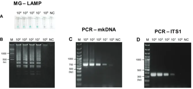
Sensitivity of MG-LAMP versus PCR-mkDNA - MG-LAMP and PCR-mkDNA were successfully performed to amplify L. martiniquensis using Leishmania 18S rRNA-LAMP primers (Nzelu et al. 2014) and L.MC-1S/ L.MC-1R primers (Kato et al. 2007), respectively. De-tection limit and cross-amplification of MG-LAMP and PCR-mkDNA assays were compared with PCR-ITS1 as-say, which was reported as the most sensitive method for L. siamensis detection (Hitakarun et al. 2014). The sensitivities of these assays were assessed with the serial dilutions of L. martiniquensis (MHOM/TH/2011/PG) DNA alone (equivalent to 133 to 0.013 parasites/µL), and the crude extracts of L. martiniquensis mixed with S. gemmea DNA (equivalent to 104 to 1 parasite per fly).
alone. Positive results were visually discriminated when the sample turned light blue (Figs 1A, 2A), whereas the negative control turned from green to colorless. Gel elec-trophoresis also showed results in agreement with the col-orimetric LAMP method using DNA intercalating mala-chite green dye (Fig. 1A versus 1B, and 2A versus 2B).
Specificity of MG-LAMP versus PCR-mkDNA - DNA of S. gemmea, other Leishmania species, and some vector-borne protozoan parasites including T. evansi, L. sabraze-si, and P. gallinaceum were determined for cross-ampli-fication of MG-LAMP, PCR-mkDNA, and PCR-ITS1 assays. All assays showed no cross-amplification with
Fig. 1: sensitivity of malachite green-loop-mediated isothermal amplification (MG-LAMP), polymerase chain reaction of minicircle kinetoplast DNA gene (PCR-mkDNA), and PCR-ITS1 to detect Leishmania martiniquensis. Different concentrations of L. martiniquensis from 104 to 1 promastigote (equivalent to 133 to 0.013 parasites/µL)were used as the templates. (A) Visual detection of MG-LAMP; (B) agarose gel elec-trophoresis of MG-LAMP products; (C) agarose gel elecelec-trophoresis of PCR-mkDNA products; (D) agarose gel elecelec-trophoresis of PCR-ITS1 products. M: gene ruler; DW: distilled water (negative control).
S. gemmea sand fly DNA. PCR-mkDNA assay was the most specific to amplify only Leishmania DNA, no cross-amplification with T. evansi, L. sabrazesi, and P. galli-naceum (Fig. 3C). Surprisingly, the L.MC-1S/ L.MC-1R primers, amplifying Leishmania mkDNA in this study, could discriminate between L. martiniquensis and L. siamensis with different PCR amplicon sizes, approxi-mately 650 bp for L. martiniquensis and approximately 750 bp for L. siamensis (Fig. 3C). On the other hand, MG-LAMP and PCR-ITS1 assays could amplify T. evansi but
no reactivity was detected other avian haemosporozoan DNAsamples(Fig. 3A-B, D). When amplified with oth-er Leishmania species, PCR-ITS1 using primer L5.8S/ LITSR could amplify L. (L.) major, L. (L.) amazonensis,
L. (V.) braziliensis, L. (L.) infantum, and L. (L.) donovani
with similar amplicon sizes, approximately 350 bp (Fig. 4B). PCR-mkDNA using primer L.MC-1S/ L.MC-1R was also able to amplify other Leishmania species. Similar re-sults of PCR-mkDNA product at approximately 650 bp was observed when the assay amplified L. martiniquensis
Fig. 3: specificity of malachite green-loop-mediated isothermal amplification (MG-LAMP), polymerase chain reaction of minicircle kinetoplast DNA gene (PCR-mkDNA), and PCR-ITS1 to detect Leishmania martiniquensis. (A) Visual detection of MG-LAMP; (B) agarose gel electropho-resis of MG-LAMP products; (C) agarose gel electrophoelectropho-resis of PCR-mkDNA products; (D) agarose gel electrophoelectropho-resis of PCR-ITS1 products. M: gene ruler; PG: L. martiniquensis (MHOM/TH/2011/PG); TR: L. siamensis (MHOM/TH/2010/TR); Te: Trypanosoma evansi;Ls: Leucocy-tozoon sabrazesi; Pg: Plasmodium gallinaceum; DW: distilled water (negative control).
(MHOM/TH/2013/LSCM3) that was isolated from the patient from Mae Tha District. However, different ampli-con sizes of PCR-mkDNA were found among Leishmania
species, approximately 650 bp for L. martiniquensis, ap-proximately 620 bp for L. (L.) major, L. (L.) amazonensis, and L. (V.) braziliensis, and longer than 700 bp for L. sia-mensis, L. (L.) infantum, and L. (L.) donovani (Fig. 4A).
Molecular mass screening of sand flies from a new autochthonous leishmaniasis affected area - The sen-sitivity and specificity results in a simulation model highlighted the potential of PCR-mkDNA to detect L. martiniquensis in sand flies. Therefore, PCR-mkDNA assay was consecutively applied to the mass screening of sand flies from the newly identified affected area of
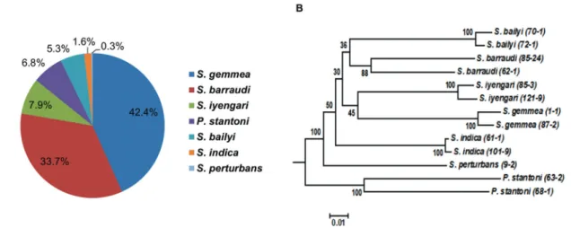
L. martiniquensis causing leishmaniasis in Tha Mae Lop Subdistrict, Mae Tha District, Lamphun Province (Chiewchanvit et al. 2015). The 380 captured female sand flies species were primarily identified microscopi-cally and overall 7 species were morphologimicroscopi-cally iden-tifiable, including P. stantoni (6.84%; 26/380), S. gem-mea (42.37%; 161/380), S. barraudi (33.68%; 128/380),
S. iyengari (7.89%; 30/380), S. bailyi (5.26%; 20/380), S. indica (1.58%; 6/380), and S. perturbans (0.26%; 1/380). Eight sand flies were morphological unidentifiable. To confirm the utility of the molecular mass screening procedure of sand flies; PCR and sequencing target-ing the mitochondrial COI gene were conducted with the 13 identified sand flies with morphological differ-ences. Phylogenetic analysis could discriminate seven groups of sand fly species, in agreement with morpho-logical identification (Fig. 5). The 13 COI sequences of seven sand fly species in the present study are availa-ble in the DNA Data Bank of Japan (DDBJ) database under the accession numbers: P. stantoni (LC136898-LC136899); S. gemmea (LC136893-LC136894); S. bar-raudi (LC136902-LC136903); S. iyengari (LC136904-LC136905); S. bailyi (LC136900-LC136901); S. indica
(LC136895-LC136896); and S. perturbans (LC136897).
After the validity of mass screening was confirmed, the crude DNA extracts of 380 sand fly individuals were then used as a template for PCR-mkDNA to identify L. martiniquensis; however, the infection was undetected.
DISCUSSION
The present study emphasised the high sensitiv-ity and specificsensitiv-ity of PCR-mkDNA to detect L. mar-tiniquensis, at least one promastigote in a sand fly, and revealed the notable ability of PCR-mkDNA assay (L.MC-1S/ L.MC-1R primers) to discriminate L. marti-niquensis from other viscerotropic Leishmania species, rendering this PCR-mkDNA assay as a promising tool for molecular mass screening of an individual sand fly for L. martiniquensis infection.
Due to various limitations in the microscopic de-tection of Leishmania in sand flies, several PCR-based techniques have been developed. The PCR-ITS1 (L5.8S/ LITSR primers) has been reported as the most accurate method to detect L. siamensis (MHOM/TH/2010/TR), as low as 0.05 parasites/µL, and used as the reference as-say to compare analytical and filed clinical sensitivity with PCR targeting the 18S rRNA, cyt b, heat shock pro-tein 70, cysteine protease B, spliced leader mini-exon, and triose-phosphate isomerase genes (Hitakarun et al. 2014). This assay has also been employed to detect L. si-amensis and L. martiniquensis DNA within the sand fly poolscaptured from outbreak areas in southern Thailand (Kanjanopas et al. 2013, Chusri et al. 2014). However, the present study showed that PCR-ITS1 had 10-fold less sensitive than PCR-mkDNA and could not discriminate
L. martiniquensis from other Leishmania species. The PCR-mkDNA may be an attractive molecular method to apply in the epidemiological study of L. martiniquensis
infection in forthcoming outbreak.
Wide applicability of LAMP in the detection of para-sitic protozoa such as Babesia, Plasmodium, Trypano-some, as well as Leishmania have been reported, due to its advantages, fast and simple amplification without the
need of an expensive thermocycler. Recently, colorimet-ric malachite green based LAMP technique based on the
18S rRNA gene was developed for the detection of L. sia-mensis, with the detection limit of at least2.5 in clinical samples, such as whole blood and saliva (Sriworarat et al. 2015). The colorimetric LAMP protocol in the pres-ent study, however, showed a higher sensitivity to detect
L. martiniquensis in a sand fly. Approximately 20-400 copies of the ITS1 and 18S rRNA gene in individual para-sites have been described, although differing somewhat among Leishmania species (Inga et al. 1998).
The PCR-mkDNA using primers L.MC-1S/ L.MC-1R can detect at least one L. (L.) major existed in a sand fly sample (Kato et al. 2007). Along the similar line, this as-say also showed the highest analytical sensitivity to detect
L. martiniquensis even when only one promastigote ex-isted in a sand fly sample, possibly due to the higher copy number of approximately 10,000 copies of the mkDNA
gene in individual parasites (Simpson 1986). Regarding the specificity, this PCR-mkDNA protocol was the most specific to detect only Leishmania parasites, comparing to MG-LAMP and ITS1. The attractive feature of PCR-mkDNA is the ability to differentiate L. martiniquensis
(MHOM/TH/2011/PG) clearly from other viscerotropic
Leishmania species, including L. siamensis, L. (L.) don-ovani, and L. (L.) infantum, rendering the applicability of PCR-mkDNA for epidemiological study of VL caused by L. martiniquensis infection in the areas where several
Leishmania species co-exist. Along similar lines, Kato et al. (2007) demonstrated that PCR-mkDNA worked on the other seven Leishmania species; L. (L.) amazonensis,
L. (L.) mexicana, L. (L.) major-like, L. (V.) panamensis,
L. (V.) braziliensis, L. (L.) guyanensis and L. (L.) major, although they have variations in their sequences. The dif-ferences in the size of amplified fragments among spe-cies may reflect the size of the dominant mkDNA in the strain because such DNA varies between 0.75 and 1 kbp in length (Brewster & Barker 2002). In addition, PCR of
kDNA gene represent the most reliable tool to detect L. infantum naturally infection in Lutzomyia longipalpis in endemic areas of Brazil, comparing with mini-exon and
18S rRNA genes (Freitas-Lidani et al. 2014). Lastly, the present study found that PCR-mkDNA showed no cross-amplification with L. sabrazesi, and P. gallinaceum, in which the potential vector of these avian hemosporidians are ceratopogonid midges. Seblova et al. (2015) suggested that Culicoides soronensis could be potential vectors of L. enriettii, relating to L. martiniquensis and L. siamensis. Validation of this PCR-mkDNA for detection of Leishma-nia in biting Culicoides midges should be further evalu-ated for research on ceratopogonid midges as the possible vector of L. martiniquensis and L. siamensis infection.
In Thailand, a few survey studies of the distribu-tion of sand fly species and their habitats have been conducted. Sergentomyia fly was the most predomi-nant genus found in the country. Until now, at least 26 species of sand fly have been reported in different provinces of Thailand, but only S. barraudi had been reported in Lumphun province (Polseela et al. 2016). The present study could provide more information of
sand fly populations in Lumphun province, especially in the area where the affected patient resides. At least seven species of sand flies were identified, including
P. stantoni, S. gemmea, S. barraudi, S. iyengari, S. bai-lyi, S. perturbans, and S. indica, of which S. gemmea
and S. barraudi were the predominant species. Vari-ous studies demonstrated Leishmania DNA in Sergen-tomyia sand flies, e.g. L. (L.) donovani in S. babu in India (Mukherjee et al. 1997), L. (L.) major DNA in
S. minuta in Portugal (Campino et al. 2013), as well as
L. siamensis in S. gemmea (Kanjanopas et al. 2013), and L. martiniquensis in S. gemmea and S. barraudi in Thailand (Chusri et al. 2014). Moreover, L. (L.) major
was also isolated from S. garnhami and successfully cultured in NNN medium (Mutinga et al. 1994). In this study, Leishmania DNA was not detected, prob-ably due to the very low infection rate (0.01-1%) among sand fly populations even in endemic areas (Kato et al. 2016). Further surveillance of larger populations us-ing the present mass screenus-ing approach will provide more information about sand flies in each endemic area. The abundance of S. gemmea and S. barraudi in the local environment of the affected patient may raise awareness of public health concerns for prevention and control of leishmaniasis among policy- and decision-makers, physicians and the general public. Further sur-veillance of larger populations using mass screening will provide more information.
In conclusion, the present study highlighted the po-tential of PCR-mkDNA method as a promising tool to detect L. martiniquensis in sand flies due to its high sensitivity and specificity. Above all, PCR-mkDNA has the valuable ability to discriminate between L. marti-niquensis and other viscerotropic Leishmania species; L. siamensis, L. (L.) donovani, and L. (L.) infantum, which may encourage researchers to adopt this approach for epidemiological studies of VL in such areas where many
Leishmania species are circulating. Identifying the po-tential vector for L. martiniquensis still remains an ur-gent needed. The molecular mass screening of individu-al sand fly for Leishmania infection by PCR-mkDNA is applicable to provide informative data on the vector and vector-Leishmania relationship in outbreak areas where several Leishmania and sand fly species co-exist and the species of potential vectors remain unknown.
ACKNOWLEDGEMENTS
AUTHORS’ CONTRIBUTION
ST conceived and designed, maintained the parasites, iden-tified sand flies species morphologically, performed molecular techniques, analysed the data, wrote the manuscript; HK con-ceived and designed, performed molecular techniques, wrote the manuscript; RY performed molecular techniques, analysed the data; AM performed molecular techniques, analysed the data; RP identified sand flies species morphologically; SL conceived and designed, maintained the parasites, wrote the manuscript.
REFERENCES
Brewster S, Barker DC. Analysis of minicircle classes in Leishmania (Vi-annia) species. Trans R Soc Trop Med Hyg. 2002; 96(Suppl. 1): 55-63.
BVBD/MoPH - Bureau of Vector Borne Disease/Ministry of Public HealthThailand. The current situation of vector borne diseases in 2013. In: Annual report 2013-Bureau of Vector Borne Disease. Bangkok: 2013. p. 20-22 (in Thai).
Campino L, Cortes S, Dionísio L, Neto L, Afonso MO, Maia C. The first detection of Leishmania major in naturally infected Sergentomyia minuta in Portugal. Mem Inst Oswaldo Cruz. 2013; 108(4): 516-8.
Chiewchanvit S, Tovanabutra N, Jariyapan N, Bates MD, Mahanu-pab P, Chuamanochan M, et al. Chronic generalized fibrotic skin lesions from disseminated leishmaniasis caused by Leishmania martiniquensis in two patients from northern Thailand infected with HIV. Br J Dermatol. 2015; 173(3): 663-70.
Chusri S, Thammapalo S, Silpapojakul K, Siriyasatien P. Animal reser-voirs and potential vectors of Leishmania siamensis in southern Thai-land. Southeast Asian J Trop Med Public Health. 2014; 45(1): 13-9.
Dedet JP, Roche B, Pratlong F, Cales-Quist D, Jouannelle J, Beni-chou JC, et al. Diffuse cutaneous infection caused by a presumed monoxenous trypanosomatid in a patient infected with HIV. Trans R Soc Trop Med Hyg. 1995; 89(6): 644-6.
Desbois N, Pratlong F, Quist D, Dedet JP. Leishmania (Leishmania) martiniquensis n. sp. (Kinetoplastida: Trypanosomatidae), de-scription of the parasite responsible for cutaneous leishmaniasis in Martinique Island (French West Indies). Parasite. 2014; 21: 12.
El Tai NO, El Fari M, Mauricio I, Miles MA, Oskam L, El Safi SH, et al. Leishmania donovani: intraspecific polymorphisms of Suda-nese isolates revealed by PCR-based analyses and DNA sequenc-ing. Exp Parasitol. 2001; 97(1): 35-44.
Freitas-Lidani KC, de Messias-Reason IJ, Ishikawa EAY. A compari-son of molecular markers to detect Lutzomyia longipalpis natu-rally infected with Leishmania(Leishmania)infantum. Mem Inst Oswaldo Cruz. 2014; 109(4): 442-7.
Hitakarun A, Tan-Ariya P, Siripattanapipong S, Mungthin M, Piyaraj P, Naaglor T, et al. Comparison of PCR methods for detection of Leishmania siamensis infection. Parasit Vectors. 2014; 7: 458.
Inga R, de Doncker S, Gomez J, Lopez M, Garcia R, Le Ray D, et al. Relation between variation in copy number of ribosomal RNA en-coding genes and size of harbouring chromosomes in Leishmania of subgenus Viannia. Mol Biochem Parasitol. 1998; 92(2): 219-28.
Kanjanopas K, Siripattanapipong S, Ninsaeng U, Hitakarun A, Jit-kaew S, Kaewtaphaya P, et al. Sergentomyia (Neophlebotomus) gemmea, a potential vector of Leishmania siamensis in southern Thailand. BMC Infect Dis. 2013; 13: 333.
Kato H, Cáceres AG, Hashiguchi Y. First evidence of a hybrid of Leishmania (Viannia) braziliensis/L. (V.) peruviana DNA de-tected from the phlebotomine sand fly Lutzomyia tejadai in Peru. PLoS Negl Trop Dis. 2016; 10(1): e0004336.
Kato H, Gomez EA, Caceres AG, Uezato H, Mimori T, Hashiguchi Y. Molecular epidemiology for vector research on leishmaniasis. Int J Environ Res Public Health. 2010; 7(3): 814-26.
Kato H, Uezato H, Gomez EA, Terayama Y, Calvopina M, Iwata H, et al. Establishment of a mass screening method of sand fly vectors for Leishmania infection by molecular biological methods. Am J Trop Med Hyg. 2007; 77(2): 324-9.
Leelayoova S, Siripattanapipong S, Hitakarun A, Kato H, Tan-ariya P, Siriyasatien P, et al. Multilocus characterization and phylo-genetic analysis of Leishmania siamensis isolated from autoch-thonous visceral leishmaniasis cases, southern Thailand. BMC Microbiol. 2013; 13: 60.
Lewis DJ. The phlebotomine sandflies (Diptera: Psychodidae) of the Oriental Region. Bull Br Mus Nat Hist (Ent). 1978; 37: 217-343.
Mukherjee S, Hassan MQ, Ghosh A, Ghosh KN, Bhattacharya A, Ad-hya S. Short report: Leishmania DNA in Phlebotomus and Ser-gentomyia species during a kala-azar epidemic. Am J Trop Med Hyg. 1997; 57(4): 423-5.
Mutinga MJ, Massamba NN, Basimike M, Kamau CC, Amimo FA, Onyido AE, et al. Cutaneous leishmaniasis in Kenya: Sergento-myia garnhami (Diptera Psychodidae), a possible vector of Leish-mania major in Kitui District: a new focus of the disease. East Afr Med J. 1994; 71(7): 424-8.
Noyes H, Pratlong F, Chance M, Ellis J, Lanotte G, Dedet JP. A previ-ously unclassified trypanosomatid responsible for human cutaneous lesions in Martinique (French West Indies) is the most divergent member of the genus Leishmania ss. Parasitology. 2002; 124: 17-24.
Nzelu CO, Caceres AG, Arrunategui-Jimenez MJ, Lanas-Rosas MF, Yanez-Trujillano HH, Luna-Caipo DV, et al. DNA barcoding for identification of sand fly species (Diptera: Psychodidae) from leishmaniasis-endemic areas of Peru. Acta Trop. 2015; 145: 45-51.
Nzelu CO, Gomez EA, Caceres AG, Sakurai T, Martini-Robles L, Uezato H, et al. Development of a loop-mediated isothermal amplification method for rapid mass-screening of sand flies for Leishmania infection. Acta Trop. 2014; 132: 1-6.
Phumee A, Kraivichian K, Chusri S, Noppakun N, Vibhagool A, Sanprasert V, et al. Detection of Leishmania siamensis DNA in saliva by polymerase chain reaction. Am J Trop Med Hyg. 2013; 89(5): 899-905.
Polseela R, Jaturas N, Thanwisai A, Sing KW, Wilson JJ. Towards monitoring the sandflies (Diptera: Psychodidae) of Thailand: DNA barcoding the sandflies of Wihan Cave, Uttaradit. Mito-chondrial DNA A DNA Mapp Seq Anal. 2016; 27(5):3795-801.
Pothirat T, Tantiworawit A, Chaiwarith R, Jariyapan N, Wannasan A, Siriyasatien P, et al. First isolation of Leishmania from Northern Thailand: case report, identification as Leishmania martiniquen-sis and phylogenetic position within the Leishmania enriettii complex. PLoS Negl Trop Dis. 2014; 8: e3339.
Seblova V, Sadlova J, Vojtkova B, Votypka J, Carpenter S, Bates PA, et al. The biting midge Culicoides sonorensis (Diptera: Cerato-pogonidae) is capable of developing late stage infections of Leish-mania enriettii. PLoS Negl Trop Dis. 2015; 9: e0004060.
Simpson L. Kinetoplast DNA in trypanosomid flagellates. Int Rev Cytol. 1986; 99: 119-79.
Sriworarat C, Phumee A, Mungthin M, Leelayoova S, Siriyasatien P. Development of loop-mediated isothermal amplification (LAMP) for simple detection of Leishmania infection. Parasit Vectors. 2015; 8: 591.
Tamura K, Stecher G, Peterson D, Filipski A, Kumar S. MEGA6: Molecular Evolutionary Genetics Analysis version 6.0. Mol Biol Evol. 2013; 30(12): 2725-9.