Simple Genome Editing of Rodent Intact
Embryos by Electroporation
Takehito Kaneko1*, Tomoji Mashimo2
1Institute of Laboratory Animals, Graduate School of Medicine, Kyoto University, Kyoto, 606–8501, Japan,
2Institute of Experimental Animal Sciences, Faculty of Medicine, Osaka University, Osaka, 565–0871,
Japan
Abstract
The clustered regularly interspaced short palindromic repeat (CRISPR)/CRISPR-associ-ated (Cas) system is a powerful tool for genome editing in animals. Recently, new technol-ogy has been developed to genetically modify animals without using highly skilled
techniques, such as pronuclear microinjection of endonucleases. Technique for animal knockout system by electroporation (TAKE) method is a simple and effective technology that produces knockout rats by introducing endonuclease mRNAs into intact embryos using electroporation. Using TAKE method and CRISPR/Cas system, the present study success-fully produced knockout and knock-in mice and rats. The mice and rats derived from embryos electroporated with Cas9 mRNA, gRNA and single-stranded oligodeoxynucleotide (ssODN) comprised the edited targeted gene as a knockout (67% of mice and 88% of rats) or knock-in (both 33%). The TAKE method could be widely used as a powerful tool to pro-duce genetically modified animals by genome editing.
Introduction
Many types of genetically modified (GM) animals have been produced to study human diseases [1–3]. Mice and rats have been used widely as important human diseases model animals [4–6]. Engineered endonucleases, including zinc-finger nucleases (ZFN), transcription activator-like effector nucleases (TALEN) and the clustered regularly interspaced short palindromic repeat (CRISPR)/CRISPR-associated (Cas) system, are recently developed high-impact technologies for the production of GM animals [7–12]. Engineered endonucleases have made the rapid pro-duction of GM animals possible without using embryonic stem (ES) cells and induced pluripo-tent stem cells. However, microinjection of endonucleases into pronuclear-stage embryos is still used routinely, which requires a high skill level to reduce cell damage, and injecting endo-nucleases into embryos one by one using a micromanipulator is time consuming. Thus, this process has prevented the rapid production of GM animals. Recently, we developed a new tech-nology to produce GM animals without using microinjection of endonucleases into embryos. Technique for animal knockout system by electroporation (TAKE) method [13] is a simple and effective technology that can produce knockout rats by the introduction of endonuclease
a11111
OPEN ACCESS
Citation:Kaneko T, Mashimo T (2015) Simple Genome Editing of Rodent Intact Embryos by Electroporation. PLoS ONE 10(11): e0142755. doi:10.1371/journal.pone.0142755
Editor:Vincenzo Coppola, Ohio State University Comprehensive Cancer Center, UNITED STATES
Received:September 10, 2015
Accepted:October 26, 2015
Published:November 10, 2015
Copyright:© 2015 Kaneko, Mashimo. This is an open access article distributed under the terms of the
Creative Commons Attribution License, which permits unrestricted use, distribution, and reproduction in any medium, provided the original author and source are credited.
Data Availability Statement:All relevant data are within the paper and its Supporting Information files.
Funding:This work was supported by a Grant-in-Aid for Scientific Research from Japan Society for the Promotion of Science (No. 26290033).
mRNAs into intact embryos using a three-step electrical pulse electroporation. Electroporation of ZFNs resulted in an embryonic survival rate (91%) and a genome-editing rate (73%) that was more than 2-fold higher than the corresponding rates from conventional microinjection [13]. Previous study reported that the knockout rats were also obtained by electroporation of the CRISPR/Cas system. However, suitable conditions must be demonstrated for the produc-tion of knockout and knock-in in rats and other species. This study determined the condiproduc-tions suitable for the efficient production of knockout and knock-in mice and rats using the TAKE method and the CRISPR/Cas system.
Materials and Methods
Animals
C57BL/6J Jcl mice and Jcl:Wistar rats were purchased from CLEA Japan Inc. (Tokyo, Japan). Male mice aged older than 11 weeks and female mice aged from 8 to 16 weeks were used as sperm and oocyte donors, respectively. Female Jcl ICR mice and Jcl:Wistar rats from 8 to 16 weeks old (CLEA Japan Inc.) were used as recipients for embryo transfer. All animals were maintained in an air-conditioned (temperature 24 ± 2°C, humidity 50 ± 10%) and light-controlled room (illuminated from 07:00 to 19:00). All the animal care and procedures per-formed in this study conper-formed to the Guidelines for Animal Experiments of Kyoto University, and were approved by the Animal Research Committee of Kyoto University.
Preparation of Cas9 mRNA, gRNA and ssODN
The plasmid expressing hCas9 (ID#41815) was obtained from the Addgene repository (www. addgene.org/CRISPR), and was modified by addition of the T7 promoter and DNA encoding an SV40 nuclear localisation signals at the N-terminal of hCas9, using an In-Fusion HD clon-ing kit (Takara Bio Inc., Shiga, Japan), accordclon-ing to the manufacturer’s protocol. Cas9 mRNA was transcribedin vitrousing an mMESSAGE mMACHINE T7 Ultra Kit (Thermo Fisher Sci-entific Inc., Waltham, MA, USA) and was polyadenylated using an A-Plus™Poly(A) polymer-ase tailing kit (Epicentre, Madison, WI, USA). RNA was then purified using a MEGAClear™kit (Thermo Fisher Scientific Inc.).
gRNA was designed to target exon 2 of the mouse and rat interleukin 2-receptor gamma (Il2rg) gene (S1 Fig) [14,15]. gRNAs expression plasmids were transcribedin vitrousing a MEGAshortscript T7 Transcription Kit (Thermo Fisher Scientific Inc.). RNA was then purified using a MEGAClear™kit (Thermo Fisher Scientific Inc.). The sequences of the single-stranded oligodeoxynucleotide (ssODN), which were obtained from IDT (Integrated DNA Technologies Inc., Coralville, IA, USA), are shown inS1 Table.
Cas9 mRNA/gRNA/ssODN for electroporation were resuspended in phosphate buffered saline (PBS with calcium and magnesium free) at 400/600/300, 200/200/200 and 100/100/ 100μg/mL, respectively.
Collection of embryos
Mouse and rat pronuclear-stage embryos were collected using the method described previously by Kaneko [16]. Mouse pronuclear-stage embryos were obtained using anin vitrofertilization technique. Cauda epididymis sperm were collected from mature males that euthanized by CO2
and cervical dislocation. Sperm were then pre-incubated for 1 h at 37°C under 5% CO 2 and 95% air in 200μl drops of fertilization medium covered with sterile mineral oil to induce
followed by an intraperitoneal injection of 7.5 IU human chorionic gonadotropin (hCG) (ASKA Pharmaceutical Co. Ltd.) 48 h later. Cumulus-oocytes complexes were collected from oviducts of females that euthanized by CO2and cervical dislocation at 13–15 h after hCG
injec-tion. These were then transferred in another 200μl drops of fertilization medium covered with
sterile mineral oil. Capacitated sperm that pre-incubated for 1 h were added into HTF medium drops with oocytes. The oocytes were then co-cultured with sperm at 37°C under 5% CO 2 and 95% air. Pronuclear-stage embryos were collected at 6 h after insemination.
Rat pronuclear-stage embryos were produced by natural mating. Female rats were induced to superovulate by an intraperitoneal injection of 300 IU/Kg PMSG (ASKA Pharmaceutical Co. Ltd.), followed by an intraperitoneal injection of 300 IU/Kg hCG (ASKA Pharmaceutical Co. Ltd.) 48 h later. Female rats were mated with male rats overnight after hCG injection. The oviducts of female rats with vaginal plugs were removed after euthanasia by CO2and cervical
dislocation and oocytes were flushed out from the ampullae with culture medium. Pronuclear-stage embryos were collected and stored in culture medium before introduction of mRNA.
Introduction of Cas9 mRNA, gRNA and ssODN into intact embryos by
the TAKE method
The TAKE method [13] was used to introduce Cas9 mRNA, gRNA and ssODN into intact mouse and rat embryos. This method introduced endonucleases into intact embryos using a super electroporator NEPA 21 (NEPA GENE Co. Ltd., Chiba, Japan). Briefly, pronuclear-stage embryos were placed in a line on the glass chamber between 5mm gap platinum metal plates (CUY520P5, NEPA GENE Co. Ltd.) that were filled with 100μL PBS containing Cas9 mRNA,
gRNA and ssODN at various concentrations. The poring pulse was set to voltage: 225 V, pulse width: 2.5 ms, pulse interval: 50 ms, and number of pulses: +4. The first and second transfer pulse were set to voltage: 20 V, pulse width: 50 ms, pulse interval: 50 ms, and number of pulses: ±5. After electroporation, all embryos were cultured before embryo transfer.
Embryo transfer
Embryos that developed to the two-cell stage after the introduction of RNA and ssODN were transferred into the oviducts of female surrogate that anesthetized using isoflurane [17]. Females were mated with vasectomised male rats the day before the embryo transfer. The num-ber of offspring that were born naturally was counted at 19–21 days of gestation.
Analysis of gene editing in offspring
Editing of the targeted gene was analyzed using genomic DNA extracted from blood that was adhered to FTA cards [18]. PCR templates were then prepared by punching out discs from the FTA card in conjunction with Ampdirect Plus buffer (Shimadzu Co., Kyoto, Japan). PCR was performed in a total volume of 15μL under the following conditions: 1 cycle of 94°C for 3 min;
35 cycles of 94°C for 30 s, 60°C for 1 min and 72°C for 45 s; and 1 cycle of 72°C for 3 min. The final reaction mixture contained 200μM dNTPs, 1.0 mM MgCl2, 0.66μM of each primer
(amplifying the mouseIl2rglocus: 234 bp, ratIl2rglocus: 292 bp,S1 Table) and 0.4 U Taq DNA polymerase (Thermo Fisher Scientific Inc.). The PCR products were directly sequenced using the BigDye terminator v3.1 cycle sequencing mix and the standard protocol for an Applied Biosystems 3130 DNA Sequencer (Thermo Fisher Scientific Inc.).
Analysis of germ-line transmission
rats after they had matured. The edited gene in the offspring of the next generation was then analysed.
Data analysis
All data were analysed using Chi-squared tests using Yates’correction for continuity.
Results
Introduction of RNA and ssODN into intact mouse pronuclear-stage
embryos
Cas9 mRNA, gRNA and ssODN was electroporated into intact mouse pronuclear-stage embryos. The gRNA that targeted mouseIl2rggene was used. Intact mouse pronuclear-stage embryos were electroporated with final concentrations of Cas9 mRNA/gRNA/ssODN of 400/ 600/300, 200/200/200 and 100/100/100μg/mL, respectively. After electroporation, the embryos
that developed to the two-cell stage were transferred into the oviducts of pseudopregnant female mice.
Of the embryos produced by electroporation with 400/600/300, 200/200/200 and 100/100/ 100μg/mL of Cas9 mRNA/gRNA/ssODN, 84, 73 and 98% of embryos survived, respectively.
Thereafter, 43, 48 and 24% of the embryos, respectively, developed into offspring, and 67, 31 and 32%, respectively, of these offspring had an editedIl2rglocus (knockout). Furthermore, 33, 9 and 18% of the offspring had a one base exchange (knock-in) at theIl2rglocus (Table 1).
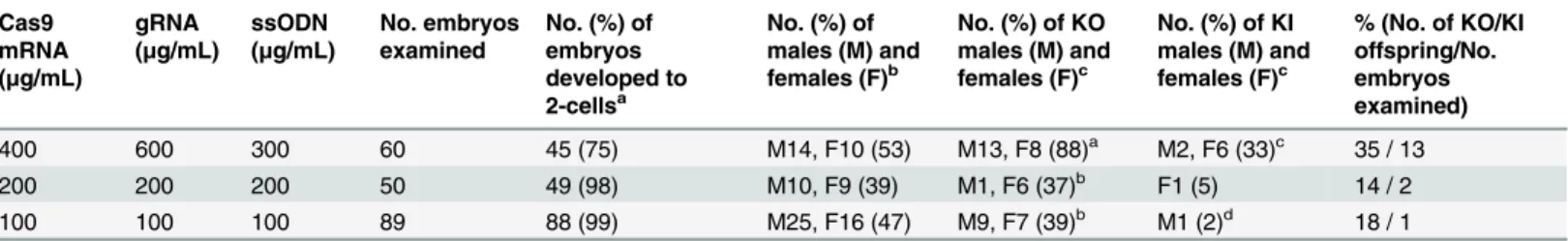
Introduction of RNA and ssODN into intact rat pronuclear-stage embryos
Intact rat pronuclear-stage embryos were electroporated Cas9 mRNA, gRNA and ssODN using the same RNA/ssODN concentration and electrical pulse conditions as the mouse embryos. The gRNA that targeted ratIl2rggene was used. Of the embryos produced by electro-poration with 400/600/300, 200/200/200 and 100/100/100μg/mL of Cas9 mRNA/gRNA/
ssODN, 75, 98 and 99% of embryos survived, respectively. Thereafter, 53, 39 and 47%, respec-tively, of the embryos developed into offspring, and 88, 37 and 39%, respecrespec-tively, of these off-spring showed knockout editing of theIl2rglocus (Table 2). Furthermore, 33, 5 and 2%, respectively, of the offspring showed knock-in with one base exchange at theIl2rglocus. Germ-line transmission of the induced mutations was confirmed in the next generation (S2 Table).
Table 1. Development of mouse embryos with introduced RNAs and ssODN using the TAKE method.
Cas9 mRNA (μg/mL)
gRNA (μg/mL)
ssODN (μg/mL)
No. embryos examined
No. (%) of embryos developed to 2-cellsa
No. (%) of males (M) and females (F)b
No. (%) of KO males (M) and females (F)c
No. (%) of KI males (M) and females (F)c
% (No. of KO/KI offspring/No. embryos examined)
400 600 300 100 84 (84) M22, F14 (43)a M11, F13 (67)c M4, F8 (33)e 24 / 12
200 200 200 100 73 (73) M21, F14 (48)a M6, F5 (31)d M2, F1 (9)f 11 / 3
100 100 100 120 117 (98) M16, F12 (24)b M5, F4 (32)d M2, F3 (18) 8 / 4
aCalculated from the number of embryos examined.
bCalculated from the number of embryos developed to two cells. cCalculated from the number of male and female mice. Significant differences atP<0.05; avs. b, cvs. d, evs. f.
Discussion
Here, we demonstrated the efficient use of the TAKE method to produce knockout and knock-in mice and rats usknock-ing the CRISPR/Cas system. The mice and rats derived from embryos knock- intro-duced with 400μg/mL of Cas9 mRNA, 600μg/mL of gRNA and 300μg/mL of ssODN showed
editing of the targeted gene as a knockout (67% of mice and 88% or rats) or showed knock-in with one base exchange at theIl2rglocus (both 33%), respectively (Tables1and2). The advan-tage of this method was that the mouse and rat genomes could be easily and quickly edited using same protocol. GM animals are usually generated by microinjection of endonucleases into embryos. Although this has been used routinely as a gold standard technique, the high level of skill and time required has prevented the rapid production of GM animals. Electropo-ration is an alternative method for introducing exogenous DNA/RNA into embryos. However, the conventional protocol required weakening of the zona pellucida of the embryos by treat-ment with Tyrode’s acid solution before electroporation for the efficient introduction of DNA [19,20]. Weakening the zona pellucida may have negative effects because the zona pellucida is important for subsequent embryonic developmentin vivo[21,22]. The TAKE method permits the introduction of endonucleases into intact embryos without weakening the zona pellucida. Thus, TAKE could efficiently introduce engineered endonucleases, such as ZFN, TALEN and CRISPR/Cas9 mRNA, into intact embryos using a NEPA 21 electroporator with a three-step electrical pulse system [13]. First electrical step (the poring pulse) makes microscopic holes in the zona pellucida and oolemma. In the second step (the transfer pulse), the initial pulses trans-fer mRNA into the cytoplasm. In the third step, the polarity-changed second-transtrans-fer pulse increases the chance of transferring mRNA into embryos. The TAKE method has dramatically increased the speed of production of GM animals. In previous methods, a high proportion of knockout rats with genome editing were produced by the introduction of ZFN mRNA. How-ever, suitable conditions to generate knockout rats using CRISPR/Cas9 have not been demon-strated. The results of the present study showed that the mice and rats derived from embryos had knockout and knock-in editing of theIl2rglocus. Although the success rate of GM animal production depends on the concentration of RNA, animals with edited genomes could be obtained with as little as 100μg/mL of Cas9 mRNA, 100μg/mL of gRNA and 100μg/mL of
ssODN using the TAKE method (Tables1and2).
In conclusion, we demonstrated that the TAKE method could be adapted to produce knock-out and knock-in mice and rats. This technology has the potential to produce GM animals quickly, easily and successfully without special skills such as conventional pronuclear microin-jection method. Furthermore, high success rate lead to reduction the number of females used
Table 2. Development of rat embryos with introduced RNA and ssODN using the TAKE method.
Cas9 mRNA (μg/mL)
gRNA (μg/mL)
ssODN (μg/mL)
No. embryos examined
No. (%) of embryos developed to 2-cellsa
No. (%) of males (M) and females (F)b
No. (%) of KO males (M) and females (F)c
No. (%) of KI males (M) and females (F)c
% (No. of KO/KI offspring/No. embryos examined)
400 600 300 60 45 (75) M14, F10 (53) M13, F8 (88)a M2, F6 (33)c 35 / 13
200 200 200 50 49 (98) M10, F9 (39) M1, F6 (37)b F1 (5) 14 / 2
100 100 100 89 88 (99) M25, F16 (47) M9, F7 (39)b M1 (2)d 18 / 1
a
Calculated from the number of embryos examined.
bCalculated from the number of embryos developed to two cells. c
Calculated from the number of male and female rats. Significant differences atP<0.05; avs. b, cvs. d.
as oocyte donors, which is applicable the principle of three Rs- replacement, reduction and refinement of animal use [23]. TAKE is also applicable to editing targeted genes of embryos collected from various animals. We believe that the rapid and efficient production of GM ani-mals using TAKE method with CRISPR/Cas9 system will contribute further to our under-standing of gene functions and human diseases.
Supporting Information
S1 Fig. Sequence of the mouse (A) and rat (B)Il2rglocus.Red capital letters indicate the position of the one base exchange.
(DOCX)
S1 Table. Sequences of single-stranded donor oligonucleotides (ssODNs) and primers for the Il2rg locus.
(DOCX)
S2 Table. Germ-line transmission in rat offspring with an editedIl2rglocus derived from the technique for animal knockout system by the electroporation (TAKE) method.
(DOCX)
Acknowledgments
We thank Y. Kunihiro, M Hayashi, K. Hattori and K. Yoshimi for technical support during this study.
Author Contributions
Conceived and designed the experiments: TK. Performed the experiments: TK. Analyzed the data: TK. Contributed reagents/materials/analysis tools: TK TM. Wrote the paper: TK TM.
References
1. Jaenisch R. Transgenic animals. Science. 1988; 240(4858):1468–74. PMID:3287623
2. Mullins JJ, Peters J, Ganten D. Fulminant hypertension in transgenic rats harbouring the mouse Ren-2 gene. Nature. 1990; 344(6266):541–4. PMID:2181319
3. Charreau B, Tesson L, Soulillou JP, Pourcel C, Anegon I. Transgenesis in rats: technical aspects and models. Transgenic Res. 1996; 5(4):223–34. PMID:8755162
4. Bedell MA, Largaespada DA, Jenkins NA, Copeland NG. Mouse models of human disease. Part II: recent progress and future directions. Genes Dev. 1997; 11(1):11–43. PMID:9000048
5. Jacob H. Functional genomics and rat models. Genome Res. 1999; 9(11):1013–16. PMID:10568741
6. Aitman TJ, Critser JK, Cuppen E, Dominiczak A, Fernandez-Suarez XM, Flint J, et al. Progress and prospects in rat genetics: a community view. Nat. Genet. 2008; 40(5):516–22. doi:10.1038/ng.147
PMID:18443588
7. Geurts AM, Cost GJ, Freyvert Y, Zeitler B, Miller JC, Choi VM, et al. Knockout rats produced via embryo pronuclear microinjection of designed zinc finger nucleases. Science. 2009; 325(5939):433.
8. Carbery ID, Ji D, Harrington A, Brown V, Weinstein EJ, Liaw L, et al. Targeted genome modification in mice using zinc-finger nucleases. Genetics. 2010; 186(2):451–9. doi:10.1534/genetics.110.117002
PMID:20628038
9. Tesson L, Usal C, Ménoret S, Leung E, Niles BJ, Remy S, et al. Knockout rats generated by embryo microinjection of TALENs. Nat. Biotechnol. 2011; 29(8):695–6. doi:10.1038/nbt.1940PMID:
21822240
10. Sung YH, Baek IJ, Kim DH, Jeon J, Lee J, Lee K, et al. Knockout mice created by TALEN-mediated gene targeting. Nat. Biotechnol. 2013; 31(1):23–4. doi:10.1038/nbt.2477PMID:23302927
12. Li W, Teng F, Li T, Zhou Q. Simultaneous generation and germline transmission of multiple gene muta-tions in rat using CRISPR-Cas systems. Nat. Biotechnol. 2013; 31(8):684–6. doi:10.1038/nbt.2652
PMID:23929337
13. Kaneko T, Sakuma T, Yamamoto T, Mashimo T. Simple knockout by electroporation of engineered endonucleases into intact rat embryos. Sci. Rep. 2014; 4:6382. doi:10.1038/srep06382PMID: 25269785
14. Mashimo T, Takizawa A, Kobayashi J, Kunihiro Y, Yoshimi K, Ishida S, et al. Generation and character-ization of severe combined immunodeficiency rats. Cell Rep. 2012; 2(3):685–94. doi:10.1016/j.celrep.
2012.08.009PMID:22981234
15. Sakuma T, Ochiai H, Kaneko T, Mashimo T, Tokumasu D, Sakane Y, et al. Repeating pattern of non-RVD variations in DNA-binding modules enhances TALEN activity. Sci. Rep. 2013; 3:3379. doi:10. 1038/srep03379PMID:24287550
16. Kaneko T, Mashimo T. Creating knockout and knockin rodents using engineered endonucleases via direct embryo injection. In: Shondra M, editor. Methods in Molecular Biology 1239. Springer; 2015. pp. 307–315.
17. Nagy A, Gertsenstein M, Vintersten K, Behringer R. Manipulating the mouse embryo. A laboratory man-ual. 3rd ed. Cold Spring Harbor Laboratory Press; 2003.
18. Nakanishi S, Kuramoto T, Serikawa T. Simple Genotyping Method Using AmpdirectR Plus and FTAR Technologies: Application to the Identification of Transgenic Animals and Their Routine Genetic Moni-toring. Lab. Anim. Res. 2009; 25(1):75–8.
19. Grabarek JB, Plusa B, Glover DM, Zernicka-Goetz M. Efficient delivery of dsRNA into zona-enclosed mouse oocytes and preimplantation embryos by electroporation. Genesis. 2002; 32(4):269–76. PMID:
11948914
20. Peng H, Wu Y, Zhang Y. Efficient delivery of DNA and morpholinos into mouse preimplantation embryos by electroporation. PLoS One. 2012; 7(8):e43748. doi:10.1371/journal.pone.0043748PMID: 22928027
21. Bronson RA, McLaren A. Transfer to the mouse oviduct of eggs with and without the zona pellucida. J. Reprod. Fertil. 1970; 22(1):129–37. PMID:5421101
22. Modliński JA. The role of the zona pellucida in the development of mouse eggs in vivo. J. Embryol. Exp. Morphol. 1970; 23(3):539–47. PMID:5473304