Hypoxia-Inducible Factor 2 Alpha Is Essential for Hepatic
Outgrowth and Functions via the Regulation of
leg1
Transcription in the Zebrafish Embryo
Tzung-Yi Lin1, Chi-Fu Chou1, Hsin-Yu Chung1, Chia-Yin Chiang1, Chung-Hao Li1, Jen-Leih Wu2, Han-Jia Lin1, Tun-Wen Pai3, Chin-Hwa Hu1, Wen-Shyong Tzou4*
1Institute of Bioscience and Biotechnology, National Taiwan Ocean University, Keelung, Taiwan,2Institute of Cellular and Organismic Biology, Academia Sinica, Taipei, Taiwan,3Department of Computer Science and Engineering, National Taiwan Ocean University, Keelung, Taiwan,4Department of Life Sciences, National Taiwan Ocean University, Keelung, Taiwan
Abstract
The liver plays a vital role in metabolism, detoxification, digestion, and the maintenance of homeostasis. During development, the vertebrate embryonic liver undergoes a series of morphogenic processes known as hepatogenesis. Hepatogenesis can be separated into three interrelated processes: endoderm specification, hepatoblast differentiation, and hepatic outgrowth. Throughout this process, signaling molecules and transcription factors initiate and regulate the coordination of cell proliferation, apoptosis, differentiation, intercellular adhesion, and cell migration. Hifs are already recognized to be essential in embryonic development, but their role in hepatogenesis remains unknown. Using the zebrafish embryo as a model organism, we report that the lack of Hif2-alpha but not Hif1-alpha blocks hepatic outgrowth. While Hif2-alpha is not involved in hepatoblast specification, this transcription factor regulates hepatocyte cell proliferation during hepatic outgrowth. Furthermore, we demonstrated that the lack of Hif2-alpha can reduce the expression of liver-enriched gene 1 (leg1), which encodes a secretory protein essential for hepatic outgrowth. Additionally, exogenous mRNA expression ofleg1can rescue the small liver phenotype ofhif2-alphamorphants. We also showed that Hif2-alpha directly binds to the promoter region ofleg1to controlleg1expression. Interestingly, we discovered overrepresented, high-density Hif-binding sites in the potential upstream regulatory sequences ofleg1in teleosts but not in terrestrial mammals. We concluded thathif2-alphais a key factor required for hepatic outgrowth and regulatesleg1expression in zebrafish embryos. We also proposed that thehif2-alpha-leg1axis in liver development may have resulted from the adaptation of teleosts to their environment.
Citation:Lin T-Y, Chou C-F, Chung H-Y, Chiang C-Y, Li C-H, et al. (2014) Hypoxia-Inducible Factor 2 Alpha Is Essential for Hepatic Outgrowth and Functions via the Regulation ofleg1Transcription in the Zebrafish Embryo. PLoS ONE 9(7): e101980. doi:10.1371/journal.pone.0101980
Editor:Sheng-Ping L. Hwang, Institute of Cellular and Organismic Biology, Taiwan ReceivedJanuary 21, 2014;AcceptedJune 13, 2014;PublishedJuly 7, 2014
Copyright:ß2014 Lin et al. This is an open-access article distributed under the terms of the Creative Commons Attribution License, which permits unrestricted use, distribution, and reproduction in any medium, provided the original author and source are credited.
Funding:This work was supported by the National Science Council, Taiwan, R.O.C. (Grant Nos. NSC 95-2113-M-019-003, NSC 96-2627-B-019-002, NSC 97-2627-B-019-002, NSC 98-2627-B-97-2627-B-019-002, NSC 98-2313-B-019-004-MY3, NSC 101-2311-B-019 -001, NSC 102-2627-B-019-002-, NSC 102-2633-B-019 -001) and the Center of Excellence for the Oceans, National Taiwan Ocean University. The funders had no role in study design, data collection and analysis, decision to publish, or preparation of the manuscript.
Competing Interests:The authors have declared that no competing interests exist. * Email: [email protected]
Introduction
In vertebrates, the liver is the largest internal organ and is responsible for metabolism, detoxification, digestion, and the maintenance of homeostasis. Understanding liver development not only helps us to understand the morphogenesis and development of other major organs but also provides a key to the delineation of liver carcinogenesis as well as mechanistic clues to the rational production of hepatocytes from stem cells.
In the embryo, the liver is derived from the endoderm of three germ layers. In mouse, hepatic specification starts in the ventral foregut endoderm at embryonic day 8.0 (e8.0). Subsequently, the liver diverticulum forms adjacent to the heart at e9.0. At e9.5, hepatic endoderm cells (hepatoblasts) in the liver diverticulum delaminate from the epithelium and invade the septum transver-sum mesenchyme (STM), thereby forming a liver bud. Hepato-blasts found in liver buds are bi-potential, with the parenchyma
differentiating into the liver (hepatocytes) and the cells localized next to the portal veins differentiating into the bile ducts [1,2].
In zebrafish, hepatogenesis is divided into three main stages: hepatoblast specification, hepatocyte differentiation, and hepatic outgrowth [3–5]. Cells in the anterior endodermal rod are specified into hepatoblasts at 22 hours post-fertilization (hpf) during the hepatoblast specification phase. Hepatoblasts settle on the left side of the anterior gut tube, and the liver bud begins to form approximately 26–28 hpf. Several marker genes, such as
ceruloplasmin (cp), transferrin (tfa), and liver fatty acid-binding protein
(lfabp), are expressed in the liver bud during the differentiation phase from 32 hpf onwards. The liver bud leaves the intestine at 50 hpf, and a rapid growth phase of the liver begins at 80–84 hpf [6]. Finally, the liver relocates to the right side from the left side by 5 days post-fertilization (dpf) [3].
and Wnt pathways are involved in endoderm patterning and hepatoblast specification [7–11]; however, the anatomic localiza-tion of the adjacent tissues as the source of signaling molecules is not the same between mouse and zebrafish [12,13]. Among the conserved transcription factors controlling hepatogenesis [3],hhex
and prox1are both expressed in the zebrafish hepatic bud at 22 hpf, and both play essential roles during the delamination of hepatoblasts and liver budding [13–16]. Multiple hnfs are also involved in liver development and differentiation in mammals [17–20]. The transcription factors sox17[4,21], foxa1[22], foxa2
[4,22],foxa3[4,22], andgata4–6[23–25] are expressed in both the endoderm and the liver bud. Recently, several genes, including liver-enriched gene 1 (leg1) [26], were shown to be important in the outgrowth stage. Leg1 is a secretory protein, and the knockdown ofleg1diminishes the liver expansion and can lead to a hypoplastic exocrine pancreas and intestine in the zebrafish embryo.
Hypoxia-inducible transcription factors (Hif1, Hif2, Hif3) are members of the PAS (Per-ARNT-Sim) family of basic helix-loop-helix transcription factors and are well known to be involved in metabolism, angiogenesis, erythropoiesis, cell proliferation, and apoptosis [27,28]. Loss of both hif1-alpha alleles in the mouse embryo has been shown to lead to a complete lack of cephalic vascularization, reduction in the number of somites, neural tube defects [29], the inhibition of neural crest migration [30], cardiovascular malformations, and marked cell death within the cephalic mesenchyme [31].hif2-alphais essential for catecholamine homeostasis [32] and neural [33] and hematopoietic development [34]. A knockout of hif2-alpha caused developmental defects in several organs, including the retina, heart, lung, liver, bone marrow and muscle [32,35,36]. hif has also been shown to participate in liver disease, liver regeneration, liver fibrosis, and hepatocellular carcinoma [37].
Recently, hypoxic cells were found in the mouse fetal liver (e11.0) by hypoxic probe staining [38]. Here, we hypothesized that
hifis involved in the process of liver development in the zebrafish embryo. hif2-alpha-null mice do not survive due to a circulatory failure during mid-gestational embryonic development [32]. In contrast, the zebrafish embryo is an ideal model system to study liver development because liver development in zebrafish is mostly independent of cardiovascular and blood development [6]. Here, we demonstrated that hif2-alpha controls the hepatic outgrowth phase but not the liver specification phase in zebrafish embryos. Moreover, hif2-alpha regulates hepatic outgrowth directly by binding to the hypoxia response elements (HREs) located in the promoter regions upstream of the leg1 gene. Additionally, we identified high-density clusters of HRE upstream of theleg1gene in teleosts but not in terrestrial mammals. Interestingly, when we mimicked hypoxic conditions by treating zebrafish embryos with CoCl2,we detected up-regulated expression of thehif1-alphatarget gene (igfbp-1) but not thehif2-alphatarget genes (leg1,birc5a,birc5b).
Materials and Methods
Ethics statement
This study did not involve non-human primates. All experi-ments described in this research were performed in full accordance with the guidelines for animal experiments released by the College of Life Sciences, National Taiwan Ocean University. This study was approved by the Animal Ethics Committee at the National Taiwan Ocean University (Affidavit of Approval of Animal Use Protocol code 101045).
Zebrafish maintenance
Adult zebrafish and Tg(lfabp:EGFP) transgenic fish [39] were maintained and bred as described previously [40]. The embryos were collected using natural mating and were cultured at 28.5uC.
Mopholino and cRNA injection
The morpholinos of hif-alpha, p53 [33] and leg1 [18] were designed, and the concentration used were previously described. Briefly, we injected the translational morpholinos ofhif1-alpha, 59 -CAGTGACAACTCCAGTATCCATTCC-39 (6 ng/embryo),
hif2-alpha, 59-CGCTGTTCTCGCGTAATTCCCGCAG-39 (6 ng/embryo), 59-hif3-alpha, CCTTTTCGACGTAGAGTT-CACCATC-39 (12 ng/embryo), and p53, 59 -GCGCCATTGCTTTGCAAGAATTG-39 (9 ng/embryo) at the one-cell stage. For capped-RNA synthesis, the full-length cDNAs of hif2a and leg1 were cloned into the pT7TS vector and the pCS2+
vector, respectively [39]. After linearization, the plasmids were transcribed in vitro with T7/SP6 RNA polymerase and a mMESSAGE mMACHINE kit from Ambion (Austin, TX, USA, cat. no. AM1345/AM1340). For the rescue assay, 75 pg of purifiedhif2-alphacRNA or 50 pg ofleg1cRNA were co-injected with thehif2-alphamorpholino into one-cell stage embryos.
Whole-mount immunostaining and TUNEL assay Whole-mount immunostaining with a phospho-histone H3 (pH3) antibody was performed as previously described [41]. Tg(lfabp:EGFP) transgenic fish embryos were fixed with 4% PFA overnight at 4uC. The fixed embryos were treated with methanol overnight at220uC and were washed three times with 1X PBT for five minutes. Washed samples were incubated with 3% bovine serum albumin for one hour at room temperature. Samples were incubated with primary antibodies for mouse anti-GFP (1:20 dilution, Invitrogen, Carlsbad, CA, USA, cat. no. A11120) and rabbit anti-pH3 Ser 10 (1:400 dilution, Santa Cruz Biotechnology, Inc., Dallas, Texas, USA, cat. no. sc-8656-R) overnight at 4uC. After being washed with PBT for 20 minutes, samples were further incubated with secondary antibodies of Alexa Fluor 488 conju-gated anti-mouse IgG (1:600 dilution, Invitrogen, Carlsbad, CA, USA, cat. no. A11001) and Alexa Fluor 568 conjugated anti-rabbit IgG (1:600 dilution, Invitrogen, Carlsbad, CA, USA, cat. no. A11011) for two hours at room temperature. Finally, samples were washed three times with 1X PBT for ten minutes and kept at 4uC. We performed the unpaired t-test to compare the cell numbers betweenhif-2 alphamorphants and wild-type embryos.
TUNEL assays were performed with an in situ cell death detection kit using TMR red (Roche, cat. no. 12 156 792 910). Briefly, fixed 4 dpf Tg(lfabp:EGFP) transgenic fish embryos were incubated in permeabilization solution for 15 minutes on ice. The samples were labeled with the TUNEL reaction mixture for one hour at 37uC in the dark. Finally, the labeled samples were washed three times with 1X PBT for ten minutes and kept at 4uC. All images were acquired using a fluorescence microscope (OLYM-PUS BX 51, Olympus, Tokyo, Japan).
Embryonic cell isolation and fluorescence activated cell sorting (FACS)
The counting of EGFP+
The cells were centrifuged for 3 min at 3000 rpm. After discarding the supernatant, the cell pellets were suspended in Leibovitz medium L15 containing 1% lamb serum, 0.8 mM CaCl2, penicillin 50 U/ml and streptomycin 0.05 mg/ml. The cell counting was analyzed by BD FACSCanto II.
Whole-mount in situ hybridization
Antisense digoxigenin (Roche Applied Science, Indianapolis, IN, USA, cat. no. 1 277 073) probes for hif1-alpha
(XM_005160492.1, bases 2723–3063), hif2-alpha (DQ375242, bases 2557–3137), hif3-alpha (NM_200405.1, bases 1967–2959),
lfapb (NM_001044712, bases 29–346), hhex (NM_130934, bases 10–717),prox1(NM_131405.2, bases 179–1397),ins(NM_131056, bases 71–334), try (NM_131708.1, bases 219–645), ifabp
(NM_131405.2, bases 15–524) and leg1 (NM_001100056, bases 1–1095) were generated byin vitrotranscription using T7 or SP6 RNA polymerase as described previously [40]. Fixed zebrafish embryos were treated with methanol overnight and were then rehydrated with 1X PBT. Rehydrated samples were treated with proteinase K (10mg/ml) at room temperature and fixed with 4% PFA for 30 minutes to remove enzyme activity. The secondary fixed samples were incubated with a hybridization buffer (50% deionized formamide, 5X SSC, 0.1% Tween-20, 50mg/ml heparin, 500mg/ml RNase-free tRNA) at 65uC for three hours after rinsing with 1X PBT. The samples were hybridized with
150 ng of antisense probe in hybridization buffer overnight at 65uC. The samples were washed by changing the buffer from 2X SSC to 0.2X SSC at 65uC gradually. After rinsing with 1X PBT, the samples were incubated for three hours at room temperature in 1% blocking reagent (Roche Applied Science, Indianapolis, IN, USA, cat. no. 11 096 176 001). The samples were incubated with Anti-Digoxigenin-AP, Fab fragments (1:10000 dilution, Roche Applied Science, Indianapolis, IN, USA, cat. no. 11 093 274 910) overnight at 4uC. The samples were colored with Nitroblue tetrazolium chloride (NBT) (Roche Applied Science, Indianapolis, IN, USA, cat. no. 11 383 213 001) and 5-Bromo-4-chloro-3-indolyl-phosphate, 4-toluidine salt (BCIP, 4-toluidine salt) (Roche Applied Science, Indianapolis, IN, USA, cat. no. 11 383 221 001) in the dark after 1X PBT wash. Finally, the coloring reaction was blocked by 4% PFA incubation for 20 minutes. All images were acquired using a microscope (OLYMPUS BX 51, Olympus, Tokyo, Japan).
Quantitative Reverse-Transcription Polymerase Chain Reaction (qPCR)
Total RNA from zebrafish embryos was purified and reverse transcribed to generate complementary DNA (cDNA). Quantita-tive RT-PCR was performed using a Bio-Rad, iQ5 Gradient Real Time PCR System (Bio-Rad, Hercules, CA, USA) as described previously [33]. The primer sequences are listed in Table S1 Figure 1. Gene expression pattern ofhif-1alpha, hif-2 alphaandhif-3 alphain zebrafish embryos.The expression patterns ofhif1-alpha(A– D),hif2-alpha(E–H) andhif3-alpha(I–L) were performed with anti-sense probes by WISH in zebrafish embryos at 2–5 dpf. WISH, whole-mount in situ hybridization. dpf, days post-fertilization. lv, liver.
doi:10.1371/journal.pone.0101980.g001
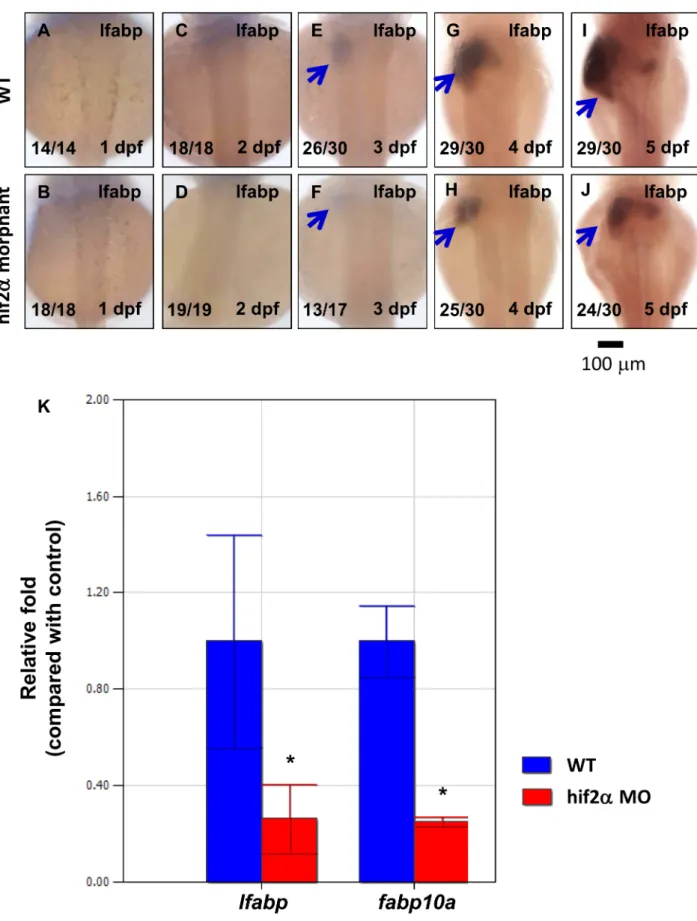
Figure 2. Knockdown of hif2-alphaconfers a small liver phenotype.(A–J) Development of the embryonic liver was monitored by the expression pattern of thelfabpgene in wild-type embryos andhif2-alphaATG-MO-injected embryos at 1–5 dpf by WISH, thereby revealing a small liver phenotype in hif2-alphamorphants. (K) Expression of thelfabpandfabp10agenes was investigated in wild-type embryos andhif2-alpha
morphants at 5 dpf by qPCR (n = 30). Expression was normalized tob-actin. WT, wild type. The experiment was performed in triplicate, error bars indicate S.D. *,p,0.05, unpairedt-test.
[33,43–46]. The qPCR reaction was as follows: 95uC for 3 minutes, 40 cycles of 95uC for 15 seconds, and 60uC for 1 minutes. The expression levels ofb-actin mRNA were used to normalize the relative mRNA abundance. Relative quantification was determined using the DDCt method: relative expres-sion = 22DDCt. The mean values and standard deviations were calculated using the iQ5 Optical System Software. The experi-ment was performed in triplicate. Data were analyzed with the unpairedt-testto compare the treated groups with the untreated group (wild type). *indicates that the significant difference wasp, 0.05.
Chromatin immunoprecipitation (ChIP)
ChIP was performed using a Magna ChIP G Chromatin Immunoprecipitation Kit (Millipore, Billerica, MA, USA, cat. no. 17–611) as described previously [33]. Adult zebrafish liver tissue was cross-linked with 1% formaldehyde, and unreacted formal-dehyde was quenched by 125 mM glycine. The liver tissue was homogenized by a tissue grinder and lysed in Cell Lysis Buffer (Millipore, Billerica, MA, USA, cat. no. CS200634) after being washed with 125 mM glycine. The cell pellets were suspended in Nuclear Lysis Buffer (Millipore, Billerica, MA, USA, cat. no. CS200623) after centrifugation and washed with 1X PBS. The cell lysates were sonicated on wet ice with twenty 30-s pulses (30-s pause between pulses) by a sonicator (MISONIX, Farmingdale, NY, USA, cat. no. S-4000), and the DNA was sheared to,200–
500 base pairs in length. Recombinant protein G covalently bound to magnetic beads was incubated with a polyclonal antibody against anti-Hif2-alpha (GeneTex, Irvine, CA, USA, ChIP grade rabbit polyclonal antibody, cat. no. GTX103707) or normal Rabbit IgG (Millipore, Billerica, MA, USA, cat. no. 12–370) as the negative control at 4uC for 3 hours before the sheared DNA was incubated with the magnetic bead-linked antibody overnight at 4uC. The beads were washed once with different wash buffers (Millipore, Billerica, MA, USA, cat. no. 20–154, cat. no. 20–155, and cat. no. 20–156) for 10 minutes. All wash procedures were performed with a magnetic separator (Millipore, Billerica, MA, USA, Magna Grip Rack (8 Well), cat. no. 20–400). The washed immunocomplexes were eluted with TE buffer, and the protein– DNA complexes were reverse crosslinked by proteinase K digestion at 62uC for 2 hours followed by 95uC for 10 minutes. Finally, DNA was purified with spin columns and eluted using elution buffer. For PCR amplification, immunoprecipitated DNA and input DNA (unimmunoprecipitated) were used as templates. The PCR primers used in the ChIP assay are listed in Table S2.
CoCl2treatment of zebrafish embryos
The coding sequences of zebrafishhif1-alphaand zebrafish hif2-alphawere subcloned into pCMV-Tag 2A (Agilent Technologies, SC, California, USA, cat. no. 211172). The templates were generated by PCR, and we synthesized capped mRNA of FLAG-tagged hif-1 alpha and FLAG-tagged hif-2 alpha with T3 RNA Figure 3.Hif2-alphaplays a major role in liver development.The specificity of knockdown by MO was demonstrated by monitoringlfabpgene expression at 5 pdf by WISH in wild-type embryos (A) and embryos injected withhif2-alphaATG-MO (B). The small liver phenotype resulting from the knockdown ofhif2-alphacan be rescued by co-injection ofhif2-alphamRNA (C). ATG-MO ofhif2-alphabut nothif1-alphacaused the small liver phenotype (E). ATG-MO ofhif3-alphaalso caused a slight effect on liver development (F).
doi:10.1371/journal.pone.0101980.g003
polymerase and an mMESSAGE mMACHINE kit from Ambion (Austin, TX, USA, cat. no. AM1348). The zebrafish embryos were injected with 2400 pg mRNA of FLAG-tagged hif-1 alpha and 2400 pg mRNA of FLAG-taggedhif-2 alphafor CoCl2treatment, respectively. The un-injected and injected zebrafish embryos were treated with 10 mM CoCl2in Petri dishes at 28.5uC from 48 hpf to 72 hpf [47]. igfbp-1 had been reported to be up-regulated following CoCl2treatment [47,48] and was therefore used as the positive control in the qPCR experiments.
Western blot
The total proteins of CoCl2-treated zebrafish embryos were extracted with SDS lysis buffer (0.05% SDS, 200 mM Tris Base, 5 mM EDTA, 6 M Urea) and were then separated by 8% SDS-PAGE. We transferred the proteins to Immun-Blot PVDF Membranes (Bio-Rad, Hercules, CA, USA, cat. no. 162–0177) at 100 volts for 2 hours. The PVDFs were blocked with 3% milk in TBST (TBS containing 0.1% Tween-20) for 1 hour at room temperature. After blocking, the PVDFs were incubated with rabbit anti-FLAG Tag antibody (1:2000 dilution, Cell Signaling, Danvers, MA, USA, DYKDDDDK Tag Antibody, cat. no. 2368S) or mouse anti-tubulin antibody (1:2000 dilution, GeneTex, Irvine, CA, USA, cat. no. GTX628802) overnight at 4uC. The PVDFs were washed with TBST and were incubated with Goat Anti-mouse IgG-HRP (1:50000 dilution, Jackson ImmunoRe-search Laboratories, Inc., West Grove, PA, USA, cat. no. 115-001-003) or Donkey anti-rabbit IgG-HRP (1:10000 dilution, GE Healthcare Life Sciences, cat. no. NA934). After washing with TBST, the membranes were visualized by WesternBright ECL Chemiluminescent HRP Substrate (advansta, Menlo Park,
Cali-fornia, USA, cat. no. K-12045-D50). The intensity of bands on the western blots was measured by ImageJ software.
HRE cluster identification
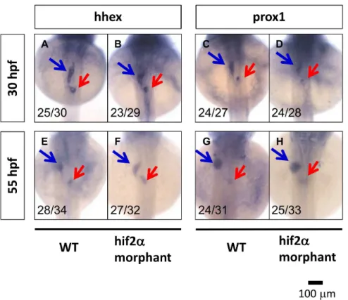
The sequences upstream of theleg1gene in terrestrial mammals (Alpaca, Bushbaby, Cat, Chimpanzee, Cow, Dog, Elephant, Ferret, Gorilla, Guinea pig, Hedgehog, Human, Horse, Hyrax, Kangaroo rat, Rhesus macaque, Marmoset, Megabat, Mouse lemur, Mouse, Opossum, Orangutan, Panda, Pig, Pika, Platypus, Rabbit, Rat, Rock hyrax, Shrew, Sloth, Squirrel, Tarsier, and Tasmanian devil), birds (Chicken, Duck, Flycatcher, Turkey, and Zebra finch), reptiles (American alligator, Chinese softshell turtle, and Lizard), and teleosts (Cod, Zebrafish, Fugu, Medaka, Platyfish, Stickleback, and Tilapia) were downloaded from the UCSC genome database (http://genome.ucsc.edu/) and the Ensembl genome database (http://www.ensembl.org/index. html). We chose a region 10 kbps upstream of the translation start site (without including the +1) of the leg1 gene for HRE mapping. We calculated the inter-HRE distances between each HRE and its first neighbor, together with the distance between each HRE and its second neighbor. We used a greedy algorithm to identify high-density HRE clusters. The greedy algorithm scans genomic sequences using a window size of 500 bps and identifies the cluster starting from the window containing the highest number of HREs in a recursive fashion. We performed Fisher’s exact test to assess if there is a significant difference between the number of HREs from the terrestrial mammals and the teleosts. Figure 4.Hif2-alphais not required for liver specification in zebrafish embryos.Liver specification inhif2-alphamorphants was detected through the expression of thehhexandprox1genes. The expression of embryonic liver specification genes,hhex(A, B, E, F) andprox1(C, D, G, H), were examined at 30 hpf (A–D) and 55 hpf (E–H) in wild-type andhif2-alphaATG-MO-injected embryos by WISH.
Results
Hif2-alphais expressed in the liver in zebrafish embryos We analyzed the gene expression pattern ofhif1-alpha,hif2-alpha, andhif3-alphaby whole-mount in situ hybridization (WISH). The results revealed that hif1-alpha was expressed in the head, notochord and intestine at 2, 3, 4 and 5 dpf and was expressed in the liver at 4 and 5 dpf (Fig. 1A–D).Hif2-alphawas expressed in the notochord and somites at 2 and 3 dpf and expressed in the pharyngeal arches, liver and intestine at 4 and 5 dpf (Fig. 1E–H).
Hif3-alphawas expressed in the head and inner ear at 2, 3, 4 and 5 dpf and was expressed in the intestine at 4 and 5 dpf (Fig. 1I–L; we also performed WISH using the sense probes ofhif1-alpha, hif2-alpha, andhif3-alpha, as shown in Fig. S1).
Knockdown ofhif2-alphaconfers a small liver phenotype Our initial goal was to understand the role hif2-alpha plays during liver development. First, we used the loss-of-function approach by injecting a morpholino molecule with a sequence matching the translation initiation site aroundhif2-alpha(hif2-alpha
ATG-MO). We found that the size of the liver was reduced in hif2-alphamorphants compared to wild-type embryos by assessing the hepatocyte marker genelfabpstarting from 3 dpf (Fig. 2A–2J). To quantify the effect on the liver by hif2-alpha, we assessed lfabp
expression levels in zebrafish embryos on 5 dpf by qPCR.lfabp
expression levels revealed a 70% down-regulation in hif2-alpha
morphants compared with wild-type embryos (Fig. 2K). These results suggest that hif2-alpha is required for the embryonic development of the zebrafish liver.
Figure 5. Knockdown ofhif2-alpha damages liver cell proliferation. WT (Tg(lfabp:EGFP)) embryos (A) and hif2-alphaATG-MO-injected embryos (B) were examined for liver cell proliferation using an anti-pH3 antibody at 4 dpf. Cell proliferation inhif2-alphaATG-MO-injected embryos was reduced compared with WT embryos. (C) Quantification of pH3-positive cells in the liver (n = 11,p,0.05). (D) The EGFP-positive cells were counted by FACS. Quantification of pH3-positive cells in the trunk (E) and tail (F) (n = 4,p,0.05).
doi:10.1371/journal.pone.0101980.g005
Hif2-alphais a major member of the hif family involved in liver development
In the previous section, we proved that hif2-alpha morphants result in a small liver phenotype (Fig. 2). Here, we performed the rescue experiment to assess the specificity of thehif2a-alpha ATG-morpholino. The small liver phenotype resulting from the knockdown of hif2-alpha(Fig. 3B) can be rescued by co-injection with hif2-alpha mRNA (Fig. 3C). The hif2-alpha over-expressing zebrafish embryos revealed no obvious side effects (Fig. 3D) compared to WT embryos (Fig. 3A). Furthermore, we employed morpholino molecules specific tohif1-alphaandhif3-alpha, respec-tively, to examine if these two members of thehiffamily are also required for liver development. Knockdown ofhif1-alphadoes not have any conspicuous effect on liver development (Fig. 3E). However, the knockdown of hif3-alpha caused a slight effect on liver development (Fig. 3F). Additionally, to examine whether the
hif2-alpha morpholino-induced effect on liver development in zebrafish embryos is mediated by p53 activity, we concurrently knocked downp53andhif2-alphausing translational morpholinos. This result demonstrates that the depletion of p53 expression did
not change the effect of the hif2-alpha morpholino on hepatic outgrowth (data no shown).
The small liver phenotype could result from a side effect of the
hif2-alpha ATG-MO. When we concurrently injected hif2-alpha
ATG-MO andhif2-alphamRNA (which was not recognized by the
hif2-alphamorpholino), the small liver phenotype was completely rescued by the ectopichif2-alpha expression, as demonstrated by
in situhybridization (Fig. 3E). This result suggests that the hif2-alphaATG-MO has minimal off target effects.
Hif2-alphais required for hepatic outgrowth but not specification
To further examine if the knockdown of hif2-alpha has any effects on hepatoblast specification, we investigated the expression pattern of the hhex and prox1 genes. At 30 hpf (beginning of budding) and 55 hpf (completion of budding), we found that the gene expression ofhhexand prox1 inhif2-alphamorphants is not affected compared with wild-type embryos (Fig. 4). These results demonstrate that hif2-alpha is required during the hepatic outgrowth phase but not in the liver specification phase in zebrafish embryos.
Knockdown ofhif2-alphaimpairs liver cell proliferation but does not increase cell apoptosis
The small liver phenotype inhif2-alphamorphants could result from decreased cell proliferation and/or cell apoptosis. We detected less proliferation (an average decrease of 62%) in the hepatocytes of hif2-alpha morphants compared to wild-type embryos using pH3 as the proliferation marker at 4 dpf (Fig. 5C, n = 11, p,0.05). Less proliferation in the liver of hif2-alpha
morphants could result in cell number changes. Therefore, we assayed the number of hepatocytes labeled with EGFP in Tg(lfabp:EGFP) by FACS. The results revealed that the EGFP-positive cells were reduced by approximately 63% on average in
hif2-alpha morphants compared to wild-type embryos (Fig. 5D, n = 4, p,0.05). We also detected proliferation in other tissues. The number of proliferating cells was lower in the trunk (an average decrease of 35%) and the tail (an average decrease of 27%) in hif2-alphamorphants compared to wild-type embryos (Fig. 5E–F, n = 4,
p,0.05 and Figure S2). However, TUNEL assays did not show an increase of cell apoptosis at 4 dpf in the hepatocytes ofhif2-alpha
morphants compared to wild-type embryos (Figure S3, n = 5). We concluded thathif2-alphareduces cell proliferation in the liver of zebrafish embryos.
Hif2-alphais required for the expansion of exocrine pancreas and intestine
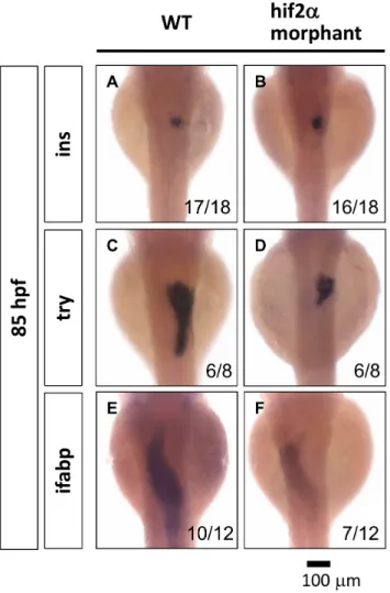
The liver, pancreas and intestine are all derived from the endoderm. We explored the effects of the depletion ofhif2-alphaon the development of the pancreas and the intestine. In the pancreas, we assessed the marker gene insulin (ins) for the endocrine pancreas and trypsin (try) for the exocrine pancreas. We found that the development of the endocrine pancreas is not affected inhif2-alphamorphants (Fig. 6A, 6B), but we detected a smaller exocrine pancreas inhif2-alpha morphants compared to wild-type embryos at 85 hpf (Fig. 6C, 6D). In the intestine, the gene expression of the intestine marker gene intestinal fatty acid binding protein (ifabp) is significantly decreased in hif2-alpha
morphants (Fig. 6E, 6F). These results suggest thathif2-alpha is involved in the organogenesis of the exocrine pancreas and the intestine.
Figure 6. Hif2-alpha is required for the expansion of the exocrine pancreas and the intestine. The development of the embryonic pancreas and the intestine was monitored by examining the gene expression pattern of the endocrine pancreas (ins) (A, B), the exocrine pancreas (try) (C, D), and the intestine (ifabp) (E, F) in wild-type embryos compared withhif2-alphaATG-MO-injected embryos at 85 hpf by WISH.
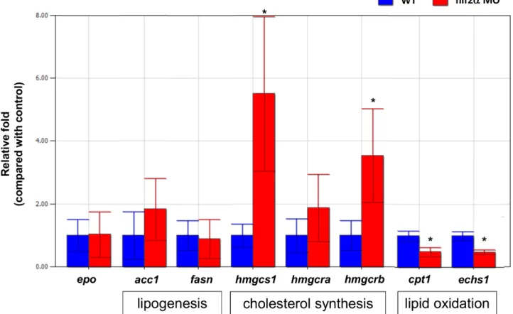
Hif2-alphaknockdown affects lipid metabolism but not EPO production in zebrafish embryos
In mammals, Hif2-alpha had been shown to affect both EPO production [49] and lipid metabolism [50]. To investigate if hif2-alphaknockdown affects EPO production and lipid metabolism in zebrafish embryos, we checked the gene expression ofacc1,fasn,
hmgcs1,hmgcra,hmgcrb, cpt1andechs1for lipid metabolism andepo
forepoproduction. We found that the gene expression ofhmgcs1
andhmgcrb(cholesterol synthesis) increased and thecpt1andechs1
(lipid oxidation) decreased significantly in hif2-alpha morphants compared to wild-type embryos at 5 dpf. However, we did not find any significant changes of the gene expression of epo, hmgcra
(cholesterol synthesis), acc1 and fasn (lipogenesis) in hif2-alpha
morphants compared to wild-type embryos at 5 dpf (Fig. 7). These results indicated thathif2-alphaaffects lipid metabolism (cholesterol synthesis in particular) in zebrafish embryos but not EPO production.
Leg1functions downstream ofHif2-alphato control hepatic outgrowth
We demonstrated that the depletion of hif2-alpha significantly decreased the size of the liver, exocrine pancreas, and intestine but not the endocrine pancreas. Zebrafish embryos injected with a morpholino againstliver-enriched gene 1(leg1) mRNA have a similar phenotype tohif2-alpha morphants; a depletion ofleg1also decreased the size of the liver, exocrine pancreas and intestine but not the endocrine pancreas [26]. The hepatoblast markers were not affected inleg1morpahants orhif2-alphamorphants (Fig. 4 and Fig. S4) [26]. Therefore, we hypothesized that hif2-alpha and leg1
function in the same pathway.
Initially, we investigated the effect of the knockdown ofleg1on hepatic outgrowth. We found that the size of the liver was significantly reduced inleg1morphants of Tg(lfabp:EGFP) at 4 dpf. We also conducted a rescue experiment by co-injecting leg1
morphants with leg1a mRNA in Tg(lfabp:EGFP) transgenic fish. Figure 7.Hif2-alphaknockdown affects lipid metabolism but not EPO production in zebrafish embryos.The expression of EPO and the genes involved in lipid metabolism were investigated in wild-type embryos andhif2-alphamorphants at 5 dpf by qPCR (n = 30). Expression was normalized tob-actin. WT, wild type. The experiment was performed in triplicate, error bars indicate S.D. *,p,0.05, unpairedt-test.
doi:10.1371/journal.pone.0101980.g007
Figure 8. leg1is required for hepatic outgrowth in zebrafish embryos.The expression pattern of thelfabpgene was examined in Tg(lfabp:EGFP) embryos (A) and compared withleg1ATG-MO-injected embryos (0.5mM) (B) as well as embryos co-injected withleg1ATG-MO (0.5mM) andleg1cRNA (C).
doi:10.1371/journal.pone.0101980.g008
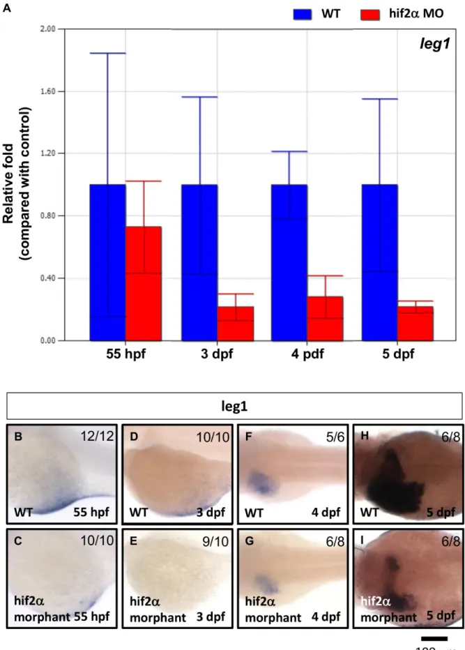
Figure 9.Leg1expression was down-regulated inhif2-alphamorphants.leg1gene expression was examined in wild-type embryos and compared withhif2-alphaATG-MO-injected embryos at 3–5 dpf by qPCR (A) and WISH (B). Expression was normalized tob-actin.
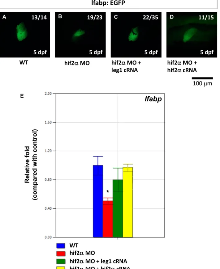
Figure 10. The small liver phenotype caused byhif2-alphaknockdown can be rescued by ectopicleg1expression.The expression pattern of thelfabpgene was examined in Tg(lfabp:EGFP) embryos (A) and compared withhif2-alphaATG-MO-injected embryos (8 ng) (B) as well as embryos co-injected withhif2-alphaATG-MO (8 ng) andleg1cRNA (C). (D)lfabpmRNA expression was detected by qPCR in wild-type embryos and compared withhif2-alphaATG-MO-injected embryos,hif2-alphaATG-MO andleg1cRNA co-injected embryos, as well ashif2-alphaATG-MO and hif2-alphacRNA co-injected embryos. Expression was normalized tob-actin.
doi:10.1371/journal.pone.0101980.g010
Figure 11. Hif2-alpha binds toleg1aandleg1bpromoters.The binding ofhif2-alphato the promoter region of theleg1a(A) andleg1b(B) genes was examined by ChIP-PCR. Seven and nine HRE-containing modules in the promoter regions ofleg1aandleg1b, respectively, are amplified from the immunocomplexes obtained by ChIP assays performed using a polyclonal antibody against anti-Hif2-alpha or a preimmune serum (IgG) as controls. (C) Schematic representation of theleg1aandleg1bpromoter regions. HRE (A/G-C-G-T-G) are annotated as dark lines. The positions of the modules analyzed in the ChIP-PCR assays are shown as grey boxes. The amplified fragments in ChIP assays, including modules 3, 4, 5 and 7 in the
Whileleg1morphants presented a small liver phenotype, we found that leg1 morphants rescued by leg1a mRNA presented a significantly restored liver size at 4 dpf, demonstrating the specificity of theleg1morpholino (Fig. 8).
Next, we found that approximately 70% ofleg1gene expression was abolished in hif2-alpha morphants compared with wild-type embryos at 3–5 dpf but not at 55 hpf (Fig. 9). This result indicates that leg1 functions downstream of hif2-alpha to control hepatic outgrowth.
To investigate ifhif2-alpharegulates hepatic outgrowth through
leg1, we conducted a rescue experiment by co-injectinghif2-alpha
morphants with leg1a mRNA in Tg(lfabp:EGFP) transgenic fish. Whilehif2-alpha morphants revealed a small liver phenotype, we found thathif2-alphamorphants rescued byleg1amRNA presented a significantly restored liver size at 5 dpf (Fig. 10A–10C). lfabp
expression, as measured by qPCR, was also significantly increased in hif2-alpha morphants rescued by leg1a mRNA (Fig. 10D, p, 0.05). Therefore, we concluded that hif2-alpha regulates hepatic outgrowth by regulatingleg1expression.
Hif2-alpha regulatesleg1 gene expression by binding to theleg1promoter
Hif2-alpha is a basic helix-loop-helix transcription factor. To further identify if Hif2-alpha regulates leg1 gene expression by directly binding to theleg1promoter region, we conducted ChIP-PCR to examine if Hif2-alpha binds to HREs in theleg1promoter. We analyzed the sequence upstream of the transcription start site
for theleg1a and leg1bgenes and grouped the sequences into 11 HRE modules and 9 modules, respectively. In the adult zebrafish liver, we found that Hif2-alpha binds HREs in module 3, module 4, module 5, and module 7 in theleg1apromoter. Hif2-alpha can also bind HREs in module 1, module 2, and module 9 in theleg1b
promoter (Fig. 11). These results suggest that Hif2-alpha regulates
leg1gene expression by binding to the HRE modules in theleg1
promoter.
The involvement of the FGF, HGF, and Wnt pathways in hepatic outgrowth regulated by Hif2-alpha
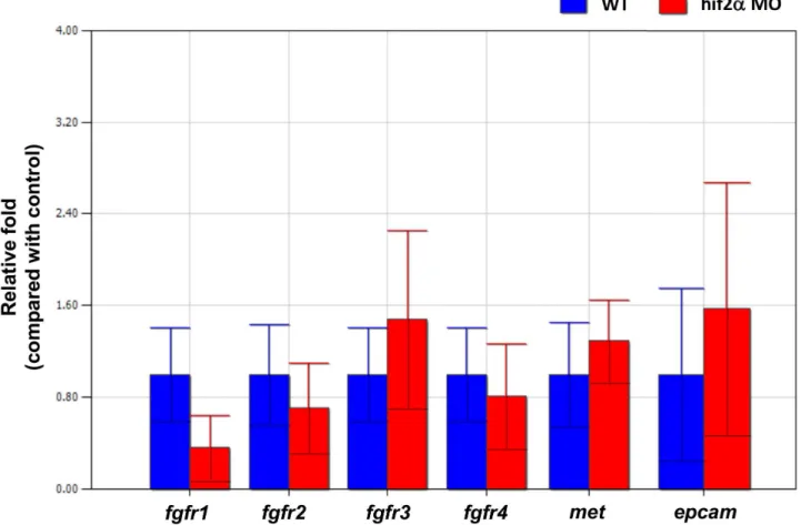
Several factors in the FGF, HGF, and Wnt pathways have been demonstrated to be involved in liver development in zebrafish [45,51,52]. To investigate if Hif2-alpha regulates hepatic out-growth through the FGF, HGF, and Wnt pathways, we checked the gene expression offgfr1,fgfr2,fgfr3,fgfr4(the FGF receptors for hepatic development in zebrafish), c-met (the HGF receptor for hepatic development in zebrafish), andepcam(the Wnt derepressor for hepatic development in zebrafish). We found no significant change of those genes inhif2-alphamorphants compared to wild-type embryos at 5 dpf by qPCR (Fig. 12). This result indicated that the regulation of hepatic outgrowth byhif2-alphawas not directly related to the FGF, HGF, and Wnt pathways.
Figure 12. The regulation of hepatic outgrowth byhif2-alphain zebrafish embryos is not through the FGF, HGF, or Wnt pathways. Gene expression offgfr1,fgfr2,fgfr3,fgfr4,met, andepcamwas examined in wild-type embryos and compared withhif2-alphaATG-MO-injected embryos at 3–5 dpf by qPCR. Expression was normalized tob-actin.
doi:10.1371/journal.pone.0101980.g012
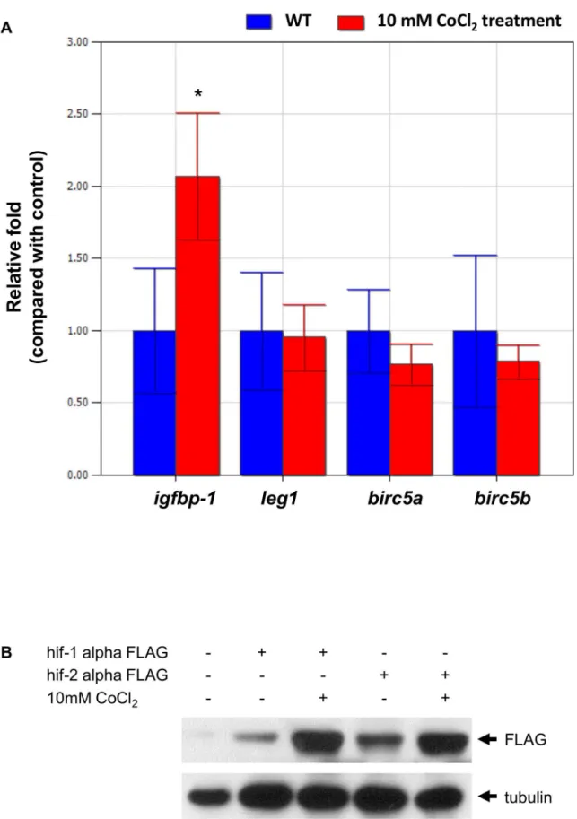
Figure 13. CoCl2treatment increased the expression of the target genes of Hif1-alpha rather than those of Hif2-alpha.(A) Gene
expression ofigfbp-1, leg1,birc5a, andbirc5bmRNA without (control, in blue) or with 10 mM CoCl2treatment (red) was examined in zebrafish
embryos at 72 hpf by qPCR. Expression was normalized tob-actin. *indicates a significant difference (p,0.05) between control embryos and CoCl2
-treated embryos by an unpairedt-test. The experiment was performed in triplicate, and error bars indicate the standard deviation. (B) The Flag-protein expression levels were performed by western blot and were normalized to tubulin.
CoCl2treatment increased the expression of the target
genes ofHif1-alpharather than those ofHif2-alpha Because we demonstrated that Hif2-alpha regulates gene expression by directly binding to the sequences upstream of both
leg1a and leg1b, a hypoxia-mimic treatment should enhance the stability of Hif2-alpha and up-regulateleg1gene expression. Here, CoCl2was used to mimic hypoxic conditions [53]. We incubated zebrafish embryos in 10 mM CoCl2from 48 hpf to 72 hpf. While the up-regulation ofigfbp-1expression [48] was evident, we did not detect a significant difference in the expression of theleg1,birc5a
andbir5bgenes. (Fig. 13A) (birc5aandbirc5bare two proven direct targets of Hif2-alpha [33]). These results suggest that a selective up-regulation of the downstream genes ofhif1-alphabut not those
of hif2-alpha in zebrafish embryos exists with hypoxia-mimic treatment.
To further prove the selective up-regulation ofHif-target genes by CoCl2, we investigated the stability of Hif1-alpha and Hif2-alpha proteins in zebrafish embryos. We injectedHif1-alpha-FLAG
and Hif2alpha-FLAG mRNA into fertilized eggs, respectively, incubated zebrafish embryos in 10 mM CoCl2from 48 hpf to 72 hpf, and extracted the protein from the embryos. We found that the stability of Hif1-alpha protein was increased after CoCl2 treatment (5.03-fold); in contrast, the stability of Hif2-alpha protein only increased slightly after CoCl2 treatment (1.98-fold) (Fig. 13B).
Table 1.HRE cluster detection in the upstream sequences of animals.
Categorya Speciesb Total speciesc Ratiod
Mammalia 6 34 18%
Aves 2 5 40%
Reptilia 1 3 33%
Teleost 6 7 86%
athe category of the animals.
bthe number of species of which upstream sequences contain no less than three HREs in a window of 500 bps. cthe species number in a category.
dthe ratio ofb/c.
doi:10.1371/journal.pone.0101980.t001
Figure 14. Distribution of HRE clustering.The distances between HRE for the first neighbored HRE (A, B) and the second neighbored HRE (C, D) were measured, and the distributions of the distance are shown in 100 bps bins.
doi:10.1371/journal.pone.0101980.g014
HRE clusters are found upstream of theleg1gene in teleosts
A homotypic binding cluster of transcription factors had been shown to be important in the tissue-specific expression of regulated genes and crucial for embryonic development. Because we showed that some HREs are bound by Hif2-alpha using ChIP-PCR, we wanted to examine if HRE clusters exist in the regulatory sequences of leg1. We mapped HREs in the upstream, potential regulatory sequences (relative to the translational start site, 10 kbps) of leg1 genes in terrestrial mammals (34 species) and teleosts (7 species). We analyzed the distribution of the distances between HREs and their first neighbors and second neighbors. We found that in the upstream sequences of the teleost leg1 gene, HREs tend to cluster and form potential high-density Hif-binding regions. However, this was not observed in the upstream sequences of the terrestrial mammalian leg1 gene (Fig. 14A–D). If we used 500 bps as the window size and counted the number of binding sites inside this window, we found that 86% of teleost sequences (including module 3, module 4, module 5, and module 7 of the leg1a gene of zebrafish) contain more than or equal to 3 HREs in comparison with 18% of terrestrial mammalian sequences (p,0.01) (Table 1, Table S3). Therefore, we concluded that high-density HREs in upstream, potential regulatory sequences are observed in teleosts but not in terrestrial mammals.
Discussion
There are three major findings in this study. First, we present the first evidence that hif2-alpha plays an important role in embryonic hepatic outgrowth. Second, we show that the ability of
hif2-alpha to drive hepatic outgrowth is mediated byleg1. Third, thehif2-alpha-leg1axis may be an adaptation in teleosts that is not observed in terrestrial mammals. We provided the evidence that
hif2-alpha is required for hepatic outgrowth but not hepatoblast specification in the zebrafish embryo. Moreover, the lack of Hif2-alpha significantly decreased leg1 expression. Exogenous leg1
mRNA can restore the liver outgrowth defect caused byhif2-alpha
knockdown. Furthermore, Hif2-alpha protein molecules can bind to HRE in the promoter sequence upstream of the leg1 gene. These observations suggest that leg1 is the gene that is downstream ofhif2-alphaand thathif2-alpharegulates hepatic development by acting onleg1. Furthermore, high-density HREs can be found in the potential regulatory sequences of leg1 in teleosts but not in terrestrial mammals.
TheHif2-alpha-leg1 axis during embryonic development in the zebrafish
The phenotype of the gene knockdown of leg1and hif2-alpha
supports the idea thatleg1is the gene that is downstream of hif2-alpha. Knockdown ofleg1has been shown to block liver expansion; however, liver initiation is not affected [26]. These results are similar to our data detailing the knockdown ofhif2-alpha.We also found that knockdown of hif2-alpha revealed a smaller exocrine pancreas and intestine (Fig. 6C, D). Again, this is similar to what was observed with the knockdown ofleg1[26]. The likelihood of theleg1gene as a downstream target ofhif2-alphais also supported by ChIP-PCR, where we demonstrated that Hif2-alpha directly binds to HRE in the promoter region of bothleg1aandleg1b.
Numerous genes have been revealed to be important during the outgrowth stage, includingalr [54], uhrf1 [43],hdac3 [55], gdf11
[55],def[56,57],delta113p53[57],pes[58],mypt1[59],npo[60], c-met[51,61],grnA[51],bms1l[62], andleg1[26]. In our study, the
Hif2-alpha-leg1axis regulates hepatic outgrowth but does not play a role in liver specification and differentiation. In addition to leg1
[26], bothdef[56,57] andnpo[60] affect hepatic outgrowth but not liver specification and differentiation. It is possible that multiple factors are required for cell proliferation and the formation of liver morphology during the hepatic outgrowth and expansion phase. These factors may interact with each other through functional association or physical interaction, and there is little redundancy in this interaction network. A lack of any factor would abolish hepatic outgrowth to varying extents. In this context,grnAhad been shown to regulate hepatic outgrowth by modulatingc-metgene expression [51]. Therefore, it is of interest to identify whether thegrna-c-met
axis [51] can exert its influence on liver development through a pathway other than thehif2-leg1axis. Further studies are required to analyze the role of each factor and their interdependence during hepatic outgrowth. It is important to note that the rescue of hif2-alphamorphants byleg1cRNA can restore the liver to a partial wild-type volume. This is consistent with the concept mentioned above, suggesting thatleg1is one of the multiple factors involved in hepatic outgrowth. Additionally, it is possible thathif2-alphamay control a distinct mechanism of hepatic outgrowth whenleg1is one of the downstream ‘‘executors’’ ofhif2-alpha.
In our previous study, we demonstrated that the knockdown of
hif2-alpha caused apoptosis in neuronal cells in the zebrafish embryo. In neuron cells, Hif2-alpha induces the gene expression of survivin (birc5A and birc5B), which regulates cell apoptosis. Interestingly, a lack of Hif2-alpha or survivin causes the cell to maintain a progenitor cell state and to continue to proliferate while inducing apoptosis in neuron cells [33]. In this study, the knockdown of hif2-alpha reduces cell proliferation but does not result in apoptosis in the developing liver of zebrafish embryos. Moreover,Hif2-alpha affects neuronal development as early as 1 dpf [33] and influences hepatic outgrowth starting at 3 dpf. This raises the possibility that Hif2-alpha employs various temporal regulatory mechanisms when comparing development in neurons and in the liver.
Hif2-alpha is a transcription factor, and its functions are restricted to the cell where it is expressed. However, in adult zebrafish, the Hif2-alpha-inducibleleg1gene encodes a secretory protein that exists in the liver, head, tail, gut and serum; however, most of the leg1 transcripts are found in adult livers [26]. Interestingly, Hif-alpha can also induce the gene expression of other secretory proteins, such as insulin growth factor–binding protein-1 (IGFBP-1) [63,64], insulin growth factor–binding protein-2 (IGFBP-2) [65], and Galectin-1 [66]. The knockdown of IGFBP-1 in zebrafish has been shown to cause growth retardation and developmental delays [63]. IGFBP-2 enhances tumor angiogenesis by promoting angiogenesis and facilitating the induction of VEGF-A in endothelial cells [65]. Galectin-1 suppresses T cell-mediated cytotoxic immune responses and promotes tumor angiogenesis [66]. Therefore, we speculate that there are additional Hif-inducible genes that encode secretory proteins that may play crucial roles during embryonic develop-ment and carcinogenesis.
The evolutionary implications of homotypic clusters of Hif-binding sites
In our previous study, we identified the unusually clustered aryl hydrocarbon receptor response element (AHRE) in the upstream regulatory sequences of the CYP3 genes in teleosts but not in terrestrial mammals [71]. Here, we also discovered high-density HREs in the potential upstream regulatory sequences ofleg1genes in teleosts but not in terrestrial mammals. We speculate that Hif2-regulated liver development throughleg1is an adaptation observed in teleosts as a result of their aquatic environment. It should be noted that the importance of the homotypic high-density transcription factor binding sites is based on an assumption that high-density homotypic DNA elements are more likely to be bound by corresponding transcription factors, thus recruiting chromatin-modification proteins and enabling a transcription-capable status for the regulated genes. Whether this assumption holds will depend on the investigation of the ability of Hif2-alpha to regulate leg1 gene expression in terrestrial mammals and teleosts.
Differential stability and different targets of Hif-alpha molecules reflect the diverse regulation of biological functions
We found that Hif2-alpha but not Hif1-alpha is required for liver development. Although Hif1-alpha and Hif2-alpha can target the same genes, they also each have their own respective unique target genes, thereby reflecting the different roles these genes play in the cellular homeostasis in hypoxia/anoxia and diseases [72]. In zebrafish embryos, Hif2-alpha is required for neuron development [33]. However, Hif1-alpha controls neural crest chemotaxis and the epithelial to mesenchymal transition by inducing the expression of the chemokine receptor Cxcr4 and Twist [30].
In addition to this difference in their target genes, the presence of Hif2-alpha protein in cells was reported not to depend on the hypoxia state; nevertheless, Hif1-alpha is ready to enhance the hypoxia-induced stability [73–75]. In our study, in the zebrafish embryo, the Hif1-alpha target gene can be induced upon treatment with CoCl2. We also demonstrated that the stability of Hif1-alpha is more significantly increased in zebrafish embryos than the stability of Hif2-alpha after treatment with CoCl2. CoCl2 has long been used to mimic hypoxia and induces Hif1-alpha expression in rabbit hepatocytes [76]. CoCl2 also binds to the oxygen-dependent degradation (ODD) domain of Hif2-alpha, thereby preventing its association with the von Hippel-Lindau protein [77]. In bovine arterial endothelial cells, treatment of CoCl2did not change the stable presence of Hif2-alpha in the cell [78]. Interestingly, a similar observation was also detected in primary mouse hepatocytes in a 1% oxygen environment [79]. Our observations are consistent with the results of the previous studies mentioned above, indicating the differential stability and control upon Hif-alpha molecules during the regulation of the various aspects of embryonic development.
The regulation of hepatic outgrowth byhif2-alphain zebrafish embryos is not through the FGF, HGF, or Wnt pathways
In mammals, FGF signaling is important for hepatic specifica-tion [7]. The fibroblast growth factors (FGF) expressed by the cardiac mesoderm induce the formation of the liver from the foregut endoderm. In zebrafish, FGF signaling is not only essential for hepatoblast specification [13] but also necessary for hepatic outgrowth [45]. When FGF signaling is blocked at 22 hpf, the gene expression levels of hepatoblast markers, such as hhexand prox1, are depressed [13]. The liver size is reduced in Tg(lfabp:dnfgfr1-egfp) zebrafish, which expresses a domain-negative fgfr1-egfp at 5 dpf
[45]. We surveyed the gene expression levels offgfr1,fgfr2, fgfr3,
fgfr4inhif2-alphamorphants compared to wild-type embryos and demonstrated that the gene expression of the FGF receptors was not affected whenhif2-alphawas knocked down.
HGF signaling is essential for hepatoblast proliferation and hepatocyte differentiation in mammals [80]. In HGF2/2mutant mice [81] and cMet2/2(the HGF receptor) mutant mice, the liver was undeveloped and smaller [82]. In zebrafish, knockdown ofmet
affected hepatic outgrowth but not hepatoblast specification [61]. Our study revealed that met expression was not changed significantly when thehif2-alpha was knocked down in zebrafish at 5 dpf.
In mammals, Wnt signaling plays two opposing roles in liver development. In endoderm patterning (the stage before hepato-blast specification), Wnt signaling represses the liver cell fate. After the hepatoblast specification stage, Wnt signaling induces hepa-tocyte differentiation and outgrowth [1]. In zebrafish, Wnt signaling is important for hepatic specification, hepatocyte differentiation and outgrowth [3,52]. The liver was smaller, and the expression of the hepatoblast markers, hhex and prox1, were decreased in wnt2bb mutant zebrafish [83]. Overexpression of
epcam (a Wnt derepressor) resulted in a larger liver in zebrafish embryos [52]. In this study, we proved that the expression level of
epcam was not altered significantly in hif2-alpha morphants compared to wild-type embryos.
These observations suggest that the FGF, HGF and Wnt pathways are upstream of the Hif pathway or that the FGF, HGF and Wnt pathways and the Hif pathway are parallel to each other in the regulation of hepatic growth.
EPO is not regulated byhif2-alphain zebrafish embryos Erythropoietin (EPO) is necessary for erythropoiesis and is regulated byhif2-alphain mice. A previous study showed that EPO production was suppressed inhif2-alphamutant mice [49]. The epo expression level was also down-regulated inhif1-alphamutant mice [84]. In zebrafish,epoexpression was up-regulated invhlmutants at 7.5 dpf [85], implying thatepowas up-regulated by the more stable Hif protein. In this study, we examined the expression levels ofepo
inhif2a-alpha morphants, and the results revealed that epogene expression was not changed whenhif2-alpha was knocked down (Fig. 7). Because the gene expression pattern data revealed that bothhif1-alphaandhif2-alphawere expressed in the liver (Fig. 1), it is possible that other family members of Hif (eg,hif1-alpha) can regulateepogene expression in the zebrafish embryo.
Expression of genes responsible for lipid metabolism can be regulated byhif2-alpha
Lipid metabolism is comprised of several processes, including lipogenesis, lipid oxidation, and cholesterol synthesis. In lipogen-esis, fasn gene expression has been shown to be suppressed in hypoxic HepG2 cells in ahif2-alpha-dependent manner [50]. In lipid oxidation, the gene expression levels ofcpt1(oxidation) have been shown to be down-regulated in mutant mice containing a stable form of Hif2-alpha. In cholesterol synthesis, hypoxia can stimulate the degradation of HMG-CoA reductase and increase the gene expression of HMG-CoA synthase and HMG-CoA reductase in CHO-7 cells [86]. Recently, the loss of Von Hippel-Lindau (VHL) was shown to increase the cholesterol level via Hif2-alpha [87].
embryos. Moreover, the expression levels of the genes responsible for cholesterol synthesis (HMG-CoA synthase (hmgcs1) and HMG-CoA reductase (hmgcrb)) were shown to be up-regulated in hif2-alphamorphants compared to wild-type embryos. Whilehif2-alpha
might be involved in the regulation of lipid oxidation and cholesterol synthesis in the zebrafish embryo, it is interesting to note that the trend of this influence is different between mammals and zebrafish embryos. Recently, more experiments have shown that lipid/cholesterol regulation is important for embryonic development [88,89]. The inclusion of metabolites and a more systematic approach for identifying the involvement of the Hif pathway shall certainly help to resolve the current inconsistency between different model organisms.
Conclusion
We demonstrated thatHif2-alphaplays an essential role in the embryonic development of hepatic outgrowth in zebrafish through directly controlling leg1 gene expression. We also speculated that the Hif2-alpha-leg1 axis may be an adaptation in teleosts because high-density Hif-binding sites in the potential upstream regulatory sequences are observed in teleosts but not in terrestrial mammals.
Supporting Information
Figure S1 Whole-mount in situ hybridization was per-formed with sense probes ofhif1-alpha, hif2-alphaand
hif3-alpha.The expression patterns ofhif1-alpha(A–D),hif2-alpha
(E–H) and hif3-alpha (I–L) were assessed with sense probes by WISH in zebrafish embryos at 2–5 dpf. WISH, whole-mount in situ hybridization. dpf, days post-fertilization.
(TIF)
Figure S2 Knockdown ofhif2-alphareduced cell prolif-eration in the trunk and tail of zebrafish embryos.Cell
apoptosis in wild-type embryos (A) and hif2-alpha ATG-MO-injected embryos (B) at 4 dpf by pH3 staining.
(TIF)
Figure S3 Knockdown ofhif2-alphadid not induce cell apoptosis in hepatocytes in the zebrafish embryos.Cell apoptosis in Tg(lfabp:EGFP) embryos (A, B, C) and hif2-alpha
ATG-MO-injected Tg(lfabp:EGFP) embryos (D, E, F) at 4 dpf by TUNEL assay. A positive control using Tg(lfabp:EGFP) embryos with DNaseI treatment is also shown (G).
(TIF)
Figure S4 Leg1is not required for liver specification in zebrafish embryos.Liver specification inleg1morphants was detected through the expression of thehhexandprox1genes. The expression of embryonic liver specification genes,hhex(A, B, E, F) andprox1(C, D, G, H), were examined at 30 hpf (A–D) and 55 hpf (E–H) in wild-type andleg1ATG-MO-injected embryos by WISH. (TIF)
Table S1 Primer sequences employed in the qPCR experiments.
(DOC)
Table S2 Primer sequences employed in the ChIP-PCR experiments.
(DOC)
Table S3 The summary of HRE clusters in the upstream sequences ofleg1comparing with ChIP-PCR assay.
(DOC)
Author Contributions
Conceived and designed the experiments: TYL CHH WST. Performed the experiments: TYL CFC CYC. Analyzed the data: TYL HYC CHL. Contributed reagents/materials/analysis tools: JLW HJL TWP. Wrote the paper: TYL CHH WST.
References
1. Zorn AM (2008) Liver development. StemBook. Cambridge (MA).
2. Si-Tayeb K, Lemaigre FP, Duncan SA (2010) Organogenesis and development of the liver. Dev Cell 18: 175–189.
3. Chu J, Sadler KC (2009) New school in liver development: lessons from zebrafish. Hepatology 50: 1656–1663.
4. Field HA, Ober EA, Roeser T, Stainier DY (2003) Formation of the digestive system in zebrafish. I. Liver morphogenesis. Dev Biol 253: 279–290. 5. Tao T, Peng J (2009) Liver development in zebrafish (Danio rerio). J Genet
Genomics 36: 325–334.
6. Korzh S, Pan X, Garcia-Lecea M, Winata CL, Pan X, et al. (2008) Requirement of vasculogenesis and blood circulation in late stages of liver growth in zebrafish. BMC Dev Biol 8: 84–98.
7. Jung J, Zheng M, Goldfarb M, Zaret KS (1999) Initiation of mammalian liver development from endoderm by fibroblast growth factors. Science 284: 1998– 2003.
8. Rossi JM, Dunn NR, Hogan BL, Zaret KS (2001) Distinct mesodermal signals, including BMPs from the septum transversum mesenchyme, are required in combination for hepatogenesis from the endoderm. Genes Dev 15: 1998–2009. 9. Calmont A, Wandzioch E, Tremblay KD, Minowada G, Kaestner KH, et al. (2006) An FGF response pathway that mediates hepatic gene induction in embryonic endoderm cells. Dev Cell 11: 339–348.
10. Thompson MD, Monga SP (2007) WNT/beta-catenin signaling in liver health and disease. Hepatology 45: 1298–1305.
11. Niu X, Shi H, Peng J (2010) The role of mesodermal signals during liver organogenesis in zebrafish. Sci China Life Sci 53: 455–461.
12. Goessling W, North TE, Lord AM, Ceol C, Lee S, et al. (2008) APC mutant zebrafish uncover a changing temporal requirement for wnt signaling in liver development. Dev Biol 320: 161–174.
13. Shin D, Shin CH, Tucker J, Ober EA, Rentzsch F, et al. (2007) Bmp and Fgf signaling are essential for liver specification in zebrafish. Development 134: 2041–2050.
14. Wallace KN, Yusuff S, Sonntag JM, Chin AJ, Pack M (2001) Zebrafish hhex regulates liver development and digestive organ chirality. Genesis 30: 141–143.
15. Ober EA, Field HA, Stainier DY (2003) From endoderm formation to liver and pancreas development in zebrafish. Mech Dev 120: 5–18.
16. Liao W, Ho CY, Yan YL, Postlethwait J, Stainier DY (2000) Hhex and scl function in parallel to regulate early endothelial and blood differentiation in zebrafish. Development 127: 4303–4313.
17. Costa RH, Kalinichenko VV, Holterman AX, Wang X (2003) Transcription factors in liver development, differentiation, and regeneration. Hepatology 38: 1331–1347.
18. Cheng W, Guo L, Zhang Z, Soo HM, Wen C, et al. (2006) HNF factors form a network to regulate liver-enriched genes in zebrafish. Dev Biol 294: 482–496. 19. Odom DT, Zizlsperger N, Gordon DB, Bell GW, Rinaldi NJ, et al. (2004)
Control of pancreas and liver gene expression by HNF transcription factors. Science 303: 1378–1381.
20. Lemaigre FP (2009) Mechanisms of liver development: concepts for under-standing liver disorders and design of novel therapies. Gastroenterology 137: 62– 79.
21. Alexander J, Stainier DY (1999) A molecular pathway leading to endoderm formation in zebrafish. Curr Biol 9: 1147–1157.
22. Odenthal J, Nusslein-Volhard C (1998) fork head domain genes in zebrafish. Dev Genes Evol 208: 245–258.
23. Reiter JF, Alexander J, Rodaway A, Yelon D, Patient R, et al. (1999) Gata5 is required for the development of the heart and endoderm in zebrafish. Genes Dev 13: 2983–2995.
24. Reiter JF, Kikuchi Y, Stainier DY (2001) Multiple roles for Gata5 in zebrafish endoderm formation. Development 128: 125–135.
25. Holtzinger A, Evans T (2005) Gata4 regulates the formation of multiple organs. Development 132: 4005–4014.
26. Chang C, Hu M, Zhu Z, Lo LJ, Chen J, et al. (2011) liver-enriched gene 1a and 1b encode novel secretory proteins essential for normal liver development in zebrafish. PLoS One 6: e22910.
28. Schofield CJ, Ratcliffe PJ (2004) Oxygen sensing by HIF hydroxylases. Nat Rev Mol Cell Biol 5: 343–354.
29. Ryan HE, Lo J, Johnson RS (1998) HIF-1 alpha is required for solid tumor formation and embryonic vascularization. EMBO J 17: 3005–3015. 30. Barriga EH, Maxwell PH, Reyes AE, Mayor R (2013) The hypoxia factor
Hif-1alpha controls neural crest chemotaxis and epithelial to mesenchymal transition. J Cell Biol 201: 759–776.
31. Iyer NV, Kotch LE, Agani F, Leung SW, Laughner E, et al. (1998) Cellular and developmental control of O2 homeostasis by hypoxia-inducible factor 1 alpha. Genes Dev 12: 149–162.
32. Tian H, Hammer RE, Matsumoto AM, Russell DW, McKnight SL (1998) The hypoxia-responsive transcription factor EPAS1 is essential for catecholamine homeostasis and protection against heart failure during embryonic development. Genes Dev 12: 3320–3324.
33. Ko CY, Tsai MY, Tseng WF, Cheng CH, Huang CR, et al. (2011) Integration of CNS survival and differentiation by HIF2alpha. Cell Death Differ 18: 1757– 1770.
34. Scortegagna M, Ding K, Zhang Q, Oktay Y, Bennett MJ, et al. (2005) HIF-2alpha regulates murine hematopoietic development in an erythropoietin-dependent manner. Blood 105: 3133–3140.
35. Scortegagna M, Ding K, Oktay Y, Gaur A, Thurmond F, et al. (2003) Multiple organ pathology, metabolic abnormalities and impaired homeostasis of reactive oxygen species in Epas12/2mice. Nat Genet 35: 331–340.
36. Compernolle V, Brusselmans K, Acker T, Hoet P, Tjwa M, et al. (2002) Loss of HIF-2alpha and inhibition of VEGF impair fetal lung maturation, whereas treatment with VEGF prevents fatal respiratory distress in premature mice. Nat Med 8: 702–710.
37. Nath B, Szabo G (2012) Hypoxia and hypoxia inducible factors: diverse roles in liver diseases. Hepatology 55: 622–633.
38. Imanirad P, Solaimani Kartalaei P, Crisan M, Vink C, Yamada-Inagawa T, et al. (2014) HIF1alpha is a regulator of hematopoietic progenitor and stem cell development in hypoxic sites of the mouse embryo. Stem Cell Res 12: 24–35. 39. Rupp RA, Snider L, Weintraub H (1994) Xenopus embryos regulate the nuclear
localization of XMyoD. Genes Dev 8: 1311–1323.
40. Westerfield M (2000) A Guide for the Laboratory Use of Zebrafish (Danio rerio). 4th edn. Eugene: University of Oregon.
41. Shepard JL, Stern HM, Pfaff KL, Amatruda JF (2004) Analysis of the cell cycle in zebrafish embryos. Methods Cell Biol 76: 109–125.
42. Covassin L, Amigo JD, Suzuki K, Teplyuk V, Straubhaar J, et al. (2006) Global analysis of hematopoietic and vascular endothelial gene expression by tissue specific microarray profiling in zebrafish. Dev Biol 299: 551–562.
43. Sadler KC, Krahn KN, Gaur NA, Ukomadu C (2007) Liver growth in the embryo and during liver regeneration in zebrafish requires the cell cycle regulator, uhrf1. Proc Natl Acad Sci U S A 104: 1570–1575.
44. Yu RM, Chu DL, Tan TF, Li VW, Chan AK, et al. (2012) Leptin-mediated modulation of steroidogenic gene expression in hypoxic zebrafish embryos: implications for the disruption of sex steroids. Environ Sci Technol 46: 9112– 9119.
45. Tsai SM, Liu DW, Wang WP (2013) Fibroblast growth factor (Fgf) signaling pathway regulates liver homeostasis in zebrafish. Transgenic Res 22: 301–314. 46. Passeri MJ, Cinaroglu A, Gao C, Sadler KC (2009) Hepatic steatosis in response to acute alcohol exposure in zebrafish requires sterol regulatory element binding protein activation. Hepatology 49: 443–452.
47. Zhang P, Lu L, Yao Q, Li Y, Zhou J, et al. (2012) Molecular, functional, and gene expression analysis of zebrafish hypoxia-inducible factor-3alpha. Am J Physiol Regul Integr Comp Physiol 303: R1165–1174.
48. Kajimura S, Aida K, Duan C (2006) Understanding hypoxia-induced gene expression in early development: in vitro and in vivo analysis of hypoxia-inducible factor 1-regulated zebra fish insulin-like growth factor binding protein 1 gene expression. Mol Cell Biol 26: 1142–1155.
49. Rankin EB, Biju MP, Liu Q, Unger TL, Rha J, et al. (2007) Hypoxia-inducible factor-2 (HIF-2) regulates hepatic erythropoietin in vivo. J Clin Invest 117: 1068–1077.
50. Rankin EB, Rha J, Selak MA, Unger TL, Keith B, et al. (2009) Hypoxia-inducible factor 2 regulates hepatic lipid metabolism. Mol Cell Biol 29: 4527– 4538.
51. Li YH, Chen MH, Gong HY, Hu SY, Li YW, et al. (2010) Progranulin A-mediated MET signaling is essential for liver morphogenesis in zebrafish. J Biol Chem 285: 41001–41009.
52. Lu H, Ma J, Yang Y, Shi W, Luo L (2013) EpCAM is an endoderm-specific Wnt derepressor that licenses hepatic development. Dev Cell 24: 543–553. 53. Piret JP, Mottet D, Raes M, Michiels C (2002) CoCl2, a chemical inducer of
hypoxia-inducible factor-1, and hypoxia reduce apoptotic cell death in hepatoma cell line HepG2. Ann N Y Acad Sci 973: 443–447.
54. Li Y, Farooq M, Sheng D, Chandramouli C, Lan T, et al. (2012) Augmenter of liver regeneration (alr) promotes liver outgrowth during zebrafish hepatogenesis. PLoS One 7: e30835.
55. Farooq M, Sulochana KN, Pan X, To J, Sheng D, et al. (2008) Histone deacetylase 3 (hdac3) is specifically required for liver development in zebrafish. Dev Biol 317: 336–353.
56. Tao T, Shi H, Huang D, Peng J (2013) Def functions as a cell autonomous factor in organogenesis of digestive organs in zebrafish. PLoS One 8: e58858.
57. Chen J, Ruan H, Ng SM, Gao C, Soo HM, et al. (2005) Loss of function of def selectively up-regulates Delta113p53 expression to arrest expansion growth of digestive organs in zebrafish. Genes Dev 19: 2900–2911.
58. Allende ML, Amsterdam A, Becker T, Kawakami K, Gaiano N, et al. (1996) Insertional mutagenesis in zebrafish identifies two novel genes, pescadillo and dead eye, essential for embryonic development. Genes Dev 10: 3141–3155. 59. Huang H, Ruan H, Aw MY, Hussain A, Guo L, et al. (2008) Mypt1-mediated
spatial positioning of Bmp2-producing cells is essential for liver organogenesis. Development 135: 3209–3218.
60. Mayer AN, Fishman MC (2003) Nil per os encodes a conserved RNA recognition motif protein required for morphogenesis and cytodifferentiation of digestive organs in zebrafish. Development 130: 3917–3928.
61. Latimer AJ, Jessen JR (2008) Hgf/c-met expression and functional analysis during zebrafish embryogenesis. Dev Dyn 237: 3904–3915.
62. Marneros AG (2013) BMS1 is mutated in aplasia cutis congenita. PLoS Genet 9: e1003573.
63. Kajimura S, Aida K, Duan C (2005) Insulin-like growth factor-binding protein-1 (IGFBP-1) mediates hypoxia-induced embryonic growth and developmental retardation. Proc Natl Acad Sci U S A 102: 1240–1245.
64. Sun CF, Tao Y, Jiang XY, Zou SM (2011) IGF binding protein 1 is correlated with hypoxia-induced growth reduce and developmental defects in grass carp (Ctenopharyngodon idellus) embryos. Gen Comp Endocrinol 172: 409–415. 65. Das SK, Bhutia SK, Azab B, Kegelman TP, Peachy L, et al. (2013) MDA-9/
syntenin and IGFBP-2 promote angiogenesis in human melanoma. Cancer Res 73: 844–854.
66. Ito K, Stannard K, Gabutero E, Clark AM, Neo SY, et al. (2012) Galectin-1 as a potent target for cancer therapy: role in the tumor microenvironment. Cancer Metastasis Rev 31: 763–778.
67. Moorman C, Sun LV, Wang J, de Wit E, Talhout W, et al. (2006) Hotspots of transcription factor colocalization in the genome of Drosophila melanogaster. Proc Natl Acad Sci U S A 103: 12027–12032.
68. Gotea V, Visel A, Westlund JM, Nobrega MA, Pennacchio LA, et al. (2010) Homotypic clusters of transcription factor binding sites are a key component of human promoters and enhancers. Genome Res 20: 565–577.
69. Lifanov AP, Makeev VJ, Nazina AG, Papatsenko DA (2003) Homotypic regulatory clusters in Drosophila. Genome Res 13: 579–588.
70. Lettice LA, Williamson I, Wiltshire JH, Peluso S, Devenney PS, et al. (2012) Opposing Functions of the ETS Factor Family Define Shh Spatial Expression in Limb Buds and Underlie Polydactyly. Developmental Cell 22: 459–467. 71. Chang CT, Chung HY, Su HT, Tseng HP, Tzou WS, et al. (2013) Regulation
of zebrafish CYP3A65 transcription by AHR2. Toxicol Appl Pharmacol 270: 174–184.
72. Patel SA, Simon MC (2008) Biology of hypoxia-inducible factor-2alpha in development and disease. Cell Death Differ 15: 628–634.
73. Hu CJ, Wang LY, Chodosh LA, Keith B, Simon MC (2003) Differential roles of hypoxia-inducible factor 1alpha (HIF-1alpha) and HIF-2alpha in hypoxic gene regulation. Mol Cell Biol 23: 9361–9374.
74. Park SK, Dadak AM, Haase VH, Fontana L, Giaccia AJ, et al. (2003) Hypoxia-induced gene expression occurs solely through the action of hypoxia-inducible factor 1alpha (HIF-1alpha): role of cytoplasmic trapping of HIF-2alpha. Mol Cell Biol 23: 4959–4971.
75. Kaelin WG Jr, Ratcliffe PJ (2008) Oxygen sensing by metazoans: the central role of the HIF hydroxylase pathway. MolCell 30: 393–402.
76. Fradette C, du Souich P (2003) Hypoxia-inducible factor-1 and activator protein-1 modulate the upregulation of CYP3A6 induced by hypoxia. Br J Pharmacol 140: 1146–1154.
77. Yuan Y, Hilliard G, Ferguson T, Millhorn DE (2003) Cobalt inhibits the interaction between hypoxia-inducible factor-alpha and von Hippel-Lindau protein by direct binding to hypoxia-inducible factor-alpha. J Biol Chem 278: 15911–15916.
78. Takahashi R, Kobayashi C, Kondo Y, Nakatani Y, Kudo I, et al. (2004) Subcellular localization and regulation of hypoxia-inducible factor-2alpha in vascular endothelial cells. Biochem Biophys Res Commun 317: 84–91. 79. Copple BL, Bustamante JJ, Welch TP, Kim ND, Moon JO (2009)
Hypoxia-inducible factor-dependent production of profibrotic mediators by hypoxic hepatocytes. Liver Int 29: 1010–1021.
80. Tanimizu N, Miyajima A (2007) Molecular mechanism of liver development and regeneration. Int Rev Cytol 259: 1–48.
81. Schmidt C, Bladt F, Goedecke S, Brinkmann V, Zschiesche W, et al. (1995) Scatter factor/hepatocyte growth factor is essential for liver development. Nature 373: 699–702.
82. Bladt F, Riethmacher D, Isenmann S, Aguzzi A, Birchmeier C (1995) Essential role for the c-met receptor in the migration of myogenic precursor cells into the limb bud. Nature 376: 768–771.
83. Ober EA, Verkade H, Field HA, Stainier DY (2006) Mesodermal Wnt2b signalling positively regulates liver specification. Nature 442: 688–691. 84. Yoon D, Pastore YD, Divoky V, Liu E, Mlodnicka AE, et al. (2006)
Hypoxia-inducible factor-1 deficiency results in dysregulated erythropoiesis signaling and iron homeostasis in mouse development. J Biol Chem 281: 25703–25711. 85. van Rooijen E, Voest EE, Logister I, Korving J, Schwerte T, et al. (2009)
Zebrafish mutants in the von Hippel-Lindau tumor suppressor display a hypoxic response and recapitulate key aspects of Chuvash polycythemia. Blood 113: 6449–6460.
86. Nguyen AD, McDonald JG, Bruick RK, DeBose-Boyd RA (2007) Hypoxia stimulates degradation of 3-hydroxy-3-methylglutaryl-coenzyme A reductase through accumulation of lanosterol and hypoxia-inducible factor-mediated induction of insigs. J Biol Chem 282: 27436–27446.
87. Ramakrishnan SK, Taylor M, Qu A, Ahn SH, Suresh MV, et al. (2014) Loss of von Hippel-Lindau protein (VHL) increases systemic cholesterol levels through
targeting hypoxia-inducible factor 2alpha and regulation of bile acid homeostasis. Mol Cell Biol 34: 1208–1220.
88. Kawasaki I, Jeong MH, Yun YJ, Shin YK, Shim YH (2013) Cholesterol-responsive metabolic proteins are required for larval development in Caenorhabditis elegans. Mol Cells 36: 410–416.