A Rapid Screening Assay Identifies
Monotherapy with Interferon-ß and
Combination Therapies with Nucleoside
Analogs as Effective Inhibitors of Ebola Virus
Stephen D. S. McCarthy1, Beata Majchrzak-Kita2, Trina Racine3, Hannah N. Kozlowski1, Darren P. Baker4, Thomas Hoenen5, Gary P. Kobinger3, Eleanor N. Fish2,6‡*, Donald R. Branch1,2,7‡
1Department of Laboratory Medicine and Pathobiology, University of Toronto, Toronto, Ontario, Canada, 2Division of Advanced Diagnostics, Infection and Immunity Group, Toronto General Research Institute, Toronto, Ontario, Canada,3Special Pathogens Program, National Microbiology Laboratory, Winnipeg, Manitoba, Canada,4Biogen, Cambridge, Massachusetts, United States of America,5Laboratory of Virology, National Institute of Allergy and Infectious Diseases, National Institutes of Health, Hamilton, Montana, United States of America,6Department of Immunology, University of Toronto, Toronto, Ontario, Canada,7Centre for Innovation, Canadian Blood Services, Toronto, Ontario, Canada
‡Senior authors contributed equally to this work. *[email protected]
Abstract
To date there are no approved antiviral drugs for the treatment of Ebola virus disease (EVD). While a number of candidate drugs have shown limited efficacyin vitroand/or in non-human primate studies, differences in experimental methodologies make it difficult to compare their therapeutic effectiveness. Using anin vitromodel ofEbola Zairereplication with transcription-competent virus like particles (trVLPs), requiring only level 2 biosafety containment, we compared the activities of the type I interferons (IFNs) IFN-αand IFN-ß, a
panel of viral polymerase inhibitors (lamivudine (3TC), zidovudine (AZT) tenofovir (TFV), favipiravir (FPV), the active metabolite of brincidofovir, cidofovir (CDF)), and the estrogen receptor modulator, toremifene (TOR), in inhibiting viral replication in dose-response and time course studies. We also tested 28 two- and 56 three-drug combinations against Ebola replication. IFN-αand IFN-ß inhibited viral replication 24 hours post-infection (IC500.038μM
and 0.016μM, respectively). 3TC, AZT and TFV inhibited Ebola replication when used
alone (50–62%) or in combination (87%). They exhibited lower IC50(0.98–6.2μM)
com-pared with FPV (36.8μM), when administered 24 hours post-infection. Unexpectedly, CDF
had a narrow therapeutic window (6.25–25μM). When dosed>50μM, CDF treatment
enhanced viral infection. IFN-ß exhibited strong synergy with 3TC (97.3% inhibition) or in tri-ple combination with 3TC and AZT (95.8% inhibition). This study demonstrates that IFNs and viral polymerase inhibitors may have utility in EVD. We identified several 2 and 3 drug combinations with strong anti-Ebola activity, confirmed in studies using fully infectious ZEBOV, providing a rationale for testing combination therapies in animal models of lethal Ebola challenge. These studies open up new possibilities for novel therapeutic options, in OPEN ACCESS
Citation:McCarthy SDS, Majchrzak-Kita B, Racine T, Kozlowski HN, Baker DP, Hoenen T, et al. (2016) A Rapid Screening Assay Identifies Monotherapy with Interferon-ß and Combination Therapies with Nucleoside Analogs as Effective Inhibitors of Ebola Virus. PLoS Negl Trop Dis 10(1): e0004364. doi:10.1371/journal.pntd.0004364
Editor:Scott C. Weaver, University of Texas Medical Branch, UNITED STATES
Received:June 13, 2015
Accepted:December 15, 2015
Published:January 11, 2016
Copyright:This is an open access article, free of all copyright, and may be freely reproduced, distributed, transmitted, modified, built upon, or otherwise used by anyone for any lawful purpose. The work is made available under theCreative Commons CC0public domain dedication.
Data Availability Statement:All data are within the paper and its Supporting Information files.
particular combination therapies, which could prevent and treat Ebola infection and poten-tially reduce drug resistance.
Author Summary
Studies to evaluate the effectiveness of candidate antiviral drugs to inhibit Ebola virus infection have been hampered by the availability and access to level 4 containment facili-ties. Using a mini-genome model system that generates Ebola virus-like particles that infect cells, we have been able to screen a panel of candidate drugs for antiviral activity, under normal level 2 containment. We compared the activities of 8 different antivirals from 3 drug classes, including drugs repurposed for the treatment of Ebola: type I interfer-ons and nucleoside analogs. Our data indicate that IFN-ß is a potent inhibitor of Ebola virus, contributing to the decision to conduct a clinical trial of IFN-ß treatment for Ebola virus disease in Guinea. Moreover, we identified that 2 and 3 drug combinations inhibit Ebola replication when administered 24 hours post-infection. Drug combinations have important implications for clinical use, since lower doses of each drug are administered, potentially decreasing side-effects and, based on different mechanisms of action, there is less likelihood for the emergence of drug resistance. These studies set the stage for both preclinical and clinical evaluation.
Introduction
As of December 13, 2015, the current outbreak of Ebola virus disease (EVD) in West Africa has resulted in 28,633 cumulative cases and 11,314 deaths [1]. Two potential vaccine candi-dates, rVSVΔG-ZEBOV and ChAd3-EBO Z, have shown durable protection from lethal Ebola challenge in mice [2] and macaques [3] respectively, and are part of the phase II/III PREVAIL trial in Liberia and Guinea (https://clinicaltrials.gov/ct2/show/NCT02344407). Other potential therapeutics, such as convalescent plasma and the antibody cocktail ZMapp [4] have been approved for an emergency phase II/III trial in Guinea (https://clinicaltrials.gov/ct2/show/ NCT02342171) and a phase I trial in Liberia (https://clinicaltrials.gov/ct2/show/
NCT02363322), respectively. However, to date there is no licensed vaccine or treatment for EVD, although improvements in supportive care are increasing survival rates [5].
Repurposing antivirals used for other viral infections, based on knowledge of mechanisms of action, has prompted accumulating interest in the application of different nucleoside/nucle-otide analogs and type I interferons (IFNs) for the treatment of Ebola virus disease (EVD). Experimental nucleoside analogs may have therapeutic efficacy for EVD, given the evidence of protection in primate and rodent disease models, 2–6 days after lethal Ebola or the related hemorrhagic Marburg virus challenges [6,7]. Favipiravir, a viral polymerase inhibitor, provides 100% protection when administered 6 days after challenge with a lethal dose of Ebola virus [6] and has been evaluated in the phase II/III JIKI trial in Guinea (https://clinicaltrials.gov/ct2/ show/NCT02329054). TKM-100802, a cocktail of siRNAs targeting VP35 and L polymerase and brincidofovir (BCV), a viral polymerase inhibitor that has activity against dsDNA viruses such as adenovirus and cytomegalovirus [8], were also considered for treatment against EVD. The brincidofovir trial was halted, ostensibly because of projections of low recruitment.
Despite infecting different target cells, Ebola and HIV-1 share many similar features early in their replication cycle. Both are RNA viruses that package a viral polymerase (L for Ebola, RT
IFN-ß and Nucleoside Analogs Inhibit Ebola
represent the view of the federal government of Canada. The funders had no role in study design, data collection and analyses, decision to publish, or preparation of the manuscript.
for HIV-1) required for early replication in the cytosol of the host cell [9]. Homology-based structural prediction of the RNA-dependant RNA polymerase of Ebola indicates the polymer-ase contains conserved structural motifs in the catalytic palm subdomain similar to viral DNA polymerases [10], supportive of nucleoside analogs potentially inhibiting Ebola replication. Inhibiting HIV-1 reverse transcription with nucleoside analogs such as lamivudine (3TC, cyti-dine analog), zidovucyti-dine (AZT, thymicyti-dine analog) or tenofovir (TFV, adenosine monophos-phate analog) is the basis for highly active antiretroviral treatment (HAART) [11,12].
Nucleoside analogs are on the WHO list of essential medicines and can be deployed in limited resource settings [13]. Moreover, AZT binds RNA through G-C and A-U bases [14], prompt-ing us to evaluate whether these nucleoside analogs might also inhibit Ebola replication.
Type I IFNs mediate diverse biological effects, including cell type-independent antiviral responses and cell type-restricted responses of immunological relevance. IFNs inhibit viral infection by preventing viral entry into target cells and by blocking different stages of the viral replication cycle for different viruses. Moreover, type I IFNs have a critical role in linking the innate and adaptive immune responses to viral challenge. IFN-α/βexpression occurs as the earliest non-specific response to viral infection. Indeed, viruses have evolved immune evasion strategies specifically targeted against an IFN response, confirming the importance of IFNs as antivirals. This immune evasion strategy is relevant when one considers the IFN response to Ebola infection [15]. Ebola proteins VP24 and VP35 inhibit host cell systems that lead to IFN production and also inhibit events associated with an IFN response [16–18]. VP24 blocks the binding of importins to phosphorylated STAT1, preventing STAT1 nuclear translocation required for transcription of interferon simulated genes [16]. VP35 binds viral dsRNA, pre-venting dsRNA degradation [17] and inhibits the phosphorylation of IRF-3 and the SUMOyla-tion of IRF-3 and IRF-7, thereby limiting IFN producSUMOyla-tion [18]. Despite these virally-encoded mechanisms to limit an IFN response to infection, different rodent and non-human primate studies provide evidence for IFN-induced partial protection: the effects of IFN-α/βtreatment in lethal Ebola virus infection reduced viremia and prolonged survival [19–21]. Thus, a poten-tial therapeutic effect for IFNs as monotherapy in EVD, or in combination with other anti-Ebola therapies, has not been resolved.
Results
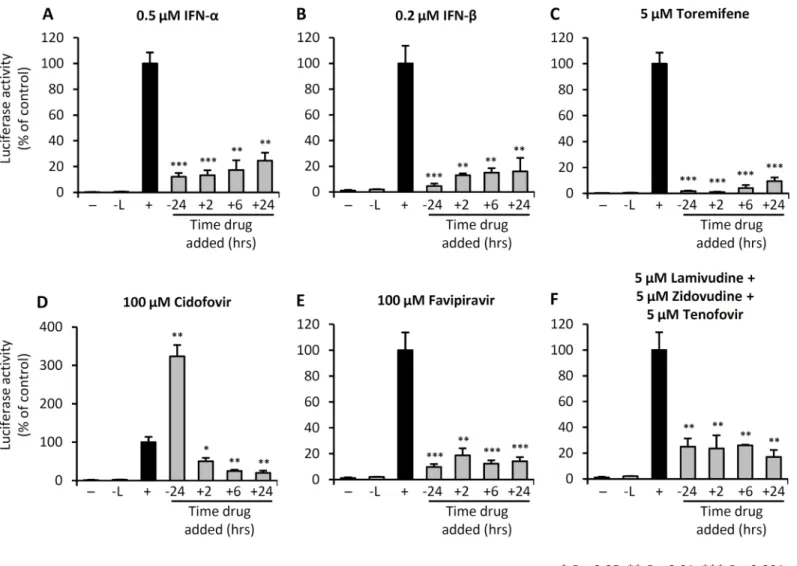
We employed an established mini-genome system to rapidly evaluate candidate drugs that could inhibitEbola Zairereplication under BSL 2 conditions [22–24]. At the outset we estab-lished the experimental conditions for infection with replication and transcription-competent virus like particles (trVLPs), by examining luciferase activity under various transfection and drug treatment conditions, which included transfection with viral support protein plasmids (S1 Fig). We included treatment with maraviroc, a CCR5 inhibitor, that would have no effect on trVLP entry and infection, thereby serving as a negative control for subsequent treatment regimens.
and FPV treatments, maximal inhibition of trVLP infection was achieved when the cells were treated prior to challenge with trVLP. By contrast, pre-treatment with CDF at 100μM, 24 hours prior to infection with trVLP, resulted in enhanced infection.
In subsequent dose-response studies, we compared the inhibitory effects of IFN-α, IFN-ß, TOR, CDF, FPV, 3TC, AZT or TFV when administered 24 hours post trVLP infection (Fig 2). The data inFig 2Isummarize the IC50dose for each drug. The IFNs exhibited the lowest IC50
values at 0.016μM for IFN-ß and 0.038μM for IFN-α. The data show a log-fold difference in IC50values for IFN-αand IFN-ß when compared in terms of U/ml, the norm for antiviral
activity measurements (Fig 2A and 2B). TOR had the next lowest IC50(0.36μM) and completely inhibited infection at doses>5μM (Fig 2C). TFV had an IC50at 0.98μM. CDF, 3TC and AZT all exhibited similar IC50values in the dose range 4.2–7.8μM, while FPV had the highest IC50of the nucleoside analogs at 36.8μM. At their IC50concentration, none of these
drugs directly inhibited luciferase reporter activity (S2 Fig). We observed a relatively small Fig 1. IFNs, toremifene, and nucleoside analogs inhibit trVLP replication.293 T cells were either left untreated (-), transfected with the mini-genome and the nucleocapsid plasmids NP, VP30, VP35 and T7 (-L), or transfected with the mini-genome and all expression plasmids to permit Ebola mini-genome entry and replication (+). (A-F) Cells were treated with the indicated drugs at the indicated doses, at each of the time points shown. Luciferase activity was measured 4 days post-trVLP infection. Values shown are the means of 4 biological replicates and are representative of 2 independent experiments. Error bars are the standard error of the mean. All drug treatment outcomes were statistically compared with the (+) control group. See alsoS1 Fig.
doi:10.1371/journal.pntd.0004364.g001
antiviral dose range for CDF (1.5–25μM) (Fig 2D), beyond which the drug appeared to enhance viral infection (S3 Fig). In cell viability assays we observe that at doses>10μM CDF affect cell viability, confounding the interpretation of the effects of CDF on viral replication.
In an orthogonal assay to confirm these findings, we next measured viral replication and transcription by qRT-PCR, following trVLP infection. trVLP-infected cells were either left untreated, or treated with the different drugs 24 hours post-infection, then viral replication and Fig 2. IFNs, toremifene and nucleoside analogs administered 24hrs post-exposure inhibit Ebola-mini-genome replication.293 T cells were either left untreated (-), transfected with the mini-genome and the nucleocapsid plasmids NP, VP30, VP35 and T7 (-L), or transfected with the mini-genome and all expression plasmids to permit Ebola mini-genome entry and replication (+), as described in Materials and Methods. 24 hours post-trVLP infection, cells were either left untreated, or treated with the indicated drugs (A-I) Dose-response plots for each of the indicated drugs. Luciferase activity (black circles) or cell viability (white squares) was measured 4 days post-infection (3 days after drug treatment). Values are the means of 4 biological replicates and are representative of 2 independent experiments. Error bars are the standard error of the mean. See alsoS2andS3Figs.
transcription evaluated 24 hours later (Fig 3). All treatments, with the exception of TOR, signif-icantly reduced the amount of genomic vRNA detected within cells (Fig 3A) and all treatments significantly reduced the synthesis of cRNA and mRNA isolated from infected cells (Fig 3B). Notably, IFN-ß treatment of trVLP-infected cells resulted in the greatest reduction in viral rep-lication and transcription.
Fig 3. IFNs, toremifene and nucleoside analogs reduce trVLP replication and transcription.293 T cells were transfected with support plasmids and infected with trVLP. 24 hours post-infection, cells were treated with the indicated drugs at their IC50doses, determined fromFig 2I. At 48hrs post-infection, total RNA was
extracted, reverse transcribed, then quantified by qPCR. Relative fold-change in -ve sense vRNA transcripts (A) and +ve sense cRNA and mRNA (B) was compared with infected, untreated cells (solvent,+ control). Technical duplicates were examined by qPCR, and means are the average of three biological replicates. Error bars are the standard error around the mean.
doi:10.1371/journal.pntd.0004364.g003
Next we examined the effectiveness of two and three drug combinations on trVLP infection. We first examined 28 two-drug combinations, using each drug at its IC50value, and used the
median-effect equation and combination index theorem [26] to determine drug synergy, addi-tive or sub-addiaddi-tive effects (Fig 4A). Synergy is defined as greater than additive effect when drugs were combined (CI<1), additive as the effect expected when combining each drug (CI = 1) and sub-additive as a smaller than expected additive effect (CI>1). When adminis-tered 24 hours post-infection, many of the two-drug combinations showed strong synergism in inhibiting trVLP replication (Fig 4J), with IFN-β+ 3TC demonstrating the greatest synergism (97.3% inhibition, CI = 0.028). 3TC was synergistic with all seven other drugs tested. Notably, when CDF was used in combination with FPV, AZT, TFV or IFN-α, it produced a sub-additive effect.
Next we tested all possible 56 three-drug combinations, using each drug at its IC50value, to
assess whether adding a third drug enhanced efficacy compared with two-drug combinations (Fig 4B–4I). This series of experiments served to validate our two-drug findings, as synergistic two-drug combinations such as IFN-β+ 3TC and IFN-β+ AZT, predicted strong synergy for the triple drug combination of IFN-ß + 3TC + AZT. As anticipated from the two-drug polygo-nogram, CDF was sub-additive when combined in three-drug combinations (Fig 4E). This was most evident even when CDF was administered in conjunction with two-drug combinations that had shown strong synergy, such as IFN-β+ 3TC or FPV + TFV, further indicating that CDF diminishes the antiviral effects of other drugs. IFN-ß, 3TC, AZT and TFV all promoted strong synergism when included in triple drug combinations, with IFN-β+ AZT specifically providing strong synergism when combined in three unique triple therapies.
From these two-drug and three-drug screens, we calculated the combination index (CI) and fractional inhibition (Fi) (Fig 4J and 4K). Many of the synergistic drug combinations (i.e. low CI) included one nucleoside analog and an IFN, while those drug combinations that were sub-additive all included CDF. IFN-βwas predominant in the most efficacious two- and three-drug combinations. In particular, IFN-β+ 3TC and IFN-β+ 3TC + AZT consistently exhibited the strongest synergism and highest Fi when administered 24 hours post-infection. Refer also toS1
andS2Tables.
In a final series of experiments, in order to validate our findings from the trVLP infection studies, we examined the antiviral effectiveness of IFN-ß, IFN-α, TOR, FVP, AZT, 3TC and TFV in 293T cells infected with ZEBOV (ZEBOV contained an eGFP reporter). CDF was excluded from these experiments. Initial dose-response studies were conducted at doses reflec-tive of those used in the trVLP experiments inFig 2. A higher dose of each drug was required to inhibit ZEBOV infection compared with trVLP infection (S4 Fig). Using the IC25of each
drug, we next evaluated 2 and 3 drug combinations for additive or synergistic effects against ZEBOV infection. All seven 2 drug combinations were synergistic (low CI) (Fig 5A), similar to the most synergistic combinations against trVLP inFig 4J. IFN-β+ 3TC proved to be the most synergistic 2 drug combination, analogous to trVLP infection. Of the most synergistic 3 drug combinations identified in the trVLP infection system, all seven exhibited synergy against ZEBOV infection, with IFN-β+ 3TC + AZT and IFN-β+ TOR + AZT exhibiting the strongest synergy (Fig 5B). The CIs determined from trVLP infection correlated well with those deter-mined using ZEBOV infection; specifically, the correlation coefficients (R2values) confirm this (Fig 5C and 5D).
Discussion
countries, ethicists, scientists, health care providers, logisticians and representatives from dif-ferent funding agencies were in attendance. A committee had been struck to evaluate the differ-ent vaccine candidates and therapeutic intervdiffer-entions being developed, which subsequdiffer-ently received an overwhelming number of submissions for consideration, and was hampered by an inability to compare antiviral effectiveness, sincein vitroand pre-clinicalin vivomodel systems Fig 4. 2 and 3 drug combinations synergistically inhibit Ebola trVLP infection.293 T cells were either left untreated, transfected with the mini-genome and the support plasmids NP, VP30, VP35, T7 but not L, or the mini-genome plus all the support plasmids. (A) Polygonogram of 28 two-drug combinations (at monotherapy IC50doses) administered at 24 hrs post-trVLP infection, then luciferase activity evaluated 3 days later. A thick red line represents strong
synergy between two drugs (CI<1), a thin black line represents additive effects (CI = 1), and a thick blue line represents strong sub-additive (less than additive) between two drugs (CI>1). (B-I) Polygonograms of 56 three-drug combinations. The backbone drug in each triple combination is listed above the heptagon, and the synergism/sub-additive effect of the additional two drugs is represented within each heptagon. (J-K) Combination index (CI) vs. fractional inhibition (Fi) plots of the most synergistic and sub-additive double (J) and triple (K) drug combinations on trVLP luciferase activity. Dotted lines identify thresholds of synergy/additive effects. Values shown are the means of 2–4 biological replicates in 2 independent experiments. Error bars are the standard error of the mean. See alsoS1andS2Tables.
doi:10.1371/journal.pntd.0004364.g004
Fig 5. Synergistic 2 and 3 drug combinations against trVLP-LUC infection inhibit fully infectious ZEBOV-GFP.(A-B) 293 T cells were infected with ZEBOV-eGFP at an MOI of 0.1, then treated with 2 and 3 drug combinations as indicated, 24 hours post-infection, at their monotherapy IC25doses. GFP
fluorescence was measured 3 days post-infection. Values are the means of 4 biological replicates, and error bars represent the standard error of the mean. Data are representative of 2 independent experiments. The combination index (CI) was plotted against the fractional inhibition (Fi) for each drug combination. (C-D) Plot of CIs for ZEBOV infections compared with CIs for trVLP infections.
vary, treatment regimens vary from prophylaxis to post-exposure administration, and direct readouts of antiviral efficacy differ. Moreover, given the virulence and high mortality associated with EVD, all of these studies have been conducted under BSL 4 conditions, limiting the num-ber of laboratories that can engage in these antiviral studies. Cognizant of these limitations, we employed the trVLP model system to compare the antiviral effectiveness of eight antiviral can-didates from three drug classes. We evaluated their antiviral activities in the context of inhibi-tion of Ebola replicainhibi-tion, using this mini-genome model that allows for rapid comparisons among compounds under BSL 2 conditions. The tetracistronic minigenome represents the most sophisticatedin vitroreplication model of Ebola virus to date. trVLPs proceed through every replication step as wild-type Ebola virus, and have been tested in multiple cell lines. Using TOR, there has been some validation of the trVLP assay. Specifically, TOR has been eval-uated in limiting Ebola virus infection of VeroE6 and HepG2 cells, and exhibited IC50values of
0.2μM and 0.03μM, respectively [27], in line with the IC50dose for TOR (0.36μM) observed with trVLP infection. Likewise, the IC50identified in the trVLP system for FPV (36.8μM), is consistent with that of 67μM recorded using Ebola virus infection [6], suggesting that this Ebola mini-genome system has relevance for screening potential antiviral compounds. Indeed, our validation studies using ZEBOV (ZEBOV-eGFP) suggest that the trVLP infection model has utility as anin vitroscreening assay when comparing different drugs as monotherapies or in 2 and 3 drug combinations.
As mentioned, the Ebola virus encodes in its genome factors that limit a type I IFN response to infection [16–18]. Yet, both rodent and non-human primate studies suggest that IFN-αand IFN-ß treatment can confer partial protection from infection, reducing viremia and prolonging survival [19–21], suggesting that it may be possible to override the inhibitory effects of the virus by treatment with IFN. At the outset, we conducted a series of experiments to compare the antiviral activities of IFN-αand IFN-ß in the trVLP infection system, and our findings sug-gest that whether treatment is administered prior to or post-infection, both IFN-αand IFN-ß exhibit antiviral activity. These findings only have relevance for thedirectantiviral activities of these IFNs, since the effects of IFN-αor IFN-ß on immune modulation for viral clearance can-not be determined using this system. Nevertheless, these data contributed to the decision to conduct a clinical trial of IFN-ß treatment for EVD in Guinea.
We provide evidence that the nucleoside/nucleotide analogs 3TC, AZT, TFV, FPV and CDF inhibit Ebola trVLP replicationin vitro. The results with 3TC are in contrast to published data that show no evidence for 3TC inhibiting Ebola virus infectionin vitro[27]. These studies examined the antiviral effectiveness of 3TC when administered one hour prior to infection, in contrast to our studies that have focused on post-exposure protection. In cells, the kinetics of 3TC phosphorylation are such that a minimum of four hours are required for optimal activity, perhaps distinguishing why our 24 hour pre-treatment, specifically a combination treatment, offered protection. Post-exposure treatment with 3TC and the other nucleoside/nucleotide analogs we examined, would more likely reveal activity against viral RNA synthesis than pre-treatment. When comparing the IC50values of each of the nucleoside analogs that we tested,
TFV exhibited the lowest IC50at ~1μM. Whether this reflects the fact that this adenosine monophosphate analog only requires two phosphorylation events to become an active drug versus three for the other nucleoside analogs, remains undetermined. Extensive published data reveal both the safety profiles [11,28,29] and the biodistribution of 3TC, AZT and TFV in the circulation and liver [30,31], the same compartments where Ebola infects monocytes, macro-phages, dendritic cells, endothelial cells and hepatocytes. Moreover, drug interactions with other nucleoside analogs have been well studied: e.g. tenofovir disoproxil fumarate, when used alone or in combination with emtricitabine effectively prevents HIV-1 infection in antiretrovi-ral pre-exposure prophylaxis (PrEP) [29].
Our studies also revealed that the active metabolite of brincidofovir, CDF, has a narrow therapeutic window of efficacy (6.25–25μM) when assessed in the trVLP assay, enhancing viral replication at higher doses when added either prior to or post-infection. In cell viability assays, CDF exhibits cytotoxicity at doses>10μM. These findings suggest that caution is required if CDF is to be considered further for the treatment of EVD, specifically that phase I/II trials define the safety profile of this drug for EVD.
Another advantage of thisin vitrosystem is that it allowed us to evaluate various 2 and 3 drug combinations and demonstrates that combination treatments limit viral replication up to 97.3%. A benefit of combination treatment is the potential to limit/avoid the emergence of drug resistance. Interestingly, IFN-ß was predominant among all the 8 antivirals considered in terms of contributing very strong synergism in combination treatments: e.g. IFN-ß + 3TC; IFN-ß + 3TC + AZT. Using this system, we observe that FPV, when administered 24 hours post-infection, has an IC50of ~ 37μM. To date, the phase II/III JIKI trial examining the efficacy of FPV against EVD has reported only modestly encouraging results. In our 2 drug combina-tion treatment studies we show that, with the excepcombina-tion of CDF, whenever FPV is included, synergy occurs, effectively reducing the CI. It may transpire that for treating EVD, FPV is most effective in a drug combination regimen.
Viewed altogether, we present anin vitroEbola trVLP screening system, that requires only level 2 biocontainment, which allowed us to compare the antiviral activities of 8 compounds, either alone or in combination. We provide evidence that IFNs are effective inhibitors of Ebola replication, with IFN-ß exhibiting greater efficacy over IFN-α, or when used in combination with nucleoside analogs. We infer from our data that whether IFN-ß treatment is administered 24 hours prior to, or up to 24 hours post-infection, reduced Ebola replication is achieved. As additional antiviral therapeutic candidates become available, we now have the capability to measure and compare their direct antiviral activities with the existing panel. This allows for rapidin vitroevaluation and the opportunity to prioritize antiviral candidates for further pre-clinical and pre-clinical trial studies.
Materials and Methods
Cell culture and trVLP infection
We employed an established mini-genome system to rapidly evaluate candidate drugs that could inhibitEbola Zairereplication under BSL 2 conditions [22]. The mini-genome encodes 3 of the 7 Ebola proteins (VP24, VP40 and GP1,2) and a luciferase reporter gene. Expression
plas-mids for the remaining four Ebola nucleocapsid proteins (L, NP, VP30 and VP35) were also included during transfection. Cell culture conditions and virus infections were performed as previously described [22]. Briefly, 80,000 producer 293 T cells (American Type Culture Collec-tion; ATCC, Rockville, USA) were seeded in individual wells of 24-well plates in 400μL Dulbec-co’s Modified Eagle Medium (DMEM) containing 10% FBS, 1% penicillin and 1%
streptomycin, and grown in 5% C02atmosphere at 37°C. Cells were transfected with the viral
replication protein plasmids (L, NP, VP30, VP35), a tetracistronic Ebola mini-genome and the T7 polymerase, using the CalPhos Mammalian Transfection Kit (Clontech Laboratories). 24 hours later, medium was replaced with 800μL DMEM with 5% FBS. The replication and tran-scription-competent virus like particles (trVLPs) were harvested 3 days later. Virus stock was frozen at -80°C.
then added to target cells. Medium was removed the following day and replaced with 800μL DMEM with 5% FBS. Four days post-infection, the medium was aspirated and cells were re-suspended in 200μL of 1xRenilaLuciferase Assay Lysis Buffer (RenillaLuciferase Assay Sys-tem, Promega). Lysates were assayed for luciferase activity.
ZEBOV-eGFP infection
We generated recombinant ZEBOV expressing enhanced green fluorescent protein (eGFP) from cDNA clones of full-length infectious ZEBOV, as previously described [32]. The eGFP reporter protein was expressed as an eighth gene, and the virus exhibited anin vitrophenotype similar to wild-type ZEBOV. Notably,in vivo, incorporation of GFP into wild-type ZEBOV results in some attenuation of disease [32]. All work with infectious ZEBOV was performed in biosafety level 4 (BSL4), at the National Microbiology Laboratory of the Public Health Agency of Canada in Winnipeg, Manitoba.
30,000 293 T cells were seeded in 96-well plates in 100μL DMEM with 10% FBS. 24 hours thereafter, the medium was replaced with 100μL DMEM with 10% FBS containing
ZEBOV-GFP at an MOI of 0.1. 24 hours post-infection, the medium was removed and replaced with 200μL of DMEM with 5% FBS, or 190μL DMEM with 5% FBS and 10μL of single or com-binations of drugs. eGFP fluorescence was measured 3 days post-infection using a Synergy HTX Multi-Mode Microplate Reader (BioTek).
Drugs
For these experiments, we used toremifiene citrate (TOR; Sigma), cidofovir hydrate (CDF; Sigma) favipiravir (FPV, T-705; Cellagen Technology), lamivudine (3TC; Sigma) zidovudine (AZT), tenofovir (TFV) maraviroc (MVC; NIH AIDS Reagent Program), Infergen (IFN alfa-con-1, Pharmunion Bsv Development Ltd.) or human interferon beta-1a (IFN-β, Avonex; Biogen).
Mini-genome RNA extraction and qRT-PCR quantification of viral RNA
Forty-eight hours after trVLP infection, medium was aspirated from 293 T cells that had either been left untreated or treated with the various drugs and total RNA extracted from cell lysates with 500μL of TRIzol (Thermo Fisher Scientific). cDNA synthesis was performed on 5μg of total RNA, using the First-Strand cDNA Synthesis Kit (GE Healthcare Life Sciences), according to the manufacturer’s instructions. A 20μl reaction also contained bulk first-strand cDNA reaction mix, DTT solution and 40 pmol of one of two trVLP specific primers [33]: vRNA for-ward (5’-GGC CTC TTC TTA TTT ATG GCG A -3’), or cRNA/mRNA reverse (5’-AGA ACC ATT ACC AGA TTT GCC TGA-3’). Both primers were synthesized by the Center for Applied Genomics (The Hospital for Sick Children, Toronto, Canada). Real-time qPCR reactions (25μl) were conducted in duplicate, using the Rotor-Gene RG-3000 thermocycler (Corbett Research, Montreal, Canada). Each reaction contained 100 ng template cDNA, 12.5μL 2 x SYBR Green PCR Master Mix (Applied Biosystems, Warrington, UK), 300 nM of both the for-ward (vRNA) and reverse (cRNA/mRNA) primers, and PCR grade H2O (Roche Diagnostics,
Indianapolis, USA). Samples lacking reverse transcriptase (No RT) during first-strand cDNA synthesis served as negative controls. Cycling parameters were as follows: initial denaturation at 95°C for 10 min, followed by 40 cycles of amplification with 95°C for 15 seconds, 56°C for 30 seconds, and 60°C for 30 seconds. Biological triplicates in the drug-treated groups were nor-malized to the average Ct of infected cells given DMSO solvent alone, by the 2-ΔCTcomparative
CTmethod.
Cell viability assay
Dose-response cytotoxicity/viability assays were conducted in 293 T cells 4 days post-infection for each of the drugs examined, either alone or in the various combinations indicated, using the MTT assay as previously described [34].
Statistics
Means were compared using a two-tailed, unpaired Student’sttest and corrected for multiple comparisons. For all figures, () denotes apvalue<0.05, () denotes apvalue<0.01 and () denotes apvalue<0.001. Error bars shown are the standard error around the mean (SEM). Synergy between two and three-drug combinations, combination index (CI) and dose-reduc-tion index (DRI) were calculated with CompuSyn Version 1.0 [26]. The coefficient of determi-nation (R2) was determined for simple linear regressions.
Supporting Information
S1 Fig. IFNs and nucleoside analogs inhibit Ebola mini-genome replicationin vitro.(A)
293 T cells were either left untreated (-), transfected with the mini-genome and the viral sup-port protein plasmids NP, VP30, VP35, T7 but not L (-L), or transfected with the mini-genome and all support plasmids that permit Ebola mini-genome entry and replication (+). At -24, 0, and +24 hours relative to infection, cells were treated with 5μM of maraviroc (MVC), lamivu-dine (3TC), zidovulamivu-dine (AZT), or tenofovir (TFV), or combinations of the three nucleoside analogs. Luciferase activity was measured four days after infection with trVLPs. (B) Luciferase activity of first passage of 293 T cells (1) treated with nucleoside analogs, and second passage of 293 T cells (2) treated with nucleoside analogs. (C) Luciferase activity of 293 T cells after treat-ing with 5μM combination of nucleoside analogs, cidofovir (CDF) or favipiravir (FPV). (D) Luciferase activity of 293 T cells treated with IFN-αor IFN-β. The values are means of four bio-logical replicates and are representative of two independent experiments. Error bars are the standard error of the mean. All drug treatment outcomes were statistically compared with the (+) control group in panels A, C and D.
(TIF)
S2 Fig. No direct effect of drugs on Renilla luciferase reporter assay.293 T cells transfected and then infected with trVLP were lysed 4 days post-infection. Cell lysate suspended in CCLR reagent was aliquoted into separate tubes and spiked with each drug at the IC50dose fromFig 2Ior DMSO solvent (+). Luciferase activity was then quantified. Values are the means of 3 independent experiments. Error bars are the standard error of the mean.
(TIF)
S3 Fig. CDF treatment increases trVLP replication.293 T cells were transfected with all expression plasmids that permit Ebola mini-genome entry and replication, then treated with the indicated doses of CDF 24hrs post-trVLP infection. Luciferase activity was measured four days after cells were infected (3 days post-CDF treatment). Values are the means of 4 biological replicates and are representative of 2 independent experiments. Error bars are the standard error of the mean. CDF treatment outcomes were compared with the (+) control group. (TIF)
quantified relative to infected, untreated cells (DMSO solvent control). Values are the means of 4 biological replicates. Error bars are the standard error of the mean.
(TIF)
S1 Table. Fi and CI values for two-drug combination treatments.Experimental details are as described in the legend toFig 4. The fractional inhibition, combination index and strength of synergy (+), additive effects (+/-) or sub-additive (-) of each 2 drug combination therapy are shown.
(TIF)
S2 Table. Fi and CI values for three-drug combination treatments.Experimental details are as described in the legend toFig 4. The fractional inhibition, combination index and strength of synergy (+), additive effects (+/-) or sub-additive (-) of each 3 drug combination therapy are shown.
(TIF)
Acknowledgments
We thank C. Tong and M. Blacquiere for assistance with experiments and Dr. R. Siemieniuk for stimulating discussions. The authors gratefully acknowledge the donation of IFN alfacon-1 by Richard Zakhia, Pharmunion Bsv Development.
Author Contributions
Conceived and designed the experiments: ENF DRB SDSM TH GPK. Performed the experi-ments: SDSM BMK TR HNK. Analyzed the data: ENF DRB SDSM. Contributed reagents/ materials/analysis tools: DRB TH GPK DPB. Wrote the paper: ENF DRB SDSM.
References
1. World Health Organization, Ebola Situation Report–16 December 2015.http://apps.who.int/ebola/en/ current-situation/ebola-situation-report-13-december-2015.
2. Wong G, Audet J, Fernando L, Fausther-Bovendo H, Alimonti JB, Kobinger GP, et al. Immunization with vesicular stomatitis virus vaccine expressing the Ebola glycoprotein provides sustained long-term protection in rodents. Vaccine. 2014; 32: 5722–9. doi:10.1016/j.vaccine.2014.08.028PMID:25173474 3. Stanley DA, Honko AN, Asiedu C, Trefry JC, Lau-Kilby AW, Johnson JC, et al. Chimpanzee adenovirus
vaccine generates acute and durable protective immunity against ebolavirus challenge. Nat Med. 2014; 20: 1126–9. doi:10.1038/nm.3702PMID:25194571
4. Qiu X, Wong G, Audet J, Bello A, Fernando L, Alimonti JB, et al. Reversion of advanced Ebola virus dis-ease in nonhuman primates with ZMapp. Nature. 2014; 514: 47–53. doi:10.1038/nature13777PMID: 25171469
5. Wong KK, Perdue CL, Malia J, Kenney JL, Peng S, Gwathney JK, et al. Supportive Care of the First 2 Ebola Virus Disease Patients at the Monrovia Medical Unit. Clin Infect Dis. 2015; 61:e47–51. doi:10. 1093/cid/civ420PMID:26021993
6. Oestereich L, Ludtke A, Wurr S, Rieger T, Munoz-Fontela C, Gunther S. Successful treatment of advanced Ebola virus infection with T-705 (favipiravir) in a small animal model. Antiviral Res. 2014; 105:17–21. doi:10.1016/j.antiviral.2014.02.014PMID:24583123
7. Warren TK, Wells J, Panchal RG, Stuthman KS, Garza NL, Van Tongeren SA, et al. Protection against filovirus diseases by a novel broad-spectrum nucleoside analogue BCX4430. Nature. 2014; 508: 402– 5. doi:10.1038/nature13027PMID:24590073
8. Florescu DF, Keck MA. Development of CMX001 (Brincidofovir) for the treatment of serious diseases or conditions caused by dsDNA viruses. Expert Rev Anti Infect Ther. 2014; 12: 1171–8. doi:10.1586/ 14787210.2014.948847PMID:25120093
9. Muhlberger E, Weik M, Volchkov VE, Klenk HD, Becker S. Comparison of the transcription and replica-tion strategies of marburg virus and Ebola virus by using artificial replicareplica-tion systems. J Virol. 1999; 73: 2333–42. PMID:9971816
10. Jácome R, Becerra A, de León SP, Lazcano A. Structural Analysis of Monomeric RNA- Dependent Polymerases: Evolutionary and Therapeutic Implications. PLoS One. 2015; 10:e0139001. doi:10. 1371/journal.pone.0139001PMID:26397100
11. Cassetti I, Madruga JV, Suleiman JM, Etzel A, Zhong L, Cheng AK, et al. The safety and efficacy of tenofovir DF in combination with lamivudine and efavirenz through 6 years in antiretroviral-naive HIV-1-infected patients. HIV Clin Trials. 2007; 8: 164–72. PMID:17621463
12. Phanuphak N, Ananworanich J, Teeratakulpisarn N, Jadwattanakul T, Kerr SJ, Chomchey N, et al. A 72-week randomized study of the safety and efficacy of a stavudine to zidovudine switch at 24 weeks compared to zidovudine or tenofovir disoproxil fumarate when given with lamivudine and nevirapine. Antivir Ther. 2012; 17: 1521–31. doi:10.3851/IMP2497PMID:23220732
13. World Health Organization, WHO Model List of Essential Medicines: 18thlist October 2013:http://www.
who.int/medicines/publications/essentialmedicines/en/.
14. Ahmed Ouameur A, Marty R, Neault JF, Tajmir-Riahi HA. AZT binds RNA at multiple sites. DNA Cell Biol. 2004; 23: 783–8. PMID:15585137
15. Cardenas WB. Evasion of the interferon-mediated antiviral response by filoviruses. Viruses. 2010; 2: 262–82. doi:10.3390/v2010262PMID:21994610
16. Xu W, Edwards MR, Borek DM, Feagins AR, Mittal A, Alinger JB, et al. Ebola virus VP24 targets a unique NLS binding site on karyopherin alpha 5 to selectively compete with nuclear import of phosphor-ylated STAT1. Cell Host Microbe. 2014; 16: 187–200. doi:10.1016/j.chom.2014.07.008PMID: 25121748
17. Fabozzi G, Nabel CS, Dolan MA, Sullivan NJ. Ebolavirus proteins suppress the effects of small interfer-ing RNA by direct interaction with the mammalian RNA interference pathway. J Virol. 2011; 85: 2512– 23. doi:10.1128/JVI.01160-10PMID:21228243
18. Chang TH, Kubota T, Matsuoka M, Jones S, Bradfute SB, Bray M, et al. Ebola Zaire virus blocks type I interferon production by exploiting the host SUMO modification machinery. PLoS Pathog. 2009; 5: e1000493. doi:10.1371/journal.ppat.1000493PMID:19557165
19. Smith LM, Hensley LE, Geisbert TW, Johnson J, Stossel A, Honko A, et al. Interferon-beta therapy pro-longs survival in rhesus macaque models of Ebola and Marburg hemorrhagic fever. J Infect Dis. 2013; 208: 310–8. doi:10.1093/infdis/jis921PMID:23255566
20. Qiu X, Wong G, Fernando L, Audet J, Bello A, Strong J, et al. mAbs and Ad-vectored IFN-alpha therapy rescue Ebola-infected nonhuman primates when administered after the detection of viremia and symp-toms. Sci Transl Med. 2013; 5: 207ra143. doi:10.1126/scitranslmed.3006605PMID:24132638 21. Qiu X, Wong G, Fernando L, Ennis J, Turner JD, Alimonti JB, et al. Monoclonal antibodies combined
with adenovirus-vectored interferon significantly extend the treatment window in Ebola virus-infected guinea pigs. J Virol. 2013; 87: 7754–7. doi:10.1128/JVI.00173-13PMID:23616649
22. Hoenen T, Watt A, Mora A, Feldmann H. Modeling The Lifecycle Of Ebola Virus Under Biosafety Level 2 Conditions With Virus-like Particles Containing Tetracistronic Minigenomes. J Vis Exp. 2014; 91: 52381. doi:10.3791/52381PMID:25285674
23. Watt A, Moukambi F, Banadyga L, Groseth A, Callison J, Herwig A, et al. A novel life cycle modeling system for Ebola virus shows a genome length-dependent role of VP24 in virus infectivity. J Virol. 2014; 88: 10511–10524. doi:10.1128/JVI.01272-14PMID:24965473
24. Hoenen T, Feldmann H. Reverse genetics systems as tools for the development of novel therapies against filoviruses. Expert Rev Anti Infect Ther. 2014; 12: 1253–1263. doi:10.1586/14787210.2014. 948848PMID:25169588
25. Johansen LM, Brannan JM, Delos SE, Shoemaker CJ, Stossel A, Lear C, et al. FDA-approved selective estrogen receptor modulators inhibit Ebola virus infection. Sci Transl Med. 2013; 5:190ra79. doi:10. 1126/scitranslmed.3005471PMID:23785035
26. Chou TC. Theoretical basis, experimental design, and computerized simulation of synergism and antagonism in drug combination studies. Pharmacol Rev. 2006; 58: 621–81. PMID:16968952 27. Johansen LM, DeWald LE, Shoemaker CJ, Hoffstrom BG, Lear-Rooney CM, Stossel A et al. A screen
of approved drugs and molecular probes identifies therapeutics with anti-Ebola virus activity. Science Transl Med. 2015; 7:290ra89.
28. Lok AS, Lai CL, Leung N, Yao GB, Cui ZY, Schiff ER, et al. Long-term safety of lamivudine treatment in patients with chronic hepatitis B. Gastroenterology. 2003; 125: 1714–22. PMID:14724824
30. Di Mascio M, Srinivasula S, Bhattacharjee A, Cheng L, Martiniova L, Herscovitch P, et al. Antiretroviral tissue kinetics: in vivo imaging using positron emission tomography. Antimicrob Agents Chemother. 2009; 53: 4086–95. doi:10.1128/AAC.00419-09PMID:19667288
31. Parang K, Wiebe LI, Knaus EE. Pharmacokinetics and tissue distribution of (+/-)-3'-azido-2',3'-dideoxy-5'-O-(2-bromomyristoyl)thymidine, a prodrug of 3'-azido-2',3'-dideoxythymidine (AZT) in mice. J Pharm Pharmacol. 1998; 50: 989–96. PMID:9811159
32. Ebihara H, Theriault S, Neumann G, Alimonti J, Geisbert J, Hensley L, et al. In vitro and in vivo charac-terization of recombinant Ebola viruses expressing enhanced green fluorescent protein. J Infect Dis. 2007; 196 Suppl 2:S313–22. PMID:17940966
33. Hoenen T, Jung S, Herwig A, Groseth A, Becker S. Both matrix proteins of Ebola virus contribute to the regulation of viral genome replication and transcription. J Virol. 2010; 403: 56–66.
34. Mehrotra S, Sharma B, Joshi S, Kroczynska B, Majchrzak B, Stein BL, et al. Essential role for the Mnk pathway in the inhibitory effects of type I interferons on myeloproliferative neoplasm (MPN) precursors. J Biol Chem. 2013; 288: 23814–22. doi:10.1074/jbc.M113.476192PMID:23814052