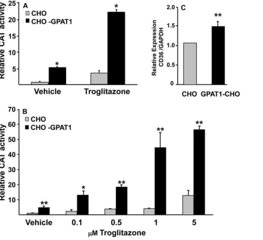
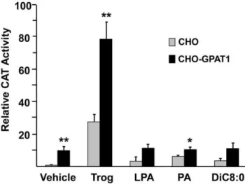
Lysophosphatidic acid activates peroxisome proliferator activated receptor-γ in CHO cells that over-express glycerol 3-phosphate acyltransferase-1.
Texto
Imagem


![Table 2. LPA and PA displace [ 3 H]rosiglitazone from PPARc- PPARc-LBD. Glycerolipid species Percent displacement of[3H]rosiglitazone sn -1-18:0-LPA 25.961.5 sn -1-16:0-LPA 29.661.2 sn-1, 2-di18:1-PA 24.2 6 0.8 sn -1-16:0, 2-18:1-PA 21.760.9](https://thumb-eu.123doks.com/thumbv2/123dok_br/17111694.237988/5.918.91.609.92.607/table-displace-rosiglitazone-glycerolipid-species-percent-displacement-rosiglitazone.webp)
Documentos relacionados
Overall, the data presented herein suggested that higher expression of PPAR c and NF- k B mRNA and protein and PPAR c up-regulated/dysregulation NF- k B expression and then
To test whether the association with membrane rafts is required for gonococcal invasion, CHO SREC-I and CHO SREC-I D CD transfected cells were treated with the membrane
In the present study, BPE treatment strongly suppressed C/EBP b mRNA and protein expression and markedly reduced the expression levels of C/EBP a and PPAR c compared with those
Expression of the kinase-dead mutant of MST2, along with SAV1 and PPAR c , showed a similar increase in the reporter activity in the absence of rosiglitazone as compared to
(C) Membrane to cytosolic ratio for the expression of ZERO or STREX proteins when co-transfected with b 1-BK Ca in cultured CHO-K1 cells.. alteration in channel protein
Thiazolidinediones, which are potent peroxisome proliferator- activated receptor- c (PPAR c ) agonists, have been shown to increase expression of UCP-2 in several tissues [32],
Variation within each of the subclones (CV = 0.5 to 0.7) was already comparable to the parental clone (CV = 0.5 at this timepoint) and did not trend with time or expression
Minimal expression and activity of PPAR c was detected in mock-infected and infected PPAR c VillinCre+ mice compared to littermate control PPAR c VillinCre- and wild-type (C57BL/6)
