Ca
Subunit Splice Variants in Resistance Arteries from Rat
Cerebral and Skeletal Muscle Vasculature
Zahra Nourian1, Min Li1, M. Dennis Leo3, Jonathan H. Jaggar3, Andrew P. Braun4, Michael A. Hill1,2*
1Dalton Cardiovascular Research Center, University of Missouri, Columbia, Missouri, United States of America,2Department of Medical Pharmacology and Physiology, University of Missouri, Columbia, Missouri, United States of America,3Department of Physiology, University of Tennessee Health Science Center, Memphis, Tennessee, United States of America,4Department of Physiology and Pharmacology, University of Calgary, Calgary, Alberta, Canada
Abstract
Previous studies report functional differences in large conductance Ca2+activated-K+channels (BK
Ca) of smooth muscle cells
(VSMC) from rat cerebral and cremaster muscle resistance arteries. The present studies aimed to determine if this complexity in BKCaactivity may, in part, be due to splice variants in the pore-forminga-subunit. BKCavariants in the intracellular C
terminus of thea-subunit, and their relative expression to totala-subunit, were examined by qPCR. Sequencing of RT-PCR products showed twoa-subunit variants, ZERO and STREX, to be identical in cremaster and cerebral arteries. Levels of STREX mRNA expression were, however, significantly higher in cremaster VSMCs (28.964.2% of total a-BKCa) compared with
cerebral vessels (16.560.9%). Further, a low level of BKCa SS4 a-subunit variant was seen in cerebral arteries, while
undetectable in cremaster arteries. Protein biotinylation assays, in expression systems and arterial preparations, were used to determine whether differences in splice variant mRNA expression affect surface membrane/cytosolic location of the channel. In AD-293 and CHO-K1 cells, rat STREX was more likely to be located at the plasma membrane compared to ZERO, although the great majority of channel protein was in the membrane in both cases. Co-expression ofb1-BKCasubunit with
STREX or ZERO did not influence the dominant membrane expression ofa-BKCasubunits, whereas in the absence ofa-BKCa,
a significant proportion ofb1-subunit remained cytosolic. Biotinylation assays of cremaster and cerebral arteries showed that differences in STREX/ZERO expression do not alter membrane/cytosolic distribution of the channel under basal conditions. These data, however, revealed that the amount ofa-BKCain cerebral arteries is approximately 20X higher than in
cremaster vessels. Thus, the data support the major functional differences in BKCaactivity in cremaster, as compared to
cerebral VSMCs, being related to totala-BKCaexpression, regardless of differences in splice variant expression.
Citation:Nourian Z, Li M, Leo MD, Jaggar JH, Braun AP, et al. (2014) Large Conductance Ca2+
-Activated K+
Channel (BKCa)a-Subunit Splice Variants in Resistance
Arteries from Rat Cerebral and Skeletal Muscle Vasculature. PLoS ONE 9(6): e98863. doi:10.1371/journal.pone.0098863
Editor:Yu Huang, The Chinese University of Hong Kong, Hong Kong
ReceivedJanuary 17, 2014;AcceptedMay 7, 2014;PublishedJune 12, 2014
Copyright:ß2014 Nourian et al. This is an open-access article distributed under the terms of the Creative Commons Attribution License, which permits unrestricted use, distribution, and reproduction in any medium, provided the original author and source are credited.
Funding:This research was supported by the National Institutes of Health (RO1 HL092241: Michael A. Hill and P01 HL095486: Gerald Meininger, P.I.) and the Natural Sciences and Engineering Research Council of Canada (RGPIN/312240: Andrew P. Braun). The funders had no role in study design, data collection and analysis, decision to publish, or preparation of the manuscript.
Competing Interests:The authors have declared that no competing interests exist.
* E-mail: [email protected]
Introduction
Potassium channels play an important role in the regulation of VSMC membrane potential and contractile activity. In particular, large conductance Ca2+
-activated, K+
channels (BKCa) are
activated in response to membrane depolarization and increases in intracellular Ca2+
to affect membrane hyperpolarization [1,2]. While BKCa channels are widely expressed in both electrically
excitable and non-excitable cells [3,4], they are relatively abundant in smooth muscle and play a key role in the regulation of vascular tone [5,6]. Structurally, the functional BKCachannel
exists as a tetramer of a-subunits forming the ion channel pore together with tissue specific auxiliaryb-subunits (b1–b4) which are typically present in a 1:1 stoichiometry [7]. The BKCaa-subunit
consists of seven transmembrane spanning domains (S0–S6) including the extracellular N-terminus, P-loop between S5 and S6 domains, and a large intracellular C terminus containing a number of regulatory sites including the regulators of conductance for K+
(RCK 1 and 2) and 2–3 Ca2+
binding sites.
The BKCaa-subunit is encoded by a single gene (KCNMA1)
containing 27 distinct exons, in contrast to eachb-subunit, which is encoded by four distinct exons [8]. BKCachannels appear to
achieve part of their functional diversity through alternative pre-mRNA splicing of the KCNMA1 gene [9,10]. Up to ten alternative splicing sites have been identified for the vertebrate BKCaa-subunit [11]. Most variation occurs in the intracellular
C-terminal part in the linker region between domains RCK1 and RCK2 and upstream of the ‘‘calcium bowl’’ [12]. Alternative splicing can modify the functional properties of BKCa channels,
including Ca2+
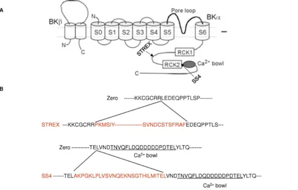
and voltage sensitivity, cell surface expression, and regulation by diverse intracellular signaling pathways. One of the most thoroughly studied a-BKCa splice variants is the STREX
exon (STRess axis regulated EXon), which derives its name from its splicing regulation by stress-axis hormones [13]. It has been shown that the STREX exon (an insertion of 58 amino acids in the C-terminal splice site 2 of thea-subunit protein) confers distinct functional phenotypes onto BKCachannels, such as altered Ca2+
signaling from stimulatory to inhibitory, compared with the ZERO variant, lacking this insert [9,14–17]. It has also been shown that BKCachannels containing the SS4 splice variant (an
insertion of a 27 amino acid segment upstream to the C-terminal Ca2+
-bowl in splice site 4 of thea-subunit) were activated more rapidly than the ZERO variant in the presence of the same voltage stimulus, and the difference in these activation kinetics was dependent on the concentration of intracellular Ca2+[18], (also
see Figure 1).
Alternatively, it has been reported that intracellular trafficking ofa-BKCamay be one of the main post-translational modifications
that can regulate the number of ion channels at the cell surface [19]. This mode of regulation can also be modulated by accessory
b-subunits. While limited studies have addressed the effects ofa -BKCasplice variants on channel trafficking to plasma membrane
[20–24], there is some discrepancy in the reported findings. For example, Kim et al (2007) reported the expression of twoa-BKCa
splice variants, termed VEDEC and QEDRL, in chick ciliary ganglion neurons that differ at the extreme C-terminus. Using HEK293T and NG108-15 cells and a cell surface biotinylation assay, QEERL channels showed markedly higher levels of constitutive expression of a-BKCa at the plasma membrane
compared with VEDEC channels, which tend to remain in the cytosol [21]. The same group further showed that co-expression of avian b1-subunits with the VEDEC isoform a-BKCa, prevented
the inhibitory effect of the VEDEC sequence on cell surface expression [20]. In contrast, studies from Toro and colleagues (2006) showed that co-expression of human b1-subunit with a human pore-forminga-subunit enhanced internalization of thea -subunit [25].
Previous electrophysiological studies from our laboratory have demonstrated that BKCa channel activity differs significantly in
VSMCs from cremaster muscle arteries compared with cerebral arteries. In particular, our functional data have revealed a
decreased Ca2+
sensitivity of cremaster BKCachannels, resulting
in more positive levels of Em being required in cremaster VSM cells to generate similar levels of outward K+
conductance [26]. Similarly, Jackson and Blair (1998) described cremaster muscle BKCachannels as being normally ‘silent’, but suggested that their
activity could be ‘recruited’ during vasoconstriction [27]. There-fore in this study, we first hypothesized that the functional differences between these two resistance vasculatures may be due, in part, to the expression of splice variants of the BKCaa-subunit.
We chose to focus on STREX and SS4 splice variants, as splice sites where they are located (i.e. 2 and 4) contain regulatory phosphorylation, palmitoylation and Ca2+
interaction sites that could functionally impact channel activity. We further hypothe-sized that an additional influence ofa-subunit splice variation may be on the surface membrane location of the channel and whether this could be affected by the accessoryb1-subunit.
Material and Methods
Tissue isolation and vessel RNA purification
All experiments and protocols were approved by the Animal Care and Use Committee, University of Missouri, USA. Our studies used male Sprague-Dawley rats weighing between 180– 280 g. Rats were anaesthetized with sodium pentobarbital (Nembutal, 100 mg/kg body weight) given by an intraperitoneal injection. Cremaster muscles were surgically removed, as previ-ously described [28], and placed in a cooled (4uC) dissection chamber. Following sacrifice by anesthetic overdose, a craniotomy was performed and the brain was removed intact and similarly placed in a cooled dissection chamber.
First- and second-order arterioles (1A/2A) from cremaster muscle and mid-cerebral arteries were isolated and rapidly subjected to total RNA purification using a Melt Total Nucleic Acid isolation system kit (Life Technologies, Carlsbad, CA, USA)
Figure 1. Location and amino acid sequences of ZERO and STREX splice variants of BKCaa-subunit. Schematic diagram illustrating (A)
sites of STREX and SS4 splicing variants ofa-BKCaand (B) the amino acid sequences of the splicing inserts (Adapted from reference [5]).
following the manufacturer’s instructions. All samples were treated by TURBO DNase digestion (Life Technologies) to minimize contamination with genomic DNA. The concentration and purity of RNA for each sample was determined by UV absorbance using a Nanodrop ND-1000 spectrophotometer (Thermo Scientific, Rockford, IL, USA) and samples were stored at 280uC until conversion to cDNA. Equal amounts of total vessel RNA extract were then reverse-transcribed into a single strand cDNA using a Superscript III First-Strand synthesis system (Life Technologies) according to the manufacturer’s instructions.
Real-time quantitative PCR
Real-time PCR was performed in triplicate, in 96-well plates, on cDNAs prepared from each sample (n = 4–5) using KAPA SYBER FAST qPCR Kit Master mix (KAPA Biosystems, Woburn, MA, USA). PCR was performed using a Mastercycler EP Realplex2
(Eppendorf-North America, Westbury, NY, USA). Reaction volume/well contained 20ml:10ml of master mix, 1ml of each
sense and antisense primers (5mM), 1ml of cDNA template and
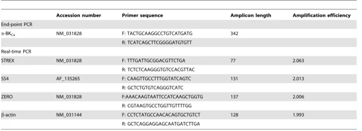
the remainder DNase-free water. Primers used in this study were based on previously published papers: ZERO [26] STREX [9], and b-actin [29]. Primers for SS4 variant were designed using Real-Time PCR primer software from Integrated DNA Technol-ogies (IDT). Details of oligo-DNA primers used to amplify BKCa a-subunits (STREX, SS4) and ZERO variants, accession numbers for the template sequences and the expected product sizes are shown in Table 1. ZERO variant primers were designed in regions of transmembrane domains in which no splice variant existed, and its expression was utilized as an indication of the total expresseda -subunit mRNA [9,13].
Real-time PCR protocols were performed as follows: pre-heating at 95uC for 2 min, 40 cycles of two-step cycling of denaturation at 95uC for 3 sec and annealing/extension steps of 25 sec at 58uC. For each qPCR determination, no enzyme and no template conditions were included to test for contamination of assay reagents. An arbitrary rat mid-cerebral artery cDNA sample was included in each plate to provide a constant calibrator point for all samples. After the final PCR cycle, a melting curve analysis was routinely performed to identify the presence of primer-dimers and to analyze the specificity of the reaction. Data were collected and analyzed using Realplex software (Eppendorf-North America).
The amplification efficiencies between targets and housekeeping genes (i.e.b-actin) were initially verified to be approximately equal (Table 1), allowing the comparative threshold (Ct) method for quantification to be used [30]. The relative expression level (R) was calculated with equations as follows: R = 22DDCt
= 22(DCt sample 2 DCt calibrator)
for the target genes in each sample set according to the published 22DDCt
method [30]. Changes in mRNA expression levels were calculated from an average of triplicate measurements and are reported as fold changes relative to the ZERO variant, after normalization tob-actin. Data were analyzed using an unpaired studentt-test: a statistically significant difference was assumed at P#0.05.
Cell surface Biotinylation assay on cultured cells
Plasmid constructs containing cDNA for full-length rat BKCa
ZERO variant or BKCaSTREX variant (gifts from Dr. Michael J.
Shipston) were transiently transfected into AD-293 cells (240085, Agilent Technology, Santa Clara, CA, USA) or CHO-K1 cells (CCL-61, ATCC, Manassas, VA, USA) with FuGENE 6 Transfection Reagent (Roche Diagnostics, Indianapolis, IN, USA). A bovineb1-BKCa plasmid DNA was also used in some
experiments as its sequence shares high homology (.95%) with rat
b1-BKCachannel protein. Cell surface biotinylation assays were
performed 24–48 hours post-transfection. In brief, live transfected cells were washed three times with Hanks’ buffered salt solution (HBSS) and then incubated on ice for 2 hours in the presence of a freshly prepared 0.5 mg/ml mixture of biotinylation reagents, EZ Link Sulfo-NHS-Lc-Lc-Biotin (21338, Thermo Scientific) and EZ Link Maleimide-PEG-Biotin (21901, Thermo Scientific). Total protein was determined to allow normalization for Avidin pull-down of biotinylated proteins after quenching of biotinylation process by ice-cold 100 mM glycine in HBSS (3x in 1 min interval incubation). Biotinylated cells were homogenized in RIPA buffer plus 1% protease inhibitor cocktail (Sigma-Aldrich, St. Louis, MO, USA), incubated on ice (30 min) and sonicated for 45 sec. Cellular debris was removed by centrifugation at 6,000 g for 10 min at 4uC. Total protein concentration was determined using the BCA protein assay kit (Thermo Scientific). Equal amounts of total biotinylated cell lysates were subsequently incubated with Mono-meric Avidin Agarose (20228, Thermo Scientific) overnight at 4uC, followed by washing with cold HBSS (3x) and one time with
Table 1.Sequence of primers used for end-point and real-time PCR.
Accession number Primer sequence Amplicon length Amplification efficiency
End-point PCR
a-BKCa NM_031828 F: TACTGCAAGGCCTGTCATGATG 342
R: TCATCAGCTTCGGGGATGTGTT
Real-time PCR
STREX NM_031828 F: TTTGATTGCGGACGTTCTGA 77 2.063
R: TCTCTCAAGGGTGTCCACGTTAC
SS4 AF_135265 F: CAAGTTGCCTTTGGTATCAGTC 131 2.013
R: GCTCTGTGTCAGGGTCATC
ZERO NM_031828 F:AAACAAGTAATTCCATCAAGCTGGTG 137 2.006
R: CGTAAGTGCCTGGTTGTTTTGG
b-actin NM_031144 F: CCTCTATGCCAACACAGTGCTGTCT 128 1.993
R: GCTCAGGAGGAGCAATGATCTTGA
water. Finally, the cytosolic fractions of cells transfected with either
a-BKCasplice variants orb1-BKCasubunit were separated from
biotinylated cell surface proteins by centrifugation (11,000 g/ 2 min/4uC). The biotinylated membrane proteins were then eluted from the beads by heating at 45uC/15 min in 26Laemmli protein sample buffer [31]. Isolated cell surface and cytosolic proteins were separated by SDS-PAGE on 4–20% TGX Precast Gels (Bio-Rad, Hercules, CA, USA), transferred onto polyvinyli-dene difluoride membranes and probed with a mouse monoclonal anti-BKCachannel (clone L6/60, 1:500, NeuroMab, Davis, CA,
USA) or an anti-BKCabsubunit antibody (ab3587, 1:500, Abcam,
Cambridge, MA, USA). Bound antibody was detected using SuperSignal West Dura ECL Chemiluminescent Substrate (34075, Thermo Scientific). Images were collected using a ChemiDoc XRS+ System (Bio-Rad) and analyzed by Image Lab software. Parallel control biotinylation assays were conducted with mock transfected cells and cells with streptavidin beads in the absence of biotin incubation. In mock transfected cells, no bands were detected related to thea-BKCasplice variants (ZERO, STREX) or
theb1 subunit (data not shown). These control studies confirmed the absence of endogenous BKCachannels in CHO-K1 cells and
the specificity of the antibodies used in this study.
Cell Surface Biotinylation assay on vessels
Biotinylation of surface proteins in intact cerebral and cremaster arteries was performed to detect the cell surface membrane expression of native a-BKCa channels in these vessel types. To
have adequate amounts of total protein, first- and second-order cremaster arterioles from four male Sprague-Dawley rats (180– 280 g) were pooled together for each separate experiment. In parallel, the whole Circle of Willis vasculature was isolated from two animals, cleaned of connective tissue and pooled to provide a cerebral artery sample. Arteries were incubated in a freshly prepared 1 mg/ml mixture of Biotin reagents, as above, in whole cell buffer solution (in mM 10 HEPES, 9 Glucose, 6 KCl, 134 NaCl, 2 CaCl2.2H2O, and 1 MgCl2.6H2O) for one hour at room
temperature while undergoing constant horizontal shaking. The arteries were then incubated at room temperature with quenching solution of 100 mM glycine in PBS for 15 min to remove any unbound biotin. The biotinylated vessels were homogenized to prepare total protein as previously described [32]. Equal amounts of total protein (,50–60mg) were incubated with Monomeric Avidin Agarose. After one hour of avidin incubation at room temperature [33,34], the non-biotinylated (cytosolic) protein fraction was separated from the biotinylated (cell membrane) protein fraction by centrifugation at 11,000 g/2 min/4uC. Biotinylated surface proteins were eluted from the avidin beads
by boiling for 3 min in 26 Laemmli buffer containing b
-mercaptoethanol (5% v/v). Western blot analysis of surface and cytosolic proteins was performed using mouse anti a-BKCa
channel (1:500, NeuroMab) or anti-BKCa b subunit (1:500)
primary antibodies. Quantification of cell surface and cytosolic protein bands was analyzed using Image Lab software (Bio-Rad) and are expressed as percentage of total protein.
Results
Identification of a-BKCasplice variants by end-point PCR
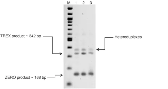
For initial identification of STREX and ZERO variants, end-point PCR was performed using primers designed to amplify the alternative splice site 2 (See Table 1 for details). Testis cDNA was used as a positive control [11]. PCR products of three separate experiments from different experimental animals were analyzed by electrophoresis and subsequently verified by sequencing. As shown
in Figure 2, two dominant bands were detected in both vasculatures. The lower band with predicted size of 168 bp was determined to be ZERO variant (a-subunit without splice insert) by direct product sequencing and the upper band (,342 bp) was confirmed to be STREX variant (a-subunit with the insertion of 174 bp at splice site 2). The third visible band likely constitutes heteroduplexes between sense and antisense strands of STREX and ZERO products consistent with earlier reports [35].
Quantification ofa-BKCasplice variants by qPCR
Identification of the SS4 variant was performed by qPCR together with subsequent quantification of expression levels of STREX variant relative to ZERO using a further set of primers (Table 1). As shown in Figure 3A and B, while a very low level of SS4 was detected in mid-cerebral arteries (0.4260.1% of totala -BKCa), the variant was undetectable in cremaster vessels. A higher
level of expression of the STREX variant was detected in cremaster arteries (28.964.2% of total a-BKCa) compared to
mid-cerebral (16.560.9% of total a-BKCa) arteries (P,0.05).
Thus, ZERO variant was calculated to be significantly (P,0.05) greater in mid-cerebral (83.160.9% of totala-BKCa) compared to
cremaster (71.164.2%) arteries (Figure 3A).
Cell Surface expression of BKCaZERO or STREX variants in
expression systems
To investigate the cell surface location ofa-BKCasplice variants
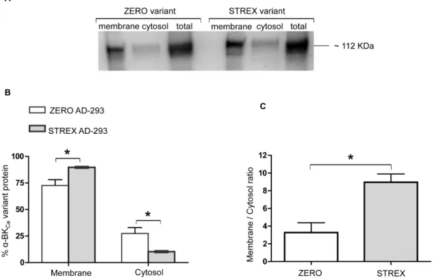
in a cell culture system, equal amounts of full-length cDNAs of rat ZERO or STREX variants were initially transfected into AD-293 cells (Figure 4A). As shown in Figure 4B, although both variants were predominantly targeted to the cell surface, the STREX variant ofa-BKCashows a significantly higher level of cell surface
expression (P = 0.02, unpaired t-test) than the ZERO variant. Conversely at the cytosolic level, the ZERO variant shows a significantly higher level of expression as compared with STREX (P = 0.02, unpaired t-test). The ratio of membrane to cytosol expression was also significantly higher for the STREX variant than ZERO (Figure 4C).
To confirm that this trafficking pattern was not cell dependent, similar experiments were conducted using CHO-K1 cells (Figure 5A). As illustrated in Figure 5B, the STREX variant again shows increased distribution at the cell membrane compared with ZERO (P = 0.002, unpaired t-test). The cytosolic expression of STREX in CHO-K1 cells shows significantly lower expression than the ZERO variant, similar to that observed in the AD-293 cell culture system. As shown in Figure 5C, the ZERO variant shows a significantly lower ratio of membrane to cytosolic expression in CHO-K1 cells. Since these two a-BKCa subunit
splice variant exhibit the same expression pattern in AD-293 and CHO-K1 cells, we used CHO-K1 cells as our cell system model for future experiments.
Cell Surface location of co-transfected BKCaZERO or
STREX variants withb1-subunit in CHO-K1 cells
As holo-BKCa channels in resistance arteries contain both a
-andb1-BKCasubunits, we hypothesized that co-expression ofb
1-subunit in CHO-K1 cells may equalize the cell surface levels of the two variants. To investigate the impact ofb1-BKCasubunit on cell
surface expression ofa-BKCasplice variants ZERO and STREX, b1-BKCa subunit was co-transfected with either the ZERO or
STREX variant (Figure 6A). In the presence ofb1-BKCasubunit,
Figure 2. Detection of ZERO and STREX variants in rat cerebral and cremaster arteries.End-point PCR products generated from cDNA derived from rat total RNA transcripts, with and without the STREX exon. Size markers are shown in the lane marked as M. Additional lanes display PCR products detected in testis (lane 1, included as a positive control), mid-cerebral arteries (lane 2) and cremaster arterioles (lane 3). Results are representative of n = 3 separate experiments.
doi:10.1371/journal.pone.0098863.g002
Figure 3. QPCR of BKCaa-subunit splice variants in rat cerebral and cremaster arteries.(A) Relative mRNA expression levels for STREX, SS4
and ZERO variants calculated as a percentage of totala-BKCamRNA detected in mid-cerebral arteries and cremaster arterioles. (B) QPCR products of
SS4, STREX and ZERO variants ofa-BKCasubunit as separated on a 2% agarose gel. Size markers shown in the lane denoted M. QPCR products
generated from cremaster arterioles are depicted in lanes 1, 3, 5 and products from mid-cerebral artery are shown in lanes 2, 4, and 6. Real-time PCR results are shown for n = 4–5 samples of each vascular bed performed in triplicate.
expression pattern (Figure 6C) as seen when the a-BKCa splice
variants were expressed without theb1-BKCasubunit (Figure 5C).
These data could indicate a dominant cell surface expression pattern ofa-BKCa splice variants of ZERO and STREX in cell
culture systems.
To determine the effect of ZERO or STREX variants on the cellular distribution of b1-BKCa subunits, biotinylated b1-BKCa
subunit proteins were also probed in cells co-transfected withb
1-BKCa and either the ZERO or STREX variant. As shown in
Figure 7A, a high level of cell surface expression of b1-BKCa
subunit (84.764.4%) was observed when co-transfected with either the ZERO or STREX variant. To assess whether over-expression ofb1-BKCasubunit, alone, is sufficient to stimulate its
surface trafficking, CHO-K1 cells were transfected with onlyb 1-BKCacDNA andb1-BKCasubunit surface location assessed by the
cell-surface biotinylation assay. Under these conditions, as shown in Figure 7B, the cell surface labeling ofb1-BKCasubunit showed
a significant decrease (P = 0.01) to approximately 60.265.2%, which was accompanied by a significant increase in cytosolic levels
of b1-BKCa subunit from approximately 15.364.5% to
39.865.2%. These data appear to indicate a stimulatory effect of a-BKCa splice variants on the surface trafficking of the
regulatory subunitb1-BKCa, while there was no apparent impact
on the surface membrane location of either ZERO or STREX by
b1-BKCasubunit.
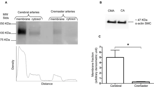
Cell surface expression of totala-BKCaprotein in cerebral
vs cremaster arteries
Previous electrophysiological studies from our laboratory have demonstrated that BKCa channel activity differs significantly in
VSMCs from cremaster muscle arteries compared with cerebral arteries [26]. Our functional data show a decreased Ca2+
sensitivity of cremaster VSMC BKCa channels compared with
those of cerebral arteries, resulting in more positive levels of Em being required for cremaster VSMCs cells to generate similar levels of outward K+
current. As a result, we hypothesized that these functional differences in channel activity could arise from differences in the molecular configuration of the channel in the two VSMC types affecting channel properties such as cell surface trafficking of a-BKCa protein. Although the lack of specific
available antibodies to distinguish ZERO from STREX variants is a technical limitation to detect a-BKCa splice variants at the
protein level, the biotinylation assay was used to determine the extent ofa-BKCaprotein at the cell surface compared with the
cytosolic fraction. Experiments were performed using homoge-nates of whole cerebral and cremaster arteries. As shown in Figure 8A and B, in both vessel types more than 90% of totala -BKCa channels were located at the cell membrane. However,
when cell surface expression ofa-BKCawas normalized to equal
amounts ofa-smooth muscle actin in each preparation, cerebral arteries show approximately 20 times higher level of total amount ofa-BKCaprotein at the cell membrane compared with cremaster
arteries (Figure 9).
Figure 4. The cell surface and cytosolic expression of BKCaa-subunit splice variants in cultured cells.Using an AD-293 cell culture
system,a-BKCaSTREX variant shows a significantly higher cell surface expression compared with the ZERO variant. Corresponding changes in the
cytosolic fraction are observed. (A) Representative Western blot showing membrane, cytosolic, and total levels ofa-BKCaprotein in AD-293 cultured
cells, transfected with eithera-BKCasplice variants ZERO or STREX. (B) Summary of membrane vs cytosolic distribution of transfected ZERO or STREX
variants ofa-BKCachannel protein (expressed as % of totala-BKCaprotein). (C) Membrane to cytosolic expression ratio of ZERO and STREX variants in
Discussion
In previous studies, we reported functional differences in BKCa
channels in VSMCs between cerebral arteries and those from cremaster muscle [26,32]. In those studies, we showed that BKCa
from cremaster VSMCs exhibit a decreased Ca2+
sensitivity and suggested that this may, in part, be due to a decrease in the amount of the b1 regulatory subunit. Regulation of BKCa is,
however, complex involving mechanisms at the levels of expression and post-translational modification, as well as its physical relationship to cellular organelles such as the sarcoplasmic reticulum [5,6,8]. On the basis of this, the aims of the present study were to examine whether differences exist in the expression of splice variants of the a-subunit of BKCa and if membrane
location of the channel differed between the two vascular beds. The major findings of the present studies were as follows: Firstly, qPCR studies demonstrate a significantly higher mRNA
expres-sion for the BKCa a-subunit STREX splice variant in rat
cremaster arteries compared with that in cerebral arteries. Secondly, we detected the predominant cell surface expression of
both the a-BKCa splice variants ZERO and STREX in cell
expression systems, with no apparent impact of b1-subunit co-expression on the degree of cell surface localization. Thirdly, although theb1-subunit expressed alone is able to reach the cell membrane, a significant proportion remains cytosolic compared with its predominant cell surface localization in the presence of either the ZERO or STREX variant. Finally, cell surface labeling revealed that the vast majority ofa-BKCachannel (.90%) in both
cremaster and cerebral arteries is located in the cell membrane fraction under basal conditions. However, a major difference between the two vascular beds was that the total amount of plasma membranea-BKCais approximately 20 times lower in cremaster
arterioles as compared with small cerebral arteries.
Although a single gene, KCNMA1, encodes the pore forming BKCa a-subunit in vertebrates, there is considerable phenotypic
diversity of these channels in different tissues. Several factors are known to contribute to this diversity including, alternative splicing [36], the co-expression of regulatoryb-subunits [37,38], and post-translational modifications including protein phosphorylation [39]. Modulation of BKCa channels by a complex network of signal
transduction pathways such as reversible protein phosphorylation has been studied extensively [39–41]. Importantly, alternate splicing of pre-mRNA leading to channel variants with differing degrees of modulation by reversible protein phosphorylation represents a potential mechanism to generate functional diversity of ion channels. In studies of cloned mouse BKCa variants,
expressed in HEK293 cells, Tian et al. (2001) demonstrated that cAMP-dependent protein kinase (PKA)-mediated phosphorylation activates BKCaZERO variant, but inhibits the STREX variant,
which could thus impact channel function including Ca2+
sensitivity [17]. The level of STREX expression also has important modulatory consequences as it has been previously shown that only one subunit within the tetrameric holo-channel needs to be of the STREX type to alter channel function [42]. It has also been shown that protein palmitoylation (a post-translational
modifica-Figure 5. Membrane and cytosolic expression of ZERO and STREX variants in CHO-K1 cells.In a CHO-K1 cell culture system, thea-BKCa
ZERO variant shows significantly lower cell surface expression than the STREX variant. Corresponding changes in the cytosolic fraction are observed. (A) Representative Western blot showing surface and cytosolic levels ofa-BKCaprotein in CHO-K1 cell culture system, transfected with eithera-BKCa
splice variants ZERO or STREX. (B) Summary of membrane vs cytosolic expression of transfected ZERO or STREX variants ofa-BKCachannel protein
(expressed as % of totala-BKCaprotein). (C) Membrane to cytosolic expression ratio of ZERO and STREXa-BKCaproteins in transfected cultured
tion affecting multiple ion channels) can regulate the activity and surface expression of BKCa channel a-subunits in native tissues
and cultured cells [19]. Specifically, Tian and colleagues described
palmitoylated BKCa channels that include plasma membrane
associated STREX variants that are inhibited by PKA-dependent phosphorylation, whereas ZERO channels are activated by PKA [43]. Therefore, the finding of lower expression of the ZERO variant and higher expression of STREX in cremaster arteries compared with mid-cerebral arteries could conceivably relate to functional alterations in BKCa Ca2+ sensitivity, as we have
previously observed for cremaster vascular smooth muscle cells [32].
However, in the present studies the cremaster vessels have been shown to express a higher level of STREX as compared to the cerebral arteries which may have been expected to convey a higher Ca2+ sensitivity [9] rather than the decreased Ca2+
sensitivity we previously reported [26]. This apparent discrepancy may relate to a number of factors including the dominant effects of differences in overall expression levels and also that the cremaster vessels were previously shown to have a lower ratio of b1:a
subunit. Importantly, theb1 subunit has previously been shown to contribute to the Ca2+
sensitivity of the channel [44]. An additional consideration is that our measurements of STREX and ZERO expression were limited to the mRNA levels. In this
previous study, we also reported a decrease in the ratio of BKCa a:bsubunits in the crude membrane fraction of cremaster vessels compared with small cerebral arteries [26]. The data from the present study suggest it is unlikely that a lower level ofb1-BKCa
would negatively influence insertion of the a-subunit into the membrane, as our data in cell expression systems showed similar levels of surface location fora-BKCain the presence and absence
of theb1-subunit. Interestingly, and in contrast, the presence of the a-subunit in the expression system resulted in an increased proportion ofb1-subunit being located at the cell surface.
In earlier studies, Jackson and Blair (1998) suggested that BKCa
is ‘silent’ in cremaster muscle vasculature under basal conditions, but may be ‘recruited’ under stimulated conditions. Such stimulation was suggested to include vasoconstriction evoked by catecholamines and high tissue PO2 levels [27]. Whether such
recruitment involves differences in splice variant expression, translocation of channel subunit proteins to the plasma membrane or post-translational modifications such as phosphorylation has not been fully elucidated. Given the high levels ofa-subunit protein found at the membrane in both vessel types, it is unlikely that a simple difference in membrane vs. cytosolic pools explains the differences observed between cremaster and cerebral vessels. This does not, however, exclude the possibility that a dynamic
Figure 6. The effect ofb1-BKCasubunit on cell surface expression ofa-BKCasplice variants.Co-transfection ofb1-BKCasubunit has little
apparent impact on cell surface expression ofa-BKCaZERO or STREX variants. (A) Representative Western blot showing levels of membrane (MF), and
cytosolic (CF) fractions ofa-BKCaprotein in CHO-K1 cultured cells, transfected with eithera-BKCasplice variants ZERO or STREX. (B) In the CHO-K1 cell
culture system,a-BKCaSTREX variant shows dominant cell surface expression when co-transfected withb1-BKCa. Results are shown for n = 5 separate
experiments and are presented as mean6SEM (*P,0.05, unpaired t-test). (C) Membrane to cytosolic ratio for the expression of ZERO or STREX proteins when co-transfected withb1-BKCain cultured CHO-K1 cells. Results are shown for n = 5 separate experiments and are presented as mean6
alteration in channel protein trafficking occurs under other conditions.
In the present study, we also found a very low level of expression of the SS4a-subunit variant relative to the ZERO variant in mid-cerebral, with no detectable expression in cremaster arteries. Although expression of the SS4 variant in vasculature (identified by RT-PCR) has been previously reported in cerebral and coronary arteries [11], its functional importance, particularly in native tissues such as small arteries, is unknown. Similarly, the functional significance of a lack of SS4 variant expression (as we report for cremaster muscle arterioles) is unclear. Using aXenopus
oocytes expression system, previous studies have suggested that ZERO and SS4 variants exhibit identical BKCachannel
charac-teristics, including single-channel conductance and voltage dependent activation [18]. Those authors did, however, show that the activation rates of SS4 channels were more rapid at a similar voltage compared with the ZERO form when [Ca2+
]iwas
higher than 5mM. In addition to a comparative lack of
information as to any functional significance of very low expression levels of the SS4 variant (approximately 0.4% of total
a-BKCamRNA) in cerebral arteries, it should also be considered
that non-smooth muscle cell contamination (for example from
neurons or adventitial cells) in whole vessel preparations could contribute to this signal.
Apart from differences in splice variant expression, it would be expected that the marked difference in total BKCachannel protein
expression would be of functional significance. This is despite the large conductance (approx 240 pS) in VSMCs from both vascular beds. Specifically, an,20-fold higher level ofa-BKCaprotein was
detected in cerebral arteries compared with cremaster arteries. Importantly, this would be reflected at the plasma membrane because a similar proportion of total BKCawas surface located in
both cerebral and cremaster arteries, as shown by our biotinyla-tion assay. While measurements were performed on homogenates of whole vessels, the majority of signal would be expected to derive from the VSMC layers. Endothelial cells of healthy arteries are thought to be devoid of BKCachannels, although this point has
been somewhat controversial [45]. Cellular capacitance measure-ments performed in our previous studies indicate that VSMC size in the two vessel types is similar, suggesting that functional effects of the expression difference would not be compensated by differences in size alone [26]. The earlier study also demonstrated, in crude membrane fractions, increaseda-BKCaprotein (approx.
3x) in cerebral vessels as compared to cremaster arterioles. Despite these measurements of marked differences in expression at the
Figure 7. Cell surface expression ofb1-BKCasubunit in the presence and absence ofa-BKCasplice variants.Co-transfection of ZERO or
STREX variants withb1-BKCasubunit enhances cell surface trafficking ofb1-BKCasubunit. The top image in panel (A) represents a Western blot for
membrane (MF) and cytosolic (CF) fractions ofb1-BKCaprotein in CHO-K1 cultured cells, co-transfected with eithera-BKCasplice variants ZERO or
STREX. The bottom Figure in panel (A) shows the membrane vs cytosolic expression of transfectedb1-BKCachannel protein when co-transfected with
eithera-BKCasplice variants of ZERO or STREX. Results are shown for n = 5 separate experiments. The top image in panel (B) shows a representative
Western blot of membrane (MF) and cytosolic (CF) fractions ofb1-BKCaprotein in CHO-K1 cultured cells, in the absence of eithera-BKCasplice variants
ZERO or STREX. The bottom Figure in panel (B) shows membrane vs cytosolic expression ofb1-BKCasubunit in cultured CHO-K1 cells in the absence
of thea-BKCasplice variants ZERO or STREX. Results are shown for n = 6 separate experiments and are presented as mean6SEM (*P,0.05, unpaired
t-test).
protein level actual K+currents only differed by approximately 2x
(at 5 mM Ca2+). Conceivably the seemingly disparate findings may
relate to the functional status of the channels including as determined by post-translational modifications such as phosphor-ylation or possibly the influence of splice variants not directly
examined in this study [46]. Another factor perhaps affecting the differences in absolute protein levels between cerebral vessels and cremaster arterioles relates to differences in adventitial structure that we have previously demonstrated [47]. It could be argued that these differences impact the access of the biotinylation reagents.
Figure 8. Membrane and cytosolic expression ofa-BKCasubunits in cerebral vs creamster arteries.a-BKCachannels are predominantly
located at the cell surface of VSMCs in both rat cerebral and cremaster arteries under basal conditions. (A) Cell surfacea-BKCaproteins were
determined using the biotinylation assay from whole cerebral (Circle of Willis pooled from 2 animals in each experiment) and cremaster arteries (first-and second-order cremaster arterioles pooled from 4 animals in each experiment). Results are shown for n = 6 separate experiments for cerebral (first-and n = 5 for cremaster arteries. Results are shown as mean6SEM. (B) Membrane to cytosolic ratio ofa-BKCachannels in cerebral vs cremaster arteries
(the corresponding Western blot showing cell surface and cytosolic levels ofa-BKCaprotein is shown in Figure 9A).
doi:10.1371/journal.pone.0098863.g008
Figure 9. Quantification of cell surface expresseda-BKCachannels in cerebral vs cremaster arteries.Total amount of membranea-BKCa
channel in cerebral arteries is approximately 20 times higher than that in cremaster arterioles. The Western blot in panel (A) shows a representative experiment depicting the distribution ofa-BKCaprotein in membrane and cytosolic fractions prepared from cerebral arteries and cremaster arterioles.
The scan beneath the blot quantifies the intensity of thea-BKCaprotein immunoreactive band in each sample, as detected by densitometry. The
We believe this to be unlikely, however, as the biotinylation reagents are small in regard to molecular weight and it was previously shown that the molecules easily penetrate the vascular wall [33]. Further, while this could theoretically effect the magnitude of the protein expression levels it would not impact theatobsubunit ratios nor the distribution between the plasma membrane and cytosol. As both the cell surface biotinylation approach and measurements performed in crude membrane fractions showed qualitatively similar same results, the conclusion that protein expression levels are greater in cerebral vessels appears robust.
In summary, significant differences exist with respect to the BKCa splice variants expressed in cremaster muscle arterioles
compared with small arteries from the cerebral vasculature. Specifically, a higher expression level of the STREX variant of the
a-subunit was observed in arterioles from cremaster muscle. While functional significance of this finding is yet to be fully demon-strated, it appears that it does not affect the plasma membrane
location of the channels as.95% ofa-subunit was found to be at the cell surface in both vessel preparations. In contrast, a marked difference in the detectable expression level was observed, with cerebral arteries expressinga-subunit protein at a level 20 times greater than that of cremaster arterioles.
Acknowledgments
Our especial thanks expressed to Dr. Michael J. Shipston for his generosity to provide DNA materials of BKCaa-subunit splice variants. Thanks are
extended to Dr. Michael J. Davis for his constructive criticism of the manuscript prior to its submission.
Author Contributions
Conceived and designed the experiments: ZN JHJ APB MAH. Performed the experiments: ZN ML MDL. Analyzed the data: ZN ML MDL JHJ APB MAH. Wrote the paper: ZN MAH. Editing of the manuscript: ZN ML MDL JHJ APB MAH.
References
1. Latorre R, Oberhauser A, Labarca P, Alvarez O (1989) Varieties of calcium-activated potassium channels. Annu Rev Physiol 51: 385–399.
2. Marty A (1981) Ca-dependent K channels with large unitary conductance in chromaffin cell membranes. Nature 291: 497–500.
3. Jan LY, Jan YN (1997) Cloned potassium channels from eukaryotes and prokaryotes. Annu Rev Neurosci 20: 91–123.
4. Yan J, Aldrich RW (2010) LRRC26 auxiliary protein allows BK channel activation at resting voltage without calcium. Nature 466: 513–516. 5. Hill MA, Yang Y, Ella SR, Davis MJ, Braun AP (2010) Large conductance,
Ca2+
-activated K+
channels (BKCa) and arteriolar myogenic signaling. FEBS
Lett 584: 2033–2042.
6. LeDoux J, Werner ME, Brayden JE, Nelson MT (2006) Calcium-activated potassium channels and the regulation of vascular tone. Physiology (Bethesda) 21: 69–78.
7. Wu RS, Marx SO (2010) The BK potassium channel in the vascular smooth muscle and kidney: alpha- and beta-subunits. Kidney Int 78: 963–974. 8. Lee US, Cui J (2010) BK channel activation: structural and functional insights.
Trends Neurosci 33: 415–423.
9. Chen L, Tian L, MacDonald SH, McClafferty H, Hammond MS, et al. (2005) Functionally diverse complement of large conductance calcium- and voltage-activated potassium channel (BK) alpha-subunits generated from a single site of splicing. J Biol Chem 280: 33599–33609.
10. Tseng-Crank J, Foster CD, Krause JD, Mertz R, Godinot N, et al. (1994) Cloning, expression, and distribution of functionally distinct Ca(2+)-activated K+channel isoforms from human brain. Neuron 13: 1315–1330.
11. Poulsen AN, Wulf H, Hay-Schmidt A, Jansen-Olesen I, Olesen J, et al. (2009) Differential expression of BK channel isoforms and beta-subunits in rat neuro-vascular tissues. Biochim Biophys Acta 1788: 380–389.
12. Poulsen AN, Jansen-Olesen I, Olesen J, Klaerke DA (2011) Neuronal fast activating and meningeal silent modulatory BK channel splice variants cloned from rat. Pflugers Arch 461: 65–75.
13. Xie J, McCobb DP (1998) Control of alternative splicing of potassium channels by stress hormones. Science 280: 443–446.
14. MacDonald SH, Ruth P, Knaus HG, Shipston MJ (2006) Increased large conductance calcium-activated potassium (BK) channel expression accompanied by STREX variant downregulation in the developing mouse CNS. BMC Dev Biol 6: 37.
15. Shipston MJ, Duncan RR, Clark AG, Antoni FA, Tian L (1999) Molecular components of large conductance calcium-activated potassium (BK) channels in mouse pituitary corticotropes. Mol Endocrinol 13: 1728–1737.
16. Tian L, Hammond MS, Florance H, Antoni FA, Shipston MJ (2001) Alternative splicing determines sensitivity of murine calcium-activated potassium channels to glucocorticoids. J Physiol 537: 57–68.
17. Tian L, Duncan RR, Hammond MS, Coghill LS, Wen H, et al. (2001) Alternative splicing switches potassium channel sensitivity to protein phosphor-ylation. J Biol Chem 276: 7717–7720.
18. Ha TS, Jeong SY, Cho SW, Jeon H, Roh GS, et al. (2000) Functional characteristics of two BKCa channel variants differentially expressed in rat brain tissues. Eur J Biochem 267: 910–918.
19. Shipston MJ (2013) Regulation of large conductance calcium- and voltage-activated potassium (BK) channels by S-palmitoylation. Biochem Soc Trans 41: 67–71.
20. Kim EY, Zou S, Ridgway LD, Dryer SE (2007) Beta1-subunits increase surface expression of a large-conductance Ca2+
-activated K+
channel isoform. J Neurophysiol 97: 3508–3516.
21. Kim EY, Ridgway LD, Zou S, Chiu YH, Dryer SE (2007) Alternatively spliced C-terminal domains regulate the surface expression of large conductance calcium-activated potassium channels. Neuroscience 146: 1652–1661. 22. Chiu YH, varez-Baron C, Kim EY, Dryer SE (2010) Dominant-negative
regulation of cell surface expression by a pentapeptide motif at the extreme COOH terminus of an Slo1 calcium-activated potassium channel splice variant. Mol Pharmacol 77: 497–507.
23. Zarei MM, Zhu N, Alioua A, Eghbali M, Stefani E, et al. (2001) A novel MaxiK splice variant exhibits dominant-negative properties for surface expression. J Biol Chem 276: 16232–16239.
24. Zarei MM, Eghbali M, Alioua A, Song M, Knaus HG, et al. (2004) An endoplasmic reticulum trafficking signal prevents surface expression of a voltage-and Ca2+
-activated K+
channel splice variant. Proc Natl Acad Sci U S A 101: 10072–10077.
25. Toro B, Cox N, Wilson RJ, Garrido-Sanabria E, Stefani E, et al. (2006) KCNMB1 regulates surface expression of a voltage and Ca2+
-activated K+
channel via endocytic trafficking signals. Neuroscience 142: 661–669. 26. Yang Y, Murphy TV, Ella SR, Grayson TH, Haddock R, et al. (2009)
Heterogeneity in function of small artery smooth muscle BKCa: involvement of the beta1-subunit. J Physiol 587: 3025–3044.
27. Jackson WF, Blair KL (1998) Characterization and function of Ca(2+)-activated K+channels in arteriolar muscle cells. Am J Physiol 274: H27–H34. 28. Meininger GA, Zawieja DC, Falcone JC, Hill MA, Davey JP (1991) Calcium
measurement in isolated arterioles during myogenic and agonist stimulation. Am J Physiol 261: H950–H959.
29. Wulf H, Hay-Schmidt A, Poulsen AN, Klaerke DA, Olesen J, et al. (2008) Molecular studies of BKCa channels in intracranial arteries: presence and localization. Cell Tissue Res 334: 359–369.
30. Livak KJ, Schmittgen TD (2001) Analysis of relative gene expression data using real-time quantitative PCR and the 2(-Delta Delta C(T)) Method. Methods 25: 402–408.
31. Chen L, Jeffries O, Rowe IC, Liang Z, Knaus HG, et al. (2010) Membrane trafficking of large conductance calcium-activated potassium channels is regulated by alternative splicing of a transplantable, acidic trafficking motif in the RCK1-RCK2 linker. J Biol Chem 285: 23265–23275.
32. Yang Y, Sohma Y, Nourian Z, Ella SR, Li M, et al. (2013) Mechanisms underlying regional differences in the Ca2+
sensitivity of BK(Ca) current in arteriolar smooth muscle. J Physiol 591: 1277–1293.
33. Bannister JP, Adebiyi A, Zhao G, Narayanan D, Thomas CM, et al. (2009) Smooth muscle cell alpha2delta-1 subunits are essential for vasoregulation by CaV1.2 channels. Circ Res 105: 948–955.
34. Crnich R, Amberg GC, Leo MD, Gonzales AL, Tamkun MM, et al. (2010) Vasoconstriction resulting from dynamic membrane trafficking of TRPM4 in vascular smooth muscle cells. Am J Physiol Cell Physiol 299: C682–C694. 35. Mahmoud SF, Bezzerides AL, Riba R, Lai GJ, Lovell PV, et al. (2002) Accurate
quantitative RT-PCR for relative expression of Slo splice variants. J Neurosci Methods 115: 189–198.
36. Shipston MJ (2001) Alternative splicing of potassium channels: a dynamic switch of cellular excitability. Trends Cell Biol 11: 353–358.
37. Brenner R, Jegla TJ, Wickenden A, Liu Y, Aldrich RW (2000) Cloning and functional characterization of novel large conductance calcium-activated potassium channel beta subunits, hKCNMB3 and hKCNMB4. J Biol Chem 275: 6453–6461.
39. Schubert R, Nelson MT (2001) Protein kinases: tuners of the BKCa channel in smooth muscle. Trends Pharmacol Sci 22: 505–512.
40. Chung SK, Reinhart PH, Martin BL, Brautigan D, Levitan IB (1991) Protein kinase activity closely associated with a reconstituted calcium-activated potassium channel. Science 253: 560–562.
41. Reinhart PH, Chung S, Martin BL, Brautigan DL, Levitan IB (1991) Modulation of calcium-activated potassium channels from rat brain by protein kinase A and phosphatase 2A. J Neurosci 11: 1627–1635.
42. Tian L, Coghill LS, McClafferty H, MacDonald SH, Antoni FA, et al. (2004) Distinct stoichiometry of BKCa channel tetramer phosphorylation specifies channel activation and inhibition by cAMP-dependent protein kinase. Proc Natl Acad Sci U S A 101: 11897–11902.
43. Tian L, Jeffries O, McClafferty H, Molyvdas A, Rowe IC, et al. (2008) Palmitoylation gates phosphorylation-dependent regulation of BK potassium channels. Proc Natl Acad Sci U S A 105: 21006–21011.
44. Bao L, Cox DH (2005) Gating and ionic currents reveal how the BKCa channel’s Ca2+sensitivity is enhanced by its beta1 subunit. J Gen Physiol 126: 393–412.
45. Sandow SL, Grayson TH (2009) Limits of isolation and culture: intact vascular endothelium and BKCa. Am J Physiol Heart Circ Physiol 297: H1–H7. 46. Rosati B, McKinnon D (2004) Regulation of ion channel expression. Circ Res
94: 874–883.