Interactions of
with
Alveolar Type II Epithelial Cells and the
Murine Respiratory Epithelium
Matthew Faron1, Joshua R. Fletcher1, Jed A. Rasmussen2, Michael A. Apicella2, Bradley D. Jones1,2*
1Graduate Program in Genetics, University of Iowa, Iowa City, Iowa, United States of America,
2Department of Microbiology, University of Iowa, Iowa City, Iowa, United States of America
Abstract
Francisella tularensisis classified as a Tier 1 select agent by the CDC due to its low infectious
dose and the possibility that the organism can be used as a bioweapon. The low dose of in-fection suggests thatFrancisellais unusually efficient at evading host defenses. Although ~50
cfu are necessary to cause human respiratory infection, the early interactions of virulent Fran-cisellawith the lung environment are not well understood. To provide additional insights into
these interactions during earlyFrancisellainfection of mice, we performed TEM analysis on
mouse lungs infected withF.tularensisstrains Schu S4, LVS and the O-antigen mutant Schu
S4waaY::TrgTn. For all three strains, the majority of the bacteria that we could detect were
observed within alveolar type II epithelial cells at 16 hours post infection. Although there were no detectable differences in the amount of bacteria within an infected cell between the three strains, there was a significant increase in the amount of cellular debris observed in the air spaces of the lungs in the Schu S4waaY::TrgTn mutant compared to either the Schu S4 or
LVS strain. We also studied the interactions ofFrancisellastrains with human AT-II cellsin vitroby characterizing the ability of these three strains to invade and replicate within these
cells. Gentamicin assay and confocal microscopy both confirmed thatF.tularensisSchu S4
replicated robustly within these cells whileF.tularensisLVS displayed significantly lower
lev-els of growth over 24 hours, although the strain was able to enter these cells at about the same level as Schu S4 (1 organism per cell), as determined by confocal imaging. The Schu S4waaY::TrgTn mutant that we have previously described as attenuated for growth in
macro-phages and mouse virulence displayed interesting properties as well. This mutant induced significant airway inflammation (cell debris) and had an attenuated growth phenotype in the human AT-II cells. These data extend our understanding of earlyFrancisellainfection by
dem-onstrating thatFrancisellaenter significant numbers of AT-II cells within the lung and that the
capsule and LPS of wild type Schu S4 helps prevent murine lung damage during infection. Furthermore, our data identified that human AT-II cells allow growth of Schu S4, but these same cells supported poor growth of the attenuated LVS strainin vitro. Collectively, these
data further our understanding of the role of AT-II cells inFrancisellainfections.
OPEN ACCESS
Citation:Faron M, Fletcher JR, Rasmussen JA, Apicella MA, Jones BD (2015) Interactions of
Francisella tularensiswith Alveolar Type II Epithelial Cells and the Murine Respiratory Epithelium. PLoS ONE 10(5): e0127458. doi:10.1371/journal. pone.0127458
Academic Editor:Alain Charbit, Université Paris Descartes, FRANCE
Received:January 22, 2015
Accepted:April 15, 2015
Published:May 26, 2015
Copyright:© 2015 Faron et al. This is an open access article distributed under the terms of the
Creative Commons Attribution License, which permits unrestricted use, distribution, and reproduction in any medium, provided the original author and source are credited.
Data Availability Statement:All relevant data are within the paper and its Supporting Information files.
Funding:This study was supported by 2PO1 AI044642 MAA, BDJ; 2U54 AI057160 BDJ.
Introduction
Francisella tularensisis a highly virulent intracellular bacterial pathogen that causes the human
infectious disease tularemia [1,2]. The most common route of infection is cutaneous, although
infection via the respiratory route is highly efficient and can cause a lethal infection in 30–60%
of patients that do not receive treatment [3]. In mice, respiratory infection with a single virulent
F.tularensisorganism is virtually always lethal while in a human as few as 50 organisms are
be-lieved to result in a potentially lethal infection [4,5]. The ability to weaponize this organism for
respiratory delivery, along with the low infective dose and the high lethality ofFrancisella
tular-ensisare the reasons why this organism is classified as a Tier 1 select agent by the Centers for Disease Control and Prevention (CDC).
In an effort to understand early events inFrancisellainfection and how they can
reproduc-ibly lead to lethal respiratory disease, it was of interest to examine the interactions between
Francisellaand the alveolar air spaces. In general, the lung is protected from microbial insult by both alveolar macrophages that reside in the extracellular alveolar air spaces and by the physical barrier composed of alveolar epithelial cells. The alveolar macrophages are loosely associated with the epithelium, and are in a relatively inactivated state where they function to
engulf particles that are inhaled during breathing [6]. Upon engulfment of a particle or
bacteri-um, alveolar macrophages increase their phagocytic activity, oxidative burst capacity and
pro-duction of pro-inflammatory cytokines [7]. These induced protective responses lead to the
release of alveolar macrophages from the airway epithelium, where they (along with their
en-gulfed cargo) are removed from the lung air spaces via the mucociliary escalator [8]. As an
early line of defense in the lungs, these activities are designed to engage and direct bacteria away from the alveolar epithelium. Since interactions with alveolar macrophages are likely to
result in the removal ofFrancisellaorganisms from airway epithelial environment, it seems
likely that the bacteria must productively interact with other cell types in order to breach the respiratory epithelium and gain access to deeper tissue and the bloodstream.
Besides alveolar macrophages, the alveolus is composed of two other cell types: alveolar epi-thelial type I (AT-I) and alveolar epiepi-thelial type II cells (AT-II), which are important compo-nents of a physical barrier to protect deeper tissues from microbes and airborne particles. AT-I cells are thin, elongated cells that comprise 95% of the alveolus surface area and are important
in maintaining the structure of the alveolus and facilitating gas exchange [9]. In contrast,
AT-II cells are smaller spherical cells that contain microvilli and lamellar bodies [10,11]. These
cells constitute the remaining 5% of the epithelial surface, but represent 60% of the alveolar
epi-thelial cells [12]. AT-II cells have diverse functions within the lung, and are involved in several
processes, including: secretion of surfactant, regeneration of the alveolar epithelium, and
pro-tecting against bacterial invasion [13]. AT-II cells protect against pathogens by sensing
patho-gens through TLR stimulation [14,15], secretion of anti-microbial peptides [16], and both
activation and deactivation of inflammation through modulation of cytokines and chemokines
[17]. However, it has been shown that pathogenic bacteria such asMycobacterium tuberculosis,
can survive these anti-microbial mechanisms and be internalized into AT-II cells, which allows
the bacteria to cross the alveolar barrier and gain access to the bloodstream [18].
Several studies have provided evidence that airway epithelial cells may be an important
ac-cess point forFrancisellato initiate disease in the lung. Using mice intranasally infected withF.
tularensisLVS, Hallet al. [19] demonstrated that GFP-labelled organisms co-localized with cells that produced pro-surfactant protein C (pSP-C), a marker for AT-II cells, and that bacte-rial counts increased rapidly in the lungs over time, suggesting that the organisms were
multi-plying within type II airway epithelial cells. Another study by Hallet al. examined the types
F.tularensisLVS, orF.tularensisSchu S4 [20]. It was observed that theseF.tularensisstrains infected a wide variety of different lung cell types that included alveolar macrophages, neutro-phils, dendritic cells, monocytes and alveolar type II cells, although the percentage of cells
in-fected varied from strain to strain [20]. We, and others, have studied the interactions ofF.
tularensiswith various types of epithelial cells [19,21,22]. These efforts have demonstrated thatF.tularensisLVS is taken up by epithelial tissue culture cells where the organisms are able to grow ~1000-fold over 24 hours, which represents a growth rate comparable to that observed
in vitroin growth media. Additionally, Gentryet al. has reported that interactions ofF.
tularen-siswith primary human AT-II cells stimulate a NF-kB-dependent cytokine release from AT-II
lung epithelial cells suggesting that these cells play important roles in recruitment of immune
cells through the pulmonary endothelium [23].
Materials and Methods
Bacterial strains and growth conditions
F.tularensisLVS,F.tularensisSchu S4 and theF.tularensis waaY::TrgTn (FTT1236) mutant in the Schu S4 background were routinely cultured on modified Mueller-Hinton agar plates (Acumedia) or in Mueller-Hinton broth supplemented with 1% glucose (wt/vol) 0.025% ferric
pyrophosphate and 2% IsoVitaleX [24]. In preparation for experiments, strains were grown in
modified Mueller-Hinton broth with aeration at 37°C. The antibiotics kanamycin and/or
spec-tinomycin were added to the growth media at a concentration of 25μg per ml when necessary.
Epithelial cell culture
Primary human alveolar type 2 cells (AT-II) that have been immortalized with SV40 were pur-chased from Applied Biological Materials, Inc. (Richmond, BC, Canada) and cultured in Pri-Grow III medium (ABM, Inc.) supplemented with 10% fetal bovine serum in collagen-coated
flasks at 37°C in 5% CO2, as directed by the supplier. When a tissue culture flask approached
80–90% confluency, the cells were lifted by trypsin-EDTA treatment, washed and then either
passaged to another collagen-coated T-75 flask, seeded into collagen-coated wells of a 24 well dish or seeded into wells containing collagen-coated coverslips. The trypsinized cells were spun
down and resuspended in 2–3 ml of fresh tissue culture media to remove the trypsin-EDTA.
Cells were quantitated with a hemacytometer and then 1 ml of cell suspension containing 3 x
105cells was seeded into the desired number of wells of a 24 well tissue culture plate the day
be-fore the cells were to be infected.
Quantitative epithelial cell growth assays
Approximately 3 x 10⁵AT-II cells were infected at a multiplicity of infection of 100:1 with mid
to late log phase (OD6000.5–0.9) cultures ofF.tularensisSchuS4,F.tularensisSchuS4waaY::
TrgTn, orF.tularensisLVS. After 4 hours, the infected AT-II cells were washed three times
with PBS before adding fresh tissue culture media containing 50μg per ml of gentamicin for
Immunofluorescence staining and microscopy
AT-II cells were seeded onto collagen-coated coverslips in 24 well dishes and infected accord-ing to the protocol described above. At 4, 16 or 24 hours post-infection the infected cells on the coverslips were fixed with 10% formalin for 30 minutes, permeabilized with acetone/meth-anol (1:1) for 15 minutes, washed with PBS and then stored in blocking buffer until immunos-taining was performed. Immunosimmunos-taining was performed using mouse anti-GFP monoclonal antibody diluted 1:10,000 (Sigma). Secondary antibodies were acquired from the University of Iowa Central Microscopy and used at a dilution of 1:500. Samples were viewed and images were captured with a Zeiss LSM 710 confocal microscope.
Murine infections
BALB/c female mice, 6–8 weeks of age, were purchased from the National Cancer Institute
(NCI). Each mouse was anesthetized by inhalation of isoflurane and then immediately infected
intranasally withF.tularensisSchuS4,F.tularensisSchuS4waaY::TrgTn orF.tularensisLVS in
a volume of 50μl. Since there are published estimates that mouse lungs contain approximately
106alveoli [25–27], approximately 108cfu of each strain (MOI of 100) were inoculated
intrana-sally to significantly increase the probability of observing early interactions of the bacteria with
lung cells. Bacterial doses were estimated from OD600readings and were confirmed by serial
di-lution and enumeration on agar plates. All work with viableF.tularensisstrains was performed
within the Carver College of Medicine Biosafety Level 3 (BSL3) Core Facility and all experi-mental protocols were reviewed for safety by the BSL3 Oversight Committee of The University
of Iowa Carver College of Medicine. Work with recombinantF.tularensisstrains was approved
by the Institutional Biosafety Committee at the University of Iowa. All animal experiments were performed according to procedures approved by The University of Iowa Institutional An-imal Care and Use committee.
Transmission electron microscopy
Mice were euthanized at either 16 or 24 hours post infection and lungs were perfused with 4% paraformaldehyde. Lungs were removed by dissection from each mouse and fixed in 4% para-formaldehyde for 2 days. After 2 days, one lung was homogenized and plated for a BSL-3 steril-ity control. The other lung samples were placed into 2.5% gluteraldehyde to await the sterilsteril-ity test result. Upon demonstration that samples were sterile, lung sections were prepared for
transmission electron microscopy as previously described [28,29]. Briefly, samples were
washed 3 times in 0.2M sodium cacodylate buffer and then fixed in a 1% osmium, 1.5% potas-sium ferrocyanide solution for 1 hour. Samples were then washed for 15 minutes in a series of increasing ethanol concentrations starting with 50% ethanol, 75% ethanol, 95% ethanol, and ending with 100% ethanol. After the ethanol washes, Epon resin was perfused into each sample by equilibrating the samples for 1 hour each first in a 2:1 (ethanol:Epon) solution, then a 1:1 so-lution and finally a 1:2 soso-lution. Samples were then placed in gel capsules and embedded in fresh Epon and placed in a 60°C oven overnight. 70 nm sections were cut from the hardened blocks using an ultramicrotome and the samples were counterstained with uranyl acetate and lead citrate. Sections were viewed on a JEM-1230 transmission electron microscope (Japan Electron Optics Laboratory Co., Peabody MA).
Statistical analysis
Two-tailed Student’s T-tests were performed with two sample equal variance using Microsoft
Results
Growth patterns of virulent
F
.
tularensis
Schu S4,
waaY
::TrgTn and LVS
in newly transformed human alveolar type 2 cells
Our research group and others have previously reported the entry and growth properties of
Francisellastrains within various tissue culture cell lines such as: A549s, HEp-2 cells, J774s,
THP-1 cells, and others. [19,21,22,30,31]. While useful for dissecting aspects of intracellular
growth, these cell lines have likely lost many of the characteristics of primary (or short passage)
cells and thus may not accurately reflect key aspects of the host-pathogen interaction [32,33].
Due to the variability of these cell lines, it is possible that important details about the growth of
F.tularensisorganisms in appropriate respiratory epithelial cells remain unknown. In order to
better understand the interactions ofFrancisellawith AT-II cells, we conducted experiments
with recently immortalized primary human AT-II cells, to compare the interactions of virulent
F.tularensisSchu S4, and theF.tularensisLVS strain in these cells. Additionally, we
character-ized the O-antigen mutant, Schu S4waaY::TrgTn (FTT_1236), which has been previously
re-ported to have 10-fold higher uptake and growth kinetics in human MDM cells [24]. The
organism grows robustly within human MDMs until 16 hours post-infection when the organ-isms cease growing and it becomes difficult to detect cells on the slide to perform microscopy.
The abilities of the threeFrancisellastrains to enter and replicate within human AT-II cells
were compared by infecting the cells with Schu S4, Schu S4waaY::TrgTn, an attenuated LPS/
capsule mutant, or LVS for either 4 or 24 hours. Both gentamicin protection assays and confo-cal microscopy were performed to acquire quantitative uptake and growth data, as well as to obtain qualitative data about the bacterial-host cell interactions. At 4 hours post-infection, the Schu S4 and LVS strains were detected entering into the primary human AT-II cells at
compa-rable levels (Fig 1A). In comparison, the SchuwaaY::TrgTn strain had uptake levels into the
human AT-II cells that were consistently ~2-fold higher than that observed for either Schu S4
or LVS; however, this increase was not significant (Fig 1A). Interestingly, at the 24 hour time
point there were significant differences in the bacterial burden in cells infected with Schu S4
compared to the cells infected with Schu S4waaY::TrgTn or LVS (P<0.05 and P<0.01,
re-spectively). Similar to work that our lab has previously reported for other tissue culture epithe-lial cell lines, Schu S4 entered and replicated ~1600-fold over the 24 hour course of the
experiment (Fig 1B). We saw no apparent sign of cell death by cells infected with Schu S4 at
24 hours although we estimated that the cells that were infected contained 2,500–5,000 cfu.
Conversely, following entry into human AT-II cells, theF.tularensisLVS strain grew
signifi-cantly less well than wild type Schu S4, as we saw only 12-fold growth over 24 hours (Fig 1B).
One possibility to explain the difference in growth in the AT-II cells was a difference in FPI
expression betweenF.tularensisSchu S4 andF.tularensisLVS. In previous work from our lab
we demonstrated that Schu S4 expresses the FPI genes 3-fold higher than LVS [30]. To
deter-mine if the poor growth of LVS observed in the human AT-II cells might be due to low FPI
ex-pression, we increased FPI gene expression by overexpression offevR. As a control, Miller
assays were performed which demonstrated that overexpression of FevR from this plasmid
in-creases aniglA-lacZreporter (4-fold) (data not shown). However, even whenfevRgene
expres-sion was substantially increased in LVS the AT-II cell growth defect was not rescued (Fig 1B).
better controls are needed to definitively test if the LVS phenotype is due to low cytosolic
amino acid concentrations (Fig 1B).
The growth pattern of the Schu S4waaY::TrgTn strain in the human AT-II cells paralleled
its growth pattern in human MDMs. Similar to MDM cells, there was significant growth within
human AT-II cells until 16 hours post-infection [24], but then the bacteria failed to maintain
that growth (Fig 1A). Since the Schu S4waaY::TrgTn stops growing after ~16 hours within the
human AT-II cells, a significant impact on the growth of the strain was observed (~90-fold
growth over 24 hours) (Fig 1B).
As a second method to examine the interactions ofFrancisellastrains with immortalized
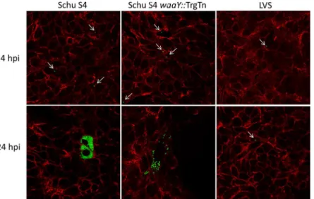
primary human AT-II cells, a similar set of experiments examined the interactions of the bacte-ria with these cells using confocal microscopy. As expected, there were no significant
observ-able differences at 4 hours post-infection between Schu S4, Schu S4waaY::TrgTn and the LVS
strains (Fig 2). Cells containing bacteria typically had one organism per cell and no apparent
replication had occurred at this time point (Fig 2). However, by 24 hours there were several
no-table differences between the three strains that correlated well with the bacterial count data
shown inFig 1. AT-II cells infected withF.tularensisSchu S4 clearly allowed vigorous
replica-tion as observed by the filling of cell cytosol with GFP-labeledF.tularensis(Fig 2). In contrast,
the majority of LVS-infected cells at 24 hours post infection still contained only a single bacte-rium, suggesting that LVS was significantly impaired in its ability to replicate within the
human AT-II cells compared to the Schu S4 strain (Fig 2). Some growth was observed within
Fig 1. Growth ofF.tularensisSchu S4,F.tularensis waaY::TrgTn andF.tularensisLVS in cultured human alveolar type II cells.Immortalized primary AT-II tissue culture cells were used to perform gentamicin protection assays. The ability of the threeFrancisellastrains to enter and replicate with these epithelial lung cells was measured after infecting at an MOI 100:1. Fold growth was calculated as the difference between the number of bacteria surviving gentamicin treatment at 4 hours and 24 hours post-infection. (A) Data are a single experiment representative of three separate experiments. (B) Data are an average of four independent experiments.
LVS infected cells, however, these cells were relatively rare and growth appeared to be limited to no more than a few rounds of replication. It is possible that these cells containing replicating LVS could have been damaged or less fit somehow, but this could not be ascertained
by the confocal microscopy performed. Cells infected withF.tularensisSchu S4waaY::TrgTn
had an intermediate growth phenotype, in that infected cells contained a range of bacteria,
from a couple of bacteria per cell, to cells nearly filled with organisms (Fig 2). The reduced
growth phenotype is likely due to host cell recognition of the mutant, which lacks capsule and O-side chain additions to the lipid A-core sugar molecules in the outer membrane, and early cell death.
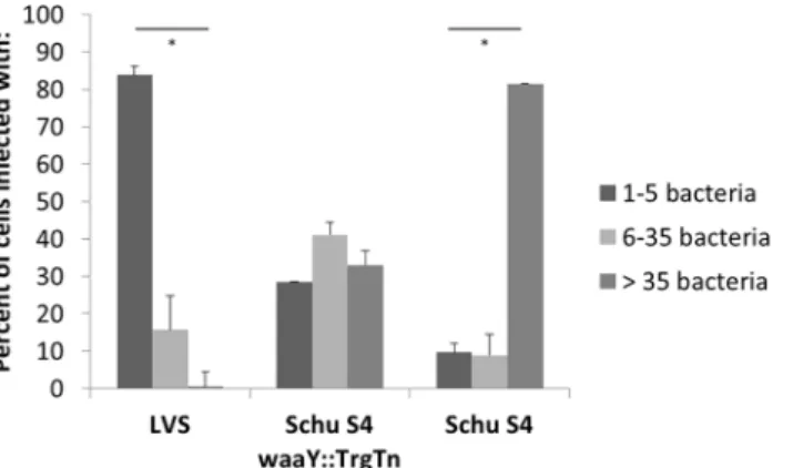
To semi-quantitatively analyze the growth patterns of each of the threeFrancisellastrains in
AT-II cells, we scored infected cells for the number of bacteria that were inside an infected cell 24 hours post-infection. Cells were placed into one of three groups: 5 or less intracellular
bacte-ria, between 6–35 intracellular bacteria, and greater than 35 intracellular bacteria. More than
300 cells for each strain were counted from three separate experiments. Using this scoring cri-teria, greater than 80% of the Schu S4-infected cells had robust growth with more than 35
bac-teria, whereas there were almost no LVS-infected cells (<1%) that had greater than 35 bacteria
(P<0.001) (Fig 3). The majority (>80%) of infected LVS cells had 1–5 bacteria per infected
cell, while cells infected with the Schu S4waaY::TrgTn strain separated into the three categories
equally (Fig 3). These data provide a clearer picture thatF.tularensisSchu S4 replicates
robust-ly in human AT-II cells whileF.tularensisLVS has a low capacity to grow in the cytosol of
these cells, although it is taken up at the same level as the wild type Schu S4. Furthermore, the
F.tularensisSchu S4waaY::TrgTn strain displays a similar growth phenotype in human AT-II cells as that observed in human MDMs, in that the strains grow well for ~16 hours or so before growth levels off. The mechanism for why this mutation results in this cell growth phenotype is unknown and is under active investigation.
Fig 2. Confocal microscopy ofF.tularensisSchu S4,F.tularensis waaY::TrgTn andF.tularensisLVS growth in cultured human alveolar type II cells.Immortalized primary AT-II cells were infected at an MOI of 100:1 and confocal microscopy was performed with cells stained for actin (red). In addition, eachFrancisella strain contained a plasmid encoding GFP and is represented as green. The thin white arrows point to single Francisellawithin cells.
Transmission electron microscopy of early
Francisella
interactions with
murine lung tissue
The interactions between AT-II cells and variousFrancisellastrains suggest that these
patho-genic bacteria have the ability to cross the epithelial barrier by the internalization ofFrancisella
into an epithelial cell. Additionally, if LVS replicates only poorly in AT-II cellsin vivothese
in-teractions may help to explain why the respiratory virulence of LVS for mice is significantly re-duced compared to the virulent Schu S4 strain. Although there has been some investigation
into the role of AT-II cellsin vivo, to our knowledge there has never been any TEM evidence
confirming internalizationin vivo. To better understand the early events that lead to
establish-ment of pneumonic tularemia and to determine ifF.tularensiscan be internalized into AT-II
cellsin vivo, mice were infected intranasally withF.tularensisSchu S4, Schu S4waaY::TrgTn,
LVS or PBS (negative control). In addition, to the intracellular growth defects observed in the
Schu S4waaY::TrgTn mutant, this strain displays decreased virulence in mice, and mice
immu-nized with Schu S4waaY::TrgTn demonstrated some protection against a wild type Schu S4
challenge [24,35]. Furthermore, through observation of the mice and histopathology, the
cause of death is different between Schu S4 and thewaaYmutant, (multiple organ failure and
suffocation due to fluid filled lungs, respectively) [35,36]. These data suggest that the Schu S4
waaY::TrgTn strain induces a host response that is distinct from that caused by wild type
or-ganisms. At 16 or 24 hours post-infection, mice were euthanized and the lungs were perfused and processed for TEM imaging. A MOI of 100 bacteria per alveolus was used to increase the probability of detecting organisms within the lung tissues.
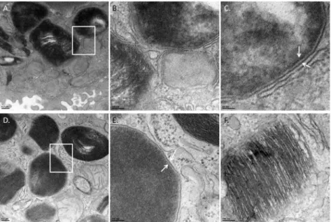
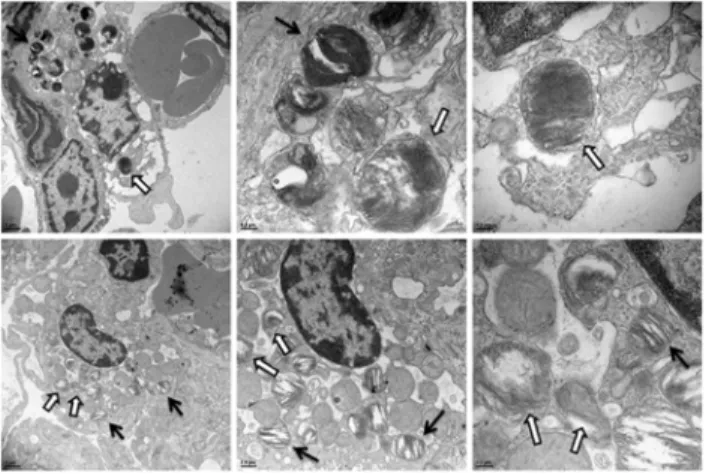
Organisms of each strain were observed in lung cells at either 16 or 24 hours post-infection, but none were observed in the lungs of mice inoculated with PBS as a control. Due to the stain-ing and TEM imagstain-ing, some of the images taken at low magnification had dark stainstain-ing that
often made distinguishingFrancisellafrom lamellar bodies (organelle that packages surfactant)
difficult. However, upon higher magnification there were stark differences observed between
the bacterium and lamellar bodies.F.tularensiswas differentiated from lamellar bodies by
dark, often smooth or bubbly staining (likely the chromosomal DNA) that was spread through-out the organism and the bacteria clearly lacked the stratified staining characteristic of the
la-mellar granules (Fig 4). Lamellar granules are organelles unique to AT-II cells that package and
secrete surfactant and are visible in TEM imaging by their stratified staining that alternates
Fig 3. Scoring of growth patterns observed in confocal imaging.Infected cells were classified into three separate categories based on the amount of bacteria each cell contained at 24 hpi. Cells infected with Schu S4 had significantly more bacteria per cell compared to cells infected with LVS 24 hpi (*P<. 001).
Percentages were calculated from greater than 300 cells containing organisms from three independent experiments.
between dark and white linear bands created from the tight packaging of surfactant membranes
[11]. Furthermore, bacteria were also characterized by identification of a double membrane
(Fig 4C). Using these characteristics, we were able to reliably identify bacteria of each strain in our TEM images.
In examining infected lung tissue we occasionally, although rarely, observed an alveolar macrophage containing an internalized organism that was within the airspace of an alveolus (5 cells total across all samples, data not shown). The most common cell type observed to contain
Francisellawere AT-II epithelial cells (Fig 5). AT-II cells were identified as the cells that pro-duced microvilli at the cell::air interface and contained lamellar granules. Multiple AT-II cells
were observed that contained 1–3 bacteria from lungs infected with each of the three
Franci-sellastrains (Fig 5). It was also interesting to note that the AT-II cells typically bordered pulmo-nary capillaries, highlighting that once the organisms entered an AT-II cell they could gain access to the bloodstream with little impediment. Furthermore, while there might be small
dif-ferences in quantitative uptake of the differentFrancisellastrains into the AT-II cells, no
differ-ences could be determined using TEM.
Observable cell death in AT-II cells with intracellular
F
.
tularensis
By 16 and 24 hpiFrancisellahad internalized into murine AT-II cellsin vivo, but at these time
points it was not possible to determine the fate of theFrancisella-infected cells (conducive for
bacterial replication or cell death). As this is anin vivoinfection it is possible that 24 hpi is not
sufficient time for the bacteria get into the alveolar space, internalize into AT-II cells, and
Fig 4. Identification ofF.tularensisin AT-II cells.TEM images of infected mouse lungs at 24 hours post infection. To differentiate between lamellar bodies and bacteria in AT-II cells, infected cells were observed under high magnification. Lamellar bodies were classified as dark stratified stained organelles whereas; bacteria were identified by smooth dark staining that lacked stratification. Panel A—Cell containing aF.
tularensisorganism magnified 8,000-fold. White rectangle marks area of interest that is shown in panels B and C. Panel B is the area of the white rectangle in Panel A magnified 25,000-fold. A bacterium and the membrane surrounding the organism are shown. Panel C—The white arrows are pointing to the double
membrane (outer and inner membrane) ofF.tularensis. (Panel D) An intracellular organism magnified 8,000-fold. The white rectangle marks an area of interest shown in Panel E. Panel E is the area of the white rectangle in Panel D magnified 25,000-fold. Panel F—25,000-fold magnification of a lamellar body
demonstrating the stratified staining from the packaged surfactant.
replicate. To better understand the outcome of infected airway epithelial cells, we infected mice
intranasally with Schu S4, Schu S4waaY::TrgTn, or LVS. Mice were sacrificed at 32 or 48 hpi
and lungs were harvested. Lung tissues were processed as described previously and TEM was performed.
Similar to infected lungs at 16 and 24 hpi, the majority of the cells containing internalized
Francisellawere AT-II cells. However, unlike cells with intracellularF.tularensisat 16 and 24
hpi, several infected AT-II cells had visible changes suggesting that the cells were dying (Fig 6).
Infected AT-II cells were observed to have loss of cytoplasmic density, swelling of mitochon-dria, and disrupted mitochondrial cristae structure, which have previously been reported in
dying cells observed by TEM imaging [37–39]. These dying cells were observed in all strains
tested and no significant differences between strains were observed by TEM imaging. In addi-tion, cells typically had only a few organisms present indicating that there was no significant replication of the bacteria within the AT-II cells. However, as TEM imaging is with sections that are 70nm thick, the lack of observable replication may be due to limitations from TEM imaging.
Fig 5. Electron microscopy images ofFrancisella tularensisstrains within murine alveolar type II epithelial cells.TEM images of infected mouse lungs at 24 hours post-infection. Mice were infected intranasally with eitherF.tularensisSchu S4 (A-F),F.tularensisSchu S4waaY::TrgTn (G-K), andF. tularensisLVS (I,L). Each image shows an AT-II cell containing organisms. The AT-II cell can be identified by the presence of microvilli at the cell-air interface and by the presence of lamellar granules. It is also worth noting that each infected AT-II cell was immediately adjacent to a pulmonary capillary. The area containing the internalized bacteria is within the white rectangle which is shown at higher magnification immediately beneath the corresponding image. The arrows identify the bacteria in each field.
The
F
.
tularensis waaY
::TrgTn mutant induces substantial airway
pathology that is not induced by wild type
F
.
tularensis
Schu S4
While we could not detect quantifiable differences in bacterial uptake into AT-II cellsin vivo,
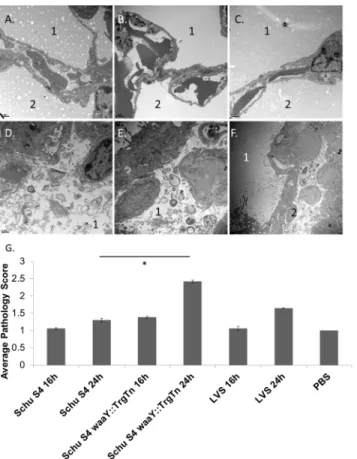
there were strain-specific differences in the pathology that was observed in the airway spaces following infection. At 16 and 24 hours post-infection, lungs infected with either PBS, Schu S4,
or LVS were essentially clear of debris within the airspace observed (Fig 7A, 7B and 7C).
How-ever, mice infected with Schu S4waaY::TrgTn at 16 hours post-infection had significant
pa-thology in the alveoli, as evidenced by large amounts of debris in the alveolar airspaces that contained both surfactant and cellular components. By 24 hours post infection with this
mu-tant, a substantial portion of the airspace was observed to contain this cellular debris (Fig 7D,
7E and 7F).
To more accurately evaluate the damage induced in the lungs by theF.tularensis waaY::
TrgTn mutant, semi-quantitation of airspace debris was scored using TEM images [40].
Indi-vidual alveolar airspaces, defined by the border of the alveolar epithelium, were evaluated for
the percent of the airspace containing cellular debris and were given a score of 1–4, with 1
hav-ing less than 25% of the airspace filled with debris and 4 behav-ing greater than 75% of the airspace filled with debris. Approximately, 50 TEM fields were evaluated per strain and time point. In the PBS control mice, there was little to no cellular debris observed at 16 or 24 hours post
infec-tion (Fig 7G). Additionally, there was no significant cellular debris observed at 16 hours
post-infection forF.tularensisSchu S4 orF.tularensisLVS compared to the control PBS infected
mice (Fig 7G). At 24 hours post-infection, both Schu S4 and LVS had some observable cellular
debris that increased the average airspace damage score from 1 to 1.3 and 1.6, respectively (Fig
7G).F.tularensis waaY::TrgTn had observable cellular debris with an average score of 1.3 at 16 hours post-infection, which was comparable to wild type Schu S4 at 24 hours post-infection. By 24 hours post-infection, there was significantly more cellular debris induced by the Schu S4
waaY::TrgTn strain than the PBS control (P<0.001), with an average score of 2.4, meaning
that the average airspace had 50–75% filled with cellular debris (Fig 7G). Furthermore, of the
airspaces scored from lungs infected with Schu S4waaY::TrgTn at 24 hpi, 38% of samples
scored had over half of the airspace filled with cellular debris and 28% of the airspaces scored had over three-fourth of the airspace filled. Comparatively, wild type Schu S4 only had 3% of
Fig 6. TEM ofF.tularensisin dying AT-II cellsin vivo.TEM images of infected mouse lungs at both 32 and 48 hpi infected with Schu S4. (A-C) AT-II cells containingFrancisellagoing through cell death characterized by lack of electron density of the cytosol and swollen mitochondria. (D-F) A separate AT-II cell undergoing cell death infected withFrancisella. The open arrows identify the bacteria in each field and the solid black arrows indicate lamellar bodies in the cells.
all scored airspaces that contained over half of the airspace filled and no airspace was observed in Schu S4 infected lungs to have greater than three-fourth of the airspace filled with cellular
debris. The mechanism by which thewaaY::TrgTn mutant induces the accumulation of debris
in the lung airspaces of mice is unknown but is likely directly related to the other properties of
this mutant, such as the induction of early cell death of MDMs and attenuationin vivo[24,35].
Discussion
F.tularensisis a Tier 1 Select Agent pathogen that is capable of causing lethal human respirato-ry infection with a verespirato-ry low inoculum. In order to cause disease, organisms in the airway must breach the epithelium and pass through the endothelium to enter the bloodstream, which al-lows the organisms to disseminate from the lung to the liver and spleen. While significant effort has been expended to study the interactions of these pathogens with various host cells, the
mechanisms by whichFrancisellapasses through the respiratory epithelia are still poorly
un-derstood. Much of the research has focused on the interactions ofFrancisellawith
macro-phagesin vitro, but it is not clear how the interactions ofFrancisellawith alveolar macrophages
would facilitate the passage of the bacteria through the respiratory epitheliumin vivo. Alveolar
macrophages are found within the airspaces, and are associated with the apical surface of the airway epithelium. These cells have high phagocytic activity and protect the lung epithelium by
Fig 7. Schu S4waaY::TrgTn increases cellular debris within the alveoli.(A-C). TEM imaging of 24 hours demonstrating clean airspace with the alveoli in lung tissue that is infected with either PBS (A), Schu S4 (B), or LVS (C) and airspace that is representative of lung tissue infected with Schu S4waaY::TrgTn (D-F). B. Graphical representation of scoring of the airspace for cellular debris. There was an increase in cellular debris in lungs infected with Schu S4waaY:TrgTn compared to PBS control (P<0.001). Averages were calculated from analysis of greater than 50 scored airspaces from 2 separate lung sections per strain and time point.
engulfing particles or organisms from the inhaled air. Following engulfment of particles, the cells detach from the airway epithelium and are typically expelled from the airway via the
mucociliary escalator [8]. Since the established role for alveolar macrophages is to engulf
parti-cles and organisms from the airspaces and remove them from the host via the mucociliary
esca-lator, it is difficult to understand howFrancisellauses alveolar macrophages to penetrate into
lung tissue although, of course, it is still possible thatFrancisellauses these cells to breach the
lung epithelium.
Type II pneumocytes, or AT-II cells, are an integral part of the alveolar epithelium,
compris-ing 15% of all lung cells [9] and have been implicated by several groups as a lung cell type in
whichFrancisellacan grow [19,21,22]. AT-II cells have a variety of biological functions
in-cluding production of surfactant that is important for biophysical stabilization of the alveolus
[41], maintenance of alveolar fluid balance [42], differentiation into AT-I cells which are
im-portant for structural integrity of the alveolus [43], and involvement with activating
compo-nents of both innate and adaptive immunity [17]. Specifically, these cells have been shown to
constitutively express several toll-like receptors, along with NLR proteins, which are involved in the production of cytokines that recruit and interact with dendritic cells, T cells, and B cells [23,44,45].
In an effort to establish a cell culture model that closely mimicsFrancisellainteractions with
lung epithelium during infection of a host, we acquired a newly available human AT-II cell line
to study the entry and growth ofFrancisellain a relevant epithelial cell line. The initial uptake
ofFrancisellaspecies (Schu S4, Schu S4waaY::TrgTn, and LVS) into these cells was compara-ble to that observed for other tissue culture non-phagocytic cell lines such as HEp-2, HeLa,
HEK-293, COS-7 and A549 cells, as observed by us and other groups [19,21,22]. Several
re-search groups have studied various properties ofF.tularensisstrains in the human lung
epithe-lial cell line A549 [46–49]. One group compared the ability ofF.tularensisSchu S4, a Schu S4
ΔpurMCDmutant,F.tularensisLVS and a LVSΔpurMCDmutant to grow in both human macrophages and A549 cells. While the work was focused on evaluating the importance of the ΔpurMCDgenes as a virulence factor, the results demonstrate no discernible growth
differ-ences betweenF.tularensisSchu S4 andF.tularensisLVS in A549 human lung epithelial cells
[46]. Others demonstrated that LVS grows well in A549 cells [48,50], but did not compare the
growth of LVS to a virulentF.tularensisstrain. Another group has attempted to develop a
more relevant cell model for studyingFrancisellainteractions by growing A549 cells in
rotat-ing-wall vessels (RWV) to stimulatein vivo-like phenotypes [49]. However, their work did not
detect any significant growth differences betweenF.tularensisSchu S4 andF.tularensisLVS in
A549 cells, although the RWV growth conditions did seem to stimulate cells to express cell
po-larity markers and other proteins indicative of cells growingin vivo.
In this work, we have observed a significant difference in the ability ofF.tularensisSchu S4
andF.tularensisLVS to grow within immortalized primary human AT-II cells after uptake. Virulent Schu S4 grew to high numbers in the cytosol of these cells (1600-fold growth was ob-served from 4 hours to 24 hours) while the LVS strain exhibited minimal growth in the human AT-II cells (~ 2- to 12-fold growth). We believe that this is a significant finding since to our knowledge these human AT-II cells are the first cells where such a significant difference in
growth exists between virulentF.tularensisand the vaccine (attenuated virulence) strainF.
better model system for studyingFrancisella-nonphagocytic cell interactions than long
estab-lished tissue culture lines, they still do not replicate all aspects that are observed ofin vivo
infec-tions. Particularly, we observed significant cell perturbations in murine AT-II cells infected
with low numbers of Schu S4 organisms (1–3 organisms visible in a given EM section) which
was not observed in the cultured human AT-II cells that contained hundreds to thousands of replicating Schu S4 organisms.
TheF.tularensis waaY::TrgTn strain also has an attenuated virulence phenotype in the human AT-II cells. Since previous work from our lab has demonstrated that this strain has a significant growth defect in human MDMs, it is possible that this strain is unable to replicate to high numbers in airway epithelial cells for the same reason. Work is currently underway to characterize the mechanism that leads to cessation of bacterial growth and cell death in both cell types. An understanding of this mechanism may help clarify how host cells combat intra-cellular bacterial pathogens.
Additionally, we performed experiments to determine what host cells are the early targets of
F.tularensisas they establish infection of the mouse lung. Murine lung tissues from mice
in-fected withF.tularensiswere examined by transmission electron microscopy to identify the
cell types that contained intracellularF.tularensis. We spent considerable effort looking for
or-ganisms within alveolar macrophages but observed very fewFrancisellaorganisms within these
cells (~5 infected alveolar macrophages in all samples imaged). It is possible that there were more alveolar macrophages that had engulfed organisms than were detected during these ex-periments since this is the primary function of the cells. However, as alveolar macrophages typ-ically detach from the epithelium after phagocytosis, it seems likely that alveolar macrophages
that had engulfedFrancisellaorganisms may have been washed out of the mouse lungs by
per-fusion. Alternatively, these alveolar macrophages could have been removed by the mucociliary escalator in the mouse lungs as part of the normal defense mechanisms before lungs were
ex-tracted and fixed. In either case, the absence of alveolar macrophages containingFrancisella
or-ganisms in the infected lung samples provides support that interactions with other cell types are important for penetration into the lung tissue.
Careful examination of the infected mouse lung tissue revealed that organisms were
consis-tently observed within AT-II cells by 16–24 hours post-infection for each of the three strains
used in our experiments (Schu S4, Schu S4waaY::TrgTn or LVS). It is not a trivial exercise to
identify intracellularFrancisellaorganisms within the AT-II cells of the alveoli by TEM, as
there are an estimated 1 x 10⁶alveoli per mouse lung and many AT-II cells per alveoli. In
addi-tion, sectioning of the lung tissue captures only a small percentage of the cellular material per alveoli for viewing, which also decreases the probability of finding evidence of intracellular bac-teria. Since we were able to find bacteria within the AT-II consistently, despite these hurdles, our data extends previous studies suggesting that these cells are an important target cell in the alveolar epithelium. The TEM images highlight the path that organisms within the AT-II cells likely follow to cause systemic disease. Following entry into the AT-II cells, the bacteria are im-mediately adjacent to alveolar capillaries. As organisms pass through or escape from an AT-II cell they will have access to the bloodstream via passage through the endothelium of the imme-diately available alveolar capillary.
and any tissue damage due to inoculum would likely cause damage to multiple cell types, in-cluding uninfected AT-II cells, which was not observed. In addition, it is well-established that
cells with intracellularF.tularensisundergo cell death by 24–48 hpi as part of the pathogenesis
of the organism. Unfortunately, we were not able to determine if there was replication within the AT-II cells by use of TEM imaging, as we were unable to examine specific cells over a suffi-cient period of time. Future efforts to address this issue would need to rely on other techniques,
such as confocal microscopy, to determine if AT-II cells are permissive for replication ofF.
tularensis in vivoor if AT-II cells primarily function as a gateway through the epithelial barrier.
The inclusion of theF.tularensisSchu S4waaY::TrgTn mutant in this work provided
im-portant contrasts to the interactions of wild typeF.tularensisSchu S4 and LVS within lung
tis-sue. This mutant strain induced significant levels of pathology in the mouse lungs in the form of debris (inflammation) that was present in the airspaces. Mouse lungs infected with Schu S4 or LVS were virtually free of inflammation at 16 hours post infection in comparison, as noted by the lack of debris-filled airspace. These data are not surprising because other work has
dem-onstrated thatF.tularensisfails to induce inflammation upon infection as it does not stimulate
pattern recognition receptors and by inducing broad immunosuppression [51]. ThewaaYgene
was identified as being important in the ability ofF.tularensisSchu S4 to grow in human
MDMs [24] and subsequent work revealed that the strain did not produce normal LPS or
cap-sule. More recently, our group has shown that the strain is defective for mouse virulence and sublethal infection of mice with this strain provides protection against lethal doses of wild type
Schu S4 [35]. This work reveals that this strain induces host cell responses in the lung in the
early stages of infection that seem to significantly alter host recognition of the strain and the course of disease. Part of this host response is likely to be initiated by early death of the host
cell, as our lab has previously shown occurs with this mutant strain [24]. An understanding of
the mechanism by which thisFrancisella waaY::TrgTn strain induces host cell death is an
on-going interest in the lab since avoidance of detection by the host is a key virulence strategy of
Francisellaand may provide important clues to developing an effective therapy against this po-tential bioweapon. In contrast to the mutant strain, the lack of cellular debris in wild type
in-fected lungs adds to the evidence that virulent wild typeFrancisellastrains establish infection
of a host using a combination of active pathogenic strategies (host cell entry, phagosome es-cape, cytosolic growth that allows systemic distribution) and stealth strategies (evasion of host detection by non-stimulatory LPS and suppression of inflammatory signals by unknown
mech-anisms) [52–54].
Taken together, the TEM data and thein vitrocell infection data have allowed us to develop
a model of howF.tularensisestablishes lung infection (Fig 8). InhaledF.tularensisorganisms
are carried to the small alveolar sacs and interact with the cell populations present in the
alve-oli: alveolar macrophages and AT-II cells. IfFrancisellafirst interacts with an alveolar
macro-phage there are at least two possible outcomes: 1-The macromacro-phage engulfs an organism, detaches from the epithelium, and is removed from the alveolus via the mucociliary escalator or 2- an organism is phagocytosed and the infected alveolar macrophage remains in the
alveo-lar space, whereFrancisellareplicates to large numbers, escapes from the cell and re-infects
ad-jacent cells in the epithelium. This re-infection may allow contact ofFrancisellawith AT-II
cells where the bacteria could pass through the epithelial barrier by entering and growing
with-in AT-II cells. It is also possible that upon with-inhalation,Francisellafirst interacts with an AT-II
cell. This interaction would also allow for the passage of the bacteria through the epithelial bar-rier via the AT-II cell. Once invaded, AT-II cells will lyse from growth of intracytoplasmic
bac-teria, releasingFrancisellaorganisms into the environment. The newly released bacteria would
have access to the endothelial vessels and could quickly disseminate to the liver and spleen
the bloodstream as extracellular organisms. In support of this model, a study by Forestalet al.
demonstrated that 75% of Schu S4 organisms in the bloodstream were extracellular [55].
Fu-ture experiments will help to clearly define the individual steps in the infection process which should help to identify targets to better treat tularemia and/or to understand the host-pathogen interactions that may help to develop a live attenuated vaccine.
Acknowledgments
We are grateful for the use of the University of Iowa Carver College of Medicine Biosafety Level 3 Core Facility as well as the DNA Core and the Central Microscopy Research Core Facil-ities. We also thank Dana Ries for her highly skilled assistance in meeting the regulations of the BSL-3.
Author Contributions
Conceived and designed the experiments: MF BDJ. Performed the experiments: MF JRF JAR BDJ. Analyzed the data: MF JRF JAR BDJ. Contributed reagents/materials/analysis tools: MF JRF JAR BDJ. Wrote the paper: MF JRF JAR MAA BDJ.
References
1. McCrumb FR. Aerosol Infection of Man with Pasteurella Tularensis. Bacteriol Rev. 1961; 25(3):262–7.
Epub 1961/09/01. PubMed PMID:16350172; PubMed Central PMCID: PMC441102.
2. Francis E, Moore D. Identity of Ohara's disease and tularemia. J Amer Med Assoc. 1926; 86:1329–32.
PubMed PMID:ISI:000201687800507.
3. Stuart BM, Pullen RL. Tularemic Pneumonia—Review of American Literature and Report of 15
Addi-tional Cases. Am J Med Sci. 1945; 210(2):223–36. doi:10.1097/00000441-194508000-00013PubMed
PMID:ISI:A1945XU80700012.
4. Saslaw S, Eigelsbach HT, Prior JA, Wilson HE, Carhart S. Tularemia vaccine study. II. Respiratory challenge. Arch Intern Med. 1961; 107:702–14. Epub 1961/05/01. PubMed PMID:13746667. Fig 8. Model of earlyFrancisellainfection within the lung.(1)Francisellaenters into the alveoli from an aerosolized infection where it either gets phagocytosed by alveolar macrophages (2a) or interacts with AT-II cells (2b). Upon uptake into alveolar macrophages there are at least two possible outcomes, either alveolar macrophages allow bacterial growth and release into the airspace (2a) or alveolar macrophages detach and are removed from the alveoli by the mucociliary escalator (2c). Growth and release from the alveolar macrophages allows reinfection with surrounding tissue including AT-II cells (3). Internalization with AT-II cells acts as a mechanism to get past the epithelial barrier and allows for interaction with endothelial cells and eventually dissemination to the liver and spleen.
5. Saslaw S, Eigelsbach HT, Wilson HE, Prior JA, Carhart S. Tularemia vaccine study. I. Intracutaneous challenge. Arch Intern Med. 1961; 107:689–701. Epub 1961/05/01. PubMed PMID:13746668. 6. Marriott HM, Dockrell DH. The role of the macrophage in lung disease mediated by bacteria. Exp Lung
Res. 2007; 33(10):493–505. Epub 2007/12/14. doi:10.1080/01902140701756562PubMed PMID: 18075824.
7. Franke-Ullmann G, Pfortner C, Walter P, Steinmuller C, Lohmann-Matthes ML, Kobzik L. Characteriza-tion of murine lung interstitial macrophages in comparison with alveolar macrophages in vitro. J Immu-nol. 1996; 157(7):3097–104. Epub 1996/10/01. PubMed PMID:8816420.
8. Laurenzi GA, Guarneri JJ, Endriga RB, Carey JP. Clearance of Bacteria by the Lower Respiratory Tract. Science. 1963; 142(3599):1572–3. Epub 1963/12/20. PubMed PMID:14075687.
9. Castranova V, Rabovsky J, Tucker JH, Miles PR. The alveolar type II epithelial cell: a multifunctional pneumocyte. Toxicol Appl Pharmacol. 1988; 93(3):472–83. Epub 1988/05/01. PubMed PMID: 3285521.
10. Jones GS, Miles PR, Lantz RC, Hinton DE, Castranova V. Ionic content and regulation of cellular vol-ume in rat alveolar type II cells. J Appl Physiol Respir Environ Exerc Physiol. 1982; 53(1):258–66. Epub
1982/07/01. PubMed PMID:7118639.
11. Perez-Gil J. Structure of pulmonary surfactant membranes and films: the role of proteins and lipid-pro-tein interactions. Biochim Biophys Acta. 2008; 1778(7–8):1676–95. Epub 2008/06/03. doi:10.1016/j. bbamem.2008.05.003PubMed PMID:18515069.
12. Crapo JD, Young SL, Fram EK, Pinkerton KE, Barry BE, Crapo RO. Morphometric characteristics of cells in the alveolar region of mammalian lungs. Am Rev Respir Dis. 1983; 128(2 Pt 2):S42–6. Epub
1983/08/01. PubMed PMID:6881707.
13. Eisele NA, Anderson DM. Host Defense and the Airway Epithelium: Frontline Responses That Protect against Bacterial Invasion and Pneumonia. J Pathog. 2011; 2011:249802. Epub 2011/01/01. doi:10. 4061/2011/249802PubMed PMID:22567325; PubMed Central PMCID: PMC3335569.
14. Wu Q, Jiang D, Minor MN, Martin RJ, Chu HW. In vivo function of airway epithelial TLR2 in host defense against bacterial infection. Am J Physiol Lung Cell Mol Physiol. 2011; 300(4):L579–86. Epub 2011/01/
18. doi:10.1152/ajplung.00336.2010PubMed PMID:21239529; PubMed Central PMCID: PMC3075102.
15. Sha Q, Truong-Tran AQ, Plitt JR, Beck LA, Schleimer RP. Activation of airway epithelial cells by toll-like receptor agonists. Am J Respir Cell Mol Biol. 2004; 31(3):358–64. Epub 2004/06/12. doi:10.1165/ rcmb.2003-0388OCPubMed PMID:15191912.
16. McCormick TS, Weinberg A. Epithelial cell-derived antimicrobial peptides are multifunctional agents that bridge innate and adaptive immunity. Periodontol 2000. 2010; 54(1):195–206. Epub 2010/08/18.
doi:10.1111/j.1600-0757.2010.00373.xPubMed PMID:20712640; PubMed Central PMCID: PMC3816379.
17. Kato A, Schleimer RP. Beyond inflammation: airway epithelial cells are at the interface of innate and adaptive immunity. Curr Opin Immunol. 2007; 19(6):711–20. Epub 2007/10/12. doi:10.1016/j.coi.2007. 08.004PubMed PMID:17928212; PubMed Central PMCID: PMC2196222.
18. Bermudez LE, Goodman J. Mycobacterium tuberculosis invades and replicates within type II alveolar cells. Infect Immun. 1996; 64(4):1400–6. Epub 1996/04/01. PubMed PMID:8606107; PubMed Central
PMCID: PMC173932.
19. Hall JD, Craven RR, Fuller JR, Pickles RJ, Kawula TH. Francisella tularensis replicates within alveolar type II epithelial cells in vitro and in vivo following inhalation. Infect Immun. 2007; 75(2):1034–9. Epub
2006/11/08. doi:10.1128/IAI.01254-06PubMed PMID:17088343; PubMed Central PMCID: PMC1828526.
20. Hall JD, Woolard MD, Gunn BM, Craven RR, Taft-Benz S, Frelinger JA, et al. Infected-host-cell reper-toire and cellular response in the lung following inhalation of Francisella tularensis Schu S4, LVS, or U112. Infect Immun. 2008; 76(12):5843–52. Epub 2008/10/15. doi:10.1128/IAI.01176-08PubMed
PMID:18852251; PubMed Central PMCID: PMC2583552.
21. Craven RR, Hall JD, Fuller JR, Taft-Benz S, Kawula TH. Francisella tularensis invasion of lung epitheli-al cells. Infect Immun. 2008; 76(7):2833–42. Epub 2008/04/23. doi:10.1128/IAI.00043-08PubMed
PMID:18426871; PubMed Central PMCID: PMC2446690.
22. Lindemann SR, McLendon MK, Apicella MA, Jones BD. An in vitro model system used to study adher-ence and invasion of Francisella tularensis live vaccine strain in nonphagocytic cells. Infect Immun. 2007; 75(6):3178–82. Epub 2007/03/07. doi:10.1128/IAI.01811-06PubMed PMID:17339345;
PubMed Central PMCID: PMC1932879.
78. Epub 2007/05/16. doi:10.1128/IAI.00157-07PubMed PMID:17502386; PubMed Central PMCID: PMC1951971.
24. Lindemann SR, Peng K, Long ME, Hunt JR, Apicella MA, Monack DM, et al. Francisella tularensis Schu S4 O-antigen and capsule biosynthesis gene mutants induce early cell death in human macro-phages. Infect Immun. 2011; 79(2):581–94. Epub 2010/11/17. doi:10.1128/IAI.00863-10PubMed
PMID:21078861; PubMed Central PMCID: PMC3028865.
25. Knust J, Ochs M, Gundersen HJ, Nyengaard JR. Stereological estimates of alveolar number and size and capillary length and surface area in mice lungs. Anat Rec (Hoboken). 2009; 292(1):113–22. Epub
2008/12/31. doi:10.1002/ar.20747PubMed PMID:19115381.
26. Osmanagic E, Sukstanskii AL, Quirk JD, Woods JC, Pierce RA, Conradi MS, et al. Quantitative assess-ment of lung microstructure in healthy mice using an MR-based 3He lung morphometry technique. J Appl Physiol (1985). 2010; 109(6):1592–9. Epub 2010/08/28. doi:10.1152/japplphysiol.00736.2010
PubMed PMID:20798272; PubMed Central PMCID: PMC3006399.
27. Townsley MI. Structure and composition of pulmonary arteries, capillaries, and veins. Compr Physiol. 2012; 2(1):675–709. Epub 2012/01/01. doi:10.1002/cphy.c100081PubMed PMID:23606929;
PubMed Central PMCID: PMC3630377.
28. Hsiao CH, Ueno N, Shao JQ, Schroeder KR, Moore KC, Donelson JE, et al. The effects of macrophage source on the mechanism of phagocytosis and intracellular survival of Leishmania. Microbes Infect. 2011; 13(12–13):1033–44. Epub 2011/07/05. doi:10.1016/j.micinf.2011.05.014PubMed PMID: 21723411; PubMed Central PMCID: PMC3185139.
29. Zhou H, Ferraro D, Zhao J, Hussain S, Shao J, Trujillo J, et al. The N-terminal region of severe acute re-spiratory syndrome coronavirus protein 6 induces membrane rearrangement and enhances virus repli-cation. J Virol. 2010; 84(7):3542–51. Epub 2010/01/29. doi:10.1128/JVI.02570-09PubMed PMID: 20106914; PubMed Central PMCID: PMC2838104.
30. Faron M, Fletcher JR, Rasmussen JA, Long ME, Allen LA, Jones BD. The Francisella tularensis migR, trmE, and cphA genes contribute to F. tularensis pathogenicity island gene regulation and intracellular growth by modulation of the stress alarmone ppGpp. Infect Immun. 2013; 81(8):2800–11. Epub 2013/
05/30. doi:10.1128/IAI.00073-13PubMed PMID:23716606; PubMed Central PMCID: PMC3719569.
31. Long ME, Lindemann SR, Rasmussen JA, Jones BD, Allen LA. Disruption of Francisella tularensis Schu S4 iglI, iglJ, and pdpC genes results in attenuation for growth in human macrophages and in vivo virulence in mice and reveals a unique phenotype for pdpC. Infect Immun. 2013; 81(3):850–61. Epub
2013/01/01. doi:10.1128/IAI.00822-12PubMed PMID:23275090; PubMed Central PMCID: PMC3584877.
32. Ferriola PC, Steigerwalt R, Robertson AT, Nettesheim P. Abnormalities in growth regulation of trans-formed rat tracheal epithelial cells. Pathobiology. 1990; 58(1):28–36. Epub 1990/01/01. PubMed PMID: 1971176.
33. Razin A, Cedar H. DNA methylation and gene expression. Microbiol Rev. 1991; 55(3):451–8. Epub
1991/09/01. PubMed PMID:1943996; PubMed Central PMCID: PMC372829.
34. Steele S, Brunton J, Ziehr B, Taft-Benz S, Moorman N, Kawula T. Francisella tularensis harvests nutri-ents derived via ATG5-independent autophagy to support intracellular growth. PLoS Pathog. 2013; 9 (8):e1003562. Epub 2013/08/24. doi:10.1371/journal.ppat.1003562PubMed PMID:23966861; PubMed Central PMCID: PMC3744417.
35. Rasmussen JA, Post DM, Gibson BW, Lindemann SR, Apicella MA, Meyerholz DK, et al. Francisella tularensis Schu S4 lipopolysaccharide core sugar and O-antigen mutants are attenuated in a mouse model of tularemia. Infect Immun. 2014; 82(4):1523–39. doi:10.1128/IAI.01640-13PubMed PMID: 24452684; PubMed Central PMCID: PMC3993386.
36. Sharma J, Mares CA, Li Q, Morris EG, Teale JM. Features of sepsis caused by pulmonary infection with Francisella tularensis Type A strain. Microb Pathog. 2011; 51(1–2):39–47. Epub 2011/03/29. doi: 10.1016/j.micpath.2011.03.007PubMed PMID:21440052; PubMed Central PMCID: PMC3090489.
37. Fujita R, Ueda H. Protein kinase C-mediated cell death mode switch induced by high glucose. Cell Death Differ. 2003; 10(12):1336–47. Epub 2003/08/23. doi:10.1038/sj.cdd.4401300PubMed PMID: 12934062.
38. Oppenheim RW, Flavell RA, Vinsant S, Prevette D, Kuan CY, Rakic P. Programmed cell death of devel-oping mammalian neurons after genetic deletion of caspases. J Neurosci. 2001; 21(13):4752–60. Epub
2001/06/27. PubMed PMID:11425902.
39. Fujita R, Ueda H. Protein kinase C-mediated necrosis-apoptosis switch of cortical neurons by condi-tioned medium factors secreted under the serum-free stress. Cell Death Differ. 2003; 10(7):782–90.
40. Gibson-Corley KN, Olivier AK, Meyerholz DK. Principles for Valid Histopathologic Scoring in Research. Vet Pathol. 2013. Epub 2013/04/06. doi:10.1177/0300985813485099PubMed PMID:23558974; PubMed Central PMCID: PMC3795863.
41. Guillot L, Nathan N, Tabary O, Thouvenin G, Le Rouzic P, Corvol H, et al. Alveolar epithelial cells: Mas-ter regulators of lung homeostasis. Int J Biochem Cell Biol. 2013; 45(11):2568–73. Epub 2013/08/31.
doi:10.1016/j.biocel.2013.08.009PubMed PMID:23988571.
42. Fehrenbach H. Alveolar epithelial type II cell: defender of the alveolus revisited. Respir Res. 2001; 2 (1):33–46. Epub 2001/11/01. PubMed PMID:11686863; PubMed Central PMCID: PMC59567. 43. Evans MJ, Hackney JD. Cell proliferation in lungs of mice exposed to elevated concentrations of
oxy-gen. Aerosp Med. 1972; 43(6):620–2. Epub 1972/06/01. PubMed PMID:5035549.
44. Schwingshackl A, Teng B, Ghosh M, Waters CM. Regulation of Monocyte Chemotactic Protein-1 se-cretion by the Two-Pore-Domain Potassium (K2P) channel TREK-1 in human alveolar epithelial cells. Am J Transl Res. 2013; 5(5):530–42. Epub 2013/08/27. PubMed PMID:23977412; PubMed Central
PMCID: PMC3745440.
45. Vir P, Gupta D, Agarwal R, Verma I. Immunomodulation of alveolar epithelial cells by Mycobacterium tuberculosis phosphatidylinositol mannosides results in apoptosis. Apmis. 2013. Epub 2013/08/08. doi:
10.1111/apm.12141PubMed PMID:23919648.
46. Pechous RD, McCarthy TR, Mohapatra NP, Soni S, Penoske RM, Salzman NH, et al. A Francisella tularensis Schu S4 purine auxotroph is highly attenuated in mice but offers limited protection against homologous intranasal challenge. PLoS One. 2008; 3(6):e2487. doi:10.1371/journal.pone.0002487
PubMed PMID:18575611; PubMed Central PMCID: PMC2429968.
47. Bradburne CE, Verhoeven AB, Manyam GC, Chaudhry SA, Chang EL, Thach DC, et al. Temporal tran-scriptional response during infection of type II alveolar epithelial cells with Francisella tularensis live vaccine strain (LVS) supports a general host suppression and bacterial uptake by macropinocytosis. J Biol Chem. 2013; 288(15):10780–91. doi:10.1074/jbc.M112.362178PubMed PMID:23322778;
PubMed Central PMCID: PMC3624459.
48. Thomas-Charles CA, Zheng H, Palmer LE, Mena P, Thanassi DG, Furie MB. FeoB-mediated uptake of iron by Francisella tularensis. Infect Immun. 2013; 81(8):2828–37. doi:10.1128/IAI.00170-13PubMed
PMID:23716605; PubMed Central PMCID: PMC3719576.
49. David J, Sayer NM, Sarkar-Tyson M. The use of a three-dimensional cell culture model to investigate host-pathogen interactions of Francisella tularensis in human lung epithelial cells. Microbes Infect. 2014; 16(9):735–45. doi:10.1016/j.micinf.2014.04.001PubMed PMID:24796635.
50. Bradburne CE, Verhoeven AB, Manyam GC, Chaudhry SA, Chang EL, Thach DC, et al. Temporal tran-scriptional response during infection of type II alveolar epithelial cells with Francisella tularensis live vaccine strain (LVS) supports a general host suppression and bacterial uptake by macropinocytosis. J Biol Chem. 2013; 288(15):10780–91. Epub 2013/01/17. doi:10.1074/jbc.M112.362178PubMed PMID: 23322778; PubMed Central PMCID: PMC3624459.
51. Bosio CM, Bielefeldt-Ohmann H, Belisle JT. Active suppression of the pulmonary immune response by Francisella tularensis Schu4. J Immunol. 2007; 178(7):4538–47. Epub 2007/03/21. PubMed PMID: 17372012.
52. Zarrella TM, Singh A, Bitsaktsis C, Rahman T, Sahay B, Feustel PJ, et al. Host-adaptation of Franci-sella tularensis alters the bacterium's surface-carbohydrates to hinder effectors of innate and adaptive immunity. PLoS One. 2011; 6(7):e22335. Epub 2011/07/30. doi:10.1371/journal.pone.0022335
PubMed PMID:21799828; PubMed Central PMCID: PMC3142145.
53. Elkins KL, Cowley SC, Bosio CM. Innate and adaptive immunity to Francisella. Ann N Y Acad Sci. 2007; 1105:284–324. Epub 2007/05/01. doi:10.1196/annals.1409.014PubMed PMID:17468235. 54. Celli J, Zahrt TC. Mechanisms of Francisella tularensis intracellular pathogenesis. Cold Spring Harb
Perspect Med. 2013; 3(4):a010314. Epub 2013/04/03. doi:10.1101/cshperspect.a010314PubMed PMID:23545572.
55. Forestal CA, Malik M, Catlett SV, Savitt AG, Benach JL, Sellati TJ, et al. Francisella tularensis has a sig-nificant extracellular phase in infected mice. The Journal of infectious diseases. 2007; 196(1):134–7.