Induces Left Ventricular Damage and Diastolic
Dysfunction in Rats
Katrin Wenzel1,2*, Gerd Wallukat2, Fatimunnisa Qadri1, Norbert Hubner2, Herbert Schulz2, Oliver Hummel2, Florian Herse1, Arnd Heuser2, Robert Fischer1,2, Harald Heidecke3, Friedrich C. Luft2,4, Dominik N. Muller2, Rainer Dietz1, Ralf Dechend2,4
1Medical Faculty of the Charite´, Berlin, Germany,2Experimental and Clinical Research Center and Max-Delbru¨ck Center for Molecular Medicine, Berlin, Germany,
3CellTrend, Luckenwalde, Germany,4HELIOS Clinic-Berlin, Franz-Volhard Clinic, Berlin, Germany
Abstract
Background: Agonistic autoantibodies to the a1-adrenergic receptor occur in nearly half of patients with refractory hypertension; however, their relevance is uncertain.
Methods/Principal Findings:We immunized Lewis rats with the second extracellular-loop peptides of the human a1A -adrenergic receptor and maintained them for one year.a1A-adrenergic antibodies (a1A-AR-AB) were monitored with a neonatal cardiomyocyte contraction assay by ELISA, and by ERK1/2 phosphorylation in humana1A-adrenergic receptor transfected Chinese hamster ovary cells. The rats were followed with radiotelemetric blood pressure measurements and echocardiography. At 12 months, the left ventricles of immunized rats had greater wall thickness than control rats. The fractional shortening and dp/dtmax demonstrated preserved systolic function. A decreased E/A ratio in immunized rats indicated a diastolic dysfunction. Invasive hemodynamics revealed increased left ventricular end-diastolic pressures and decreased dp/dtmin. Mean diameter of cardiomyocytes showed hypertrophy in immunized rats. Long-term blood pressure values and heart rates were not different. Genes encoding sarcomeric proteins, collagens, extracellular matrix proteins, calcium regulating proteins, and proteins of energy metabolism in immunized rat hearts were upregulated, compared to controls. Furthermore, fibrosis was present in immunized hearts, but not in control hearts. A subset of immunized and control rats was infused with angiotensin (Ang) II. The stressor raised blood pressure to a greater degree and led to more cardiac fibrosis in immunized, than in control rats.
Conclusions/Significance: We show thata1A-AR-AB cause diastolic dysfunction independent of hypertension, and can increase the sensitivity to Ang II. We suggest thata1A-AR-AB could contribute to cardiovascular endorgan damage.
Citation:Wenzel K, Wallukat G, Qadri F, Hubner N, Schulz H, et al. (2010)a1A-Adrenergic Receptor-Directed Autoimmunity Induces Left Ventricular Damage and Diastolic Dysfunction in Rats. PLoS ONE 5(2): e9409. doi:10.1371/journal.pone.0009409
Editor:Marcelo G. Bonini, University of Illinois at Chicago, United States of America
ReceivedOctober 15, 2009;AcceptedJanuary 16, 2010;PublishedFebruary 24, 2010
Copyright:ß2010 Wenzel et al. This is an open-access article distributed under the terms of the Creative Commons Attribution License, which permits unrestricted use, distribution, and reproduction in any medium, provided the original author and source are credited.
Funding:The Deutsche Forschungsgemeinschaft supported Ralf Dechend and Dominik N. Muller with grants in-aid. The Helmholtz Foundation also supported the studies. Dominik N. Muller is a Helmholtz Fellow. The funders had no role in study design, data collection and analysis, decision to publish, or preparation of the manuscript.
Competing Interests:For Ralf Dechends affiliation: HELIOS Clinic is a hospital which has an affiliation with the Max-Delbrueck Center (MDC). He has a contract, where he does 60% clinical duties at the HELIOS clinic and 40% research at the Max-Delbrueck Center for Molecular Medicine at the Experimental and Clinical Research Center. He also declares that there are no conflicts of interest concerning commercial affiliations and/or patents and/or data sharing with the HELIOS Clinic and the MDC. There are no restrictions on the availability or on the use of datasets.
* E-mail: [email protected]
Introduction
a1-adrenergic receptors (a1-AR) mediate vascular smooth
muscle cell (VSMC) contraction, cardiac inotropy, hypertrophy, and remodeling [1]. Others and we have described agonistic autoantibodies against the a1-AR in hypertensive patients
[2,3,4,5]. We found earlier thata1-AR-autoantibody
immunoad-sorption reduced blood pressure in patients with refractory hypertension [5]. In that study, rabbit or patient-derived a1A
-AR-autoantibodies were purified with chromatography and characterized by epitope mapping and surface plasmon resonance measurements. Phospholipase A2 group IIA (PLA2-IIA) and
L-type calcium channel (Cacna1c) genes were upregulated in cardiomyocytes and VSMC after stimulation with both purified antibodies from patients and from rabbit [5]. We showed that patient and rabbita1A-AR-antibodies result in protein kinase C
alpha activation and transient extracellular-related kinase (ERK1/ 2) phosphorylation. The antibodies also exerted acute effects on intracellular Ca2+ in cardiomyocytes and contracted mesentery artery segments [5]. In a proof-of-concept study involving theb1
Three different receptor subtypes mediate a1-adrenergic
signaling, namely a1A-, a1B-, and a1D-AR. All subtypes were
expressed in cardiac tissue but differ in the amino acid sequence of the second extracellular loop. Zhou et al immunized rats with a second extracellular loop peptide from the a1D-AR subtype
epitope [7]. The rats developed agonistic antibodies. Tail-cuff systolic blood pressure was not changed. The investigators described cardiac hypertrophy, increase in the collagen deposition, c-jun, and matrix metalloproteinase 2 (MMP2) expressions in the heart. We immunized our animals against the second extracellular loop of a1A-AR. This report is the first showing the in vivo
relevance of a1A-AR-AB (as opposed to a1D-AR-AB) to our
knowledge. We investigated the effects on blood pressure by radiotelemetry and on cardiac function by invasive hemodynamic measurements with a conductance catheter and echocardiogra-phy. Cardiac molecular pathways influenced by a1A-AR-AB
signaling were investigated by gene expression array analyses. Furthermore, we tested the hypothesis whether immunized rats react more sensitive to angiotensin (Ang) II.
Materials and Methods
Immunization
Experiments were performed in 36 male Lewis rats aged 8 weeks. We prepared a synthetic GWRQPAPEDETICQINEEP-GYVLFSAL-AmidxTFA/salt (Biosyntan GmbH, Berlin, Ger-many) peptide corresponding to the second extracellular loop of humana1A-AR. Eighteen rats were immunized by subcutaneous
injection (200mg, treated with 350mg methylated albumin)
dissolved in 1 mL saline at 0, 2, and 4 weeks. The animals were boosted monthly over 12 months. Eighteen control rats received saline. For Ang II infusion, osmotic pumps (Alzet, Cupertino, CA, USA) were implanted under isoflurane anesthesia in the animals (n = 6 per group) 12 months after first immunization. The animals received 200 ng Ang II/kg/min for 14 days (Calbiochem, La Jolla, CA, USA). Local authorities (LAGeSO, Berlin, Germany) approved the animal protocol that complied with criteria outlined by the American Physiological Society.
a1-AR-AB Detection
Rata1A-AR-AB were detected by peptide ELISA (CellTrend,
Luckenwalde, Germany). Rat sera (100mL), 3 or 12 months after
first immunization, were added (dilution 1:1000). As second antibody, we used rabbit anti rat IgG fc horseradish peroxidase (HRP) conjugated (1:35000 diluted, 100mL/well, Bethyl, Mon-tgomery, TX, USA). The reaction was detected by tetramethyl-benzidine (TMB) as substrate for the enzyme HRP.
Neonatal rat cardiomyocyte contraction assay and the detection of extracellular regulated kinase 1/2 (ERK1/2) phosphorylation in CHO cells stably transfected with humana1A-AR (CHO/a1A-AR)
were carried out as earlier described [5]. For the ERK1/2 phosphorylation experiments, 50mg of IgG purified from sera of
rats 3 months after immunization and controls were added to the CHO/a1A-AR cells for 10 min. We checked specificity by
inhibiting with 1mM of a1-AR antagonists prazosin or urapidil.
The development of AT1-AR-AB, b1-AR-AB, or b2 AR-AB
during immunization or Ang II treatment was excluded by cardiomyocyte contraction assay in presence of the antagonists.
Echocardiography, Blood Pressure and Hemodynamic Measurements
Rats were anesthetized with 2% isoflurane and kept warm on a heated platform. Temperature and ECG were continuously
monitored. Cardiac function and morphology were assessed by echocardiography with a VisualSonics Vevo 770 High-Resolution Imaging System with the use of a high-resolution (37.5 MHz) transducer. The telemetry system (Dataquest ART 4.0TM, Data Sciences International, St. Paul, MN, USA) and the implantation procedure is described in detail by Brockway et al. [8]. The radiotelemetry pressure transducers (TA11PA-C20) were implant-ed in the abdominal cavity of the rat under isoflurane anesthesia, with the transducer connected capillary tubing anchored in the lumen of the abdominal aorta. Before the implantation the zero offset was measured and the unit was soaked in 0.9% NaCl. Animals were allowed to recover for 10 days. The data from the TA11PA-C20 device were transmitted via radiofrequency signals to a receiver below the home cage and thereafter collected (sampling rate 500 Hz). The system monitors mean, systolic and diastolic blood pressure, heart and respiration rate and locomotor activity at 5-min intervals, while the rats move freely. We report mean arterial blood pressure (MAP). For hemodynamic measure-ments, rats were intubated and ventilated under isoflurane anesthesia. A 2-French conductance catheter (SPR 838 Aria, Millar Instruments, Houston, TX, USA) was inserted into the left ventricle through the right carotid artery. The stroke volume (SV), end-diastolic volume (Vol max), end-systolic volume (Vol min), and LV-pressures were measured directly in acute experiments.
Gene Expression and Immunohistochemistry
Total RNA was extracted from the cardiac apex (one-third from the whole heart including parts of left, right ventricle and septum) of three immunized and three control rats using the RNeasy Purification Kit (Qiagen GmbH, Hilden, Germany). RNA was treated by deoxyribonuclease I (Qiagen). Twomg RNA of cells were
transcribed in cRNA with One-Cycle Target labeling and Control Reagents (Affymetrix, Santa Clara, CA, USA). Non-pooled microarray experiments were performed using Rat Genome 230 2.0 Arrays (31,099 probe sets, Affymetrix). Gene expression and RT-PCR experiments were carried as previously described.[5] Primer sequences for TaqMan analyses are listed in Supplementary table S1. The MIAME-compliant microarray data are available http://www.ebi.ac.uk/arrayexpress/experiments/E-TABM-725.
Cardiac tissues were fixed in paraformaldehyde and embedded in paraffin. The 5mm thick sections were deparaffinized, rehydrated, and stained by the Trichrome-Masson-Goldner or 0.1% Sirius red saturated in picric acid. Histomorphological analysis and cardio-myocyte diameter was determined on elastica van Gieson and hematoxylin-and-eosin–stained, 4mm-thick sections of tissue placed
in 5% formalin. Cardiomyocyte diameter was determined perpen-dicular to the outer contour of the cell membrane at the nucleus level in 15 representative myocytes of the section as desribed by van Heerebeek et al [9]. Heart sections were photographed at a magnification of 10x (interstitial) or 20x (perivascular) with a Sony AVT-Horn camera using Zeiss-Axioplan 2 microscope. 25 microscopic view fields (left ventricle and septum) were evaluated. Perivascular fibrosis was analysed from cross sections of coronary arteries (12 arteries per section). Analysis and quantification of interstitial and perivascular fibrosis content were performed with ‘‘Image J’’ software public domaine (Wayne Rasband, NIH, USA). Data are presented as fractional area of fibrosis content in % of myocardial tissue. The investigators performing the analysis were not aware of the experimental groups.
Statistics
and heart rate) were analyzed by repeated measures analysis of variance with appropriate corrections.
Results
Immunization anda1A-AR-AB Detection
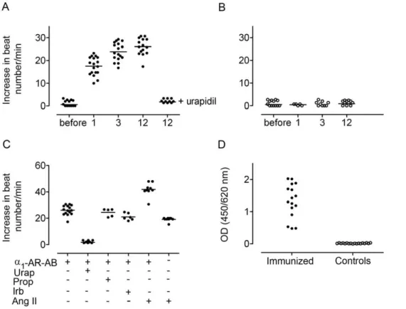
The cardiomyocyte contraction assay documented an increase of a1A-AR-AB activity one month after first peptide injection
persisting over the immunization process of 12 months. Urapidil inhibited the activity (Figure 1A). IgG fractions eluted from sera of control rats were negative in the cardiomyocyte contraction assay (Figure 1B). The development of autoantibodies against other receptors during immunization or Ang II treatment was excluded by cardiomyocyte contraction assay in presence of the specific antagonists. The combination ofa1A-AR-AB and Ang II increased
the cardiomyocyte contraction, compared to the effect ofa1A
-AR-AB or Ang II alone (Figure 1C). A high titer ofa1A-AR-AB was
detected by ELISA in the immunized rats. The controls did not show a signal (Figure 1D). The incubation of CHO/a1A-AR cells
with IgG prepared from immunized rats resulted in a stronger ERK1/2 phosphorylation than IgG fractions eluted from sera of control rats (Figure 2A). The specificity was proven by inhibition with the a1-AR receptor antagonist prazosin (62% inhibition,
P = 0.023, Figure 2B).
Cardiac Function in Immunized Rats
The diastolic interventricular septum and diastolic left ventric-ular heart wall thickness were significantly increased in 12 months
immunized rats compared to the controls (Figure 3 A). The ratio of heart/body weight (HW/BW61000) was significantly increased in the immunized rats compared to the controls (2.2160.23 vs. 1.9760.04, P = 0.0471, Supplementary table S2). Fractional shortening was similar in both groups, so that systolic function was preserved (Figure 3 B). A decreased ratio of peak flow velocity of the early rapid diastolic filling wave to peak flow velocity of the late diastolic filling wave (E/A ratio) in immunized rats indicates diastolic dysfunction (Figure 3 C). The echocardiographic findings in vivo are summarized in Supplementary table S3. Hemody-namic measurements showed higher left ventricular end-diastolic pressures (LVEDP) in immunized rat hearts (Figure 3 D). No changes in dp/dtmaxconfirmed the preserved contractility/systolic
function (Figure 3 E) and the decreased dp/dtmin indicates an
impaired relaxation/diastolic function (Figure 3 F). The analysis of the mean cardiomyocyte diameters showed a significant hyper-trophy 12 months after immunization with a1A-AR peptide,
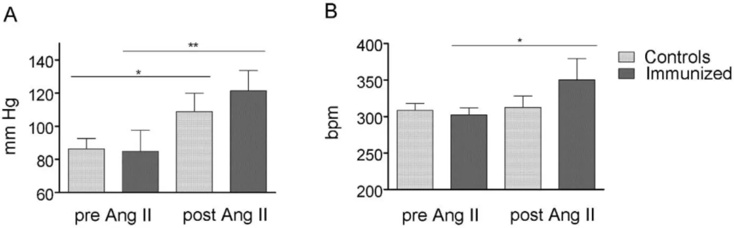
compared to controls (Figure 3 G). The blood pressure (MAP) and heart rate measured by telemetry were not different 12 months after first immunization (Figure 4).
Gene Expression Analysis and Histology
Genes coding for proteins of the sarcomere and extracellular matrix were differentially expressed in the heart (Table 1). Thus, the myosin heavy chain alpha (a-MHC) and the myosin heavy chain beta (b-MHC) were upregulated. Further, the collagen type I maintaining tissue structure and collagen type IV, a component of basal lamina that allows interaction with integrins and favors cell
Figure 1. Rat a1A-AR-AB detection.(AandB) Ordinate shows neonatal rat cardiomyocyte spontaneous beating rate; abscissa shows the immunization time point. IgGs from immunized rats increased the beating rate (A), compared to controls (B) Specificity was checked by inhibition with 1mM urapidil (Urap). (C) We excluded autoantibodies against other receptors using the b1-AR antagonist propranolol (Prop) and the AT1 receptor antagonist irbersartan (Irb). The combination ofa1A-AR-AB and Ang II increased the cardiomyocyte contraction. (D) A high titer ofa1A-AR-AB
adhesion, were increased expressed. The collagen IV binding glycoprotein laminin was also upregulated. Other genes coding for proteins involved in the Ca2+
signaling were represented. Among them were the cardiac ryanodine receptor 2 (Ryr2), the cardiac ATPase 2 Ca2+
-transporting, slow-twitch (Atp2a2), and the L-type calcium channel (Cacna1c). Important components of the energy metabolism like muscle glycogen phosphorylase and the peroxi-some proliferators-activated receptor-gamma, co-activator 1, alpha (Ppargc1a) a master regulator of metabolic function
responsible for fatty acid uptake and oxidation and oxidative phosphorylation[10] were upregulated (Table 1).
The gene expression results were validated by TaqMan analysis fora-MHC(Fold change (FC) = 3.5),b-MHC(FC = 2.8), collagen, type I, alpha 1 (FC = 1.7), and Cacna1c (FC = 2.0). The cardiac expression ofa1A-,a1B- ora1D-AR subtype was not differential in
immunized and control rats one year after immunization. Sirius red and Trichrome-Masson-Goldner staining were used for fibrosis detection. More fibrosis occurred in immunized rats
Figure 2. ERK1/2 activation in CHO/a1A-AR cells. (A) Activation after incubation with IgG from immunized and control rats or with phenylephrine (10mM) for 10 min is shown. Lane 1 and 2 represent untreated cells. (B) The specificity of ERK1/2 activation bya1A-AR-AB was checked
by prazosin inhibition (Prz, 1mM). Lane 1 represents untreated cells. Eukaryotic initiation factor 4E (elF4E) and ERK1/2 antibody were used as loading control.
doi:10.1371/journal.pone.0009409.g002
Figure 3. Echocardiography and invasive hemodynamic by conductance catheter at 12 months after first immunization. (A) Increased interventricular septum (IVS) and left ventricular heart wall (LVHW) thickness indicates hypertrophy. (B) Unchanged fractional shortening was a sign for preserved systolic function. (C) A decreased E/A ratio in immunized rats indicated a diastolic dysfunction. (D) Immunized rats had increased left ventricular end-diastolic pressure (LVEDP), (E) Unchanged dp/dtmaxand (F) diminished dp/dtmin, indicating impaired relaxation/
diastolic function. (G) Mean diameter of cardiomyocytes showed a significant hypertrophy, 12 months after immunization witha1A-AR peptide
compared to the controls (Figure 5 A, B). Perivascular fibrosis was significantly increased bya1A-AR-AB, whereas interstitial fibrosis
was significantly induced in combination with Ang II infusion (Figure 6).
Ang II Effects in Immunized and Control Rats
We tested whether or not immunized rats react more strongly to Ang II. Ang II further increased the chronotropic response ofa1A
-AR-AB, whereas phenylephrine did not result in an incremental increase in the bioassay (Supplementary figure S1). The combination ofa1A-AR-AB and Ang II caused a stronger increase
in MAP in immunized rats, compared to the Ang II treated
controls (Figure 4, P = 0.0339). The ratio of heart/body weight (HW/BW61000) was significantly increased in the immunized rats compared to the controls (2.3160.13 vs. 2.0860.12, P = 0.0109, Supplementary table S2). We used Trichrome-Masson-Goldner staining to quantify our results and found that immunized rats developed more severe fibrosis than control rats (Figure 5 A, B). The extent of fibrosis as a percent of tissue area was significant higher in the immunized rats (14.464.54% vs. 8.9062.31% in controls, P,0.00001). Ang II also induced a significant myocyte hypertrophy, which was not potentiated by
a1A-AR-AB (data not shown).
Discussion
The important findings in our study were that active immunization results in production of activating antibodies to thea1A-AR, namelya1A-AR-AB. The a1A-AR-AB in our model
did not increase telemetrically measured blood pressure. However, they clearly caused cardiac target organ damage and resulted in a model of diastolic dysfunction. Finally, stimulation of the system with an additional stressor (Ang II) resulted in worsened target-organ damage in thea1A-AR-AB-producing animals compared to
controls. The immediate question is, ‘‘how do the results reported here differ from those of Zhou et al [7]?’’ We relied on a different immunization sequence; our sequence corresponded to a1A-AR,
while Zhou et al used a sequence corresponding toa1D-AR. These
receptors are different. We investigated Ang II provocations, while they did not. We focused on target-organ damage. Finally, we monitored endorgan damage with echocardiography, invasive hemodynamics, histological analysis, radiotelemetrically deter-mined blood pressure measurements, and by gene expression array.
The immunized rats developed specific a1A-AR-AB that we
detected by both cardiomyocyte contraction assay and ELISA. The blocking experiments underscored the specificity of these antibodies. The antibodies were capable of initiating a1A-AR
signaling as documented by our phospho-ERK1/2 experiments. We documented clear-cut differences in cardiac function by two independent techniques, one relying on direct cardiac catheteri-zation that revealed a diastolic pathology. Cardiac structure was substantially different between the groups, as shown by echocar-diography and by light microscopy. Our notion that a much more sensitive blood pressure measurement than the tail-cuff technique would show a difference in blood pressure between groups, proved
Figure 4. Mean arterial blood pressure (MAP) and heart rate 12 months after first immunization, pre and post angiotensin (Ang) II treatment.a1A-AR-AB in combination with the stressor Ang II cause a greater increase in blood pressure (A) and heart rate (B) in the immunized rats
compared to the Ang II-treated controls. * indicates P,0.05, ** indicate P,0.01. doi:10.1371/journal.pone.0009409.g004
Table 1.Selected differentially expressed genes in hearts of immunized rats.
Gene Ref.-Sequence Fold
change
Sarcomere
Myosin heavy chain, alpha NM_017239 2.7 Myosin heavy chain, beta NM_017240 2.3 Collagens and extracellular matrix proteins
Collagen, type I, alpha 1 NM_053304 1.5 Collagen, type I, alpha 2 NM_053356 1.6 Procollagen, type IV, alpha 1 XM_001067473 1.7 Procollagen, type IV, alpha 2 XM_001076134 1.5 Laminin, gamma 1 XM_001071300 1.9 Ca2+
-regulation
Ryanodine receptor 2, cardiac XR_006681 1.7 ATPase, Ca2+-transporting, cardiac muscle, slow
twitch 2
NM_001110139 1.5
Ca2+-channel, voltage-dependent, L type, alpha 1C NM_012517 1.5 Energy metabolism
Peroxisome proliferators-activated receptor-gamma, coactivator 1, alpha
NM_031347 1.8
Muscle glycogen phosphorylase NM_012638 1.7 Phosphofructokinase, muscle NM_031715 1.5
not to be the case. Nonetheless, the Ang II experiments showed clearly that when a driving force for hypertension is applied, the presence of a1A-AR-AB clearly aggravates target organ damage
and blood pressure. Finally, we explored new mechanistic pathways.
Our gene expression analyses showed different compensatory mechanisms in structure and metabolism to maintain the cardiac function. In contrast to the failing rat heart with a shift in myosin isoform from a tob-MHC [11], we found instead an increased expression of both MHC isoforms. Up-regulation of b-MHC transcription can serve as an early and sensitive marker of cardiac hypertrophy [11,12] and may conserve energy [13,14]. Forced expression ofa-MHC may be beneficial in terms of increasing the myocardial contractility and may result in cardioprotection [15,16].
Myocardial remodeling implies an alteration in the extracellular matrix composition and distribution. Accordingly, we found in our array analysis the upregulation of collagen type I and IV in
immunized rat hearts. Collagen I and III maintain the tissue structure, transmit forces throughout the myocardium and contribute to the elastic properties of the myocardium [17]. The increased accumulation of collagen I and III has been associated mostly with fibrosis [18,19]. Type IV and VI collagens are components of the basal lamina and favors cell adhesion. The increased expression could be involved in the alteration of extracellular matrix cell interaction [20]. In human dilated cardiomyopathy, collagen is degraded by metalloproteinases and is replaced by fibrous intercellular deposits [21]. Zhou et al found increased MMP2 expression and activity in their a1D-AR-AB
immunization study [7]. We did not observe any increased metalloproteinase or tissue inhibitors of metalloproteinases. Other up-regulated genes encode molecular regulators of energy metabolism. The peroxisome proliferator-activated receptor gamma (PPAR-c) coactivator 1-alpha (Ppargc1a) activates multiple genes that are responsible for fatty acid uptake and oxidation and for oxidative phosphorylation [10]. The development of heart failure is accelerated byPpargc1adeficiency [22], suggesting that this coactivator may have a cardioprotective function.
Another important observation is the fact that genes coding for important Ca2+
regulating proteins, such as ATPase, Ca2+ transporting, slow-twitch (Atp2a2) coding for the sarcoplasmic/ endoplasmic reticulum calcium ATPase (SERCA2a), the cardiac ryanodine receptor 2, and the L-type calcium channel, alpha 1 C subunit (Cacnac1c) were all up-regulated. The overexpression of SERCA2a in diseased hearts has been shown to result in the recovery of contractility [23,24,25] and in improved survival, corresponding with an improvement in energy consumption [26]. Furthermore, SERCA overexpression decreases or prevents cardiac hypertrophy [27,28,29]. In our earlier study we found that acute administration of purifieda1-AR-autoantibodies from
patients or rabbita1A-AR-AB to neonatal cardiomyocytes affected
intracellular Ca2+at two different levels, namely the acute, short-term elevation of intracellular Ca2+
, and the increased transcript expression ofCacna1c[5]. In this study, we also found a long-term upregulation ofCacna1c. A link between increased L-type calcium channel, alpha 1 C subunit levels and hypertrophy has also been demonstrated for the human heart [30].
Rysa et al. performed DNA microarray analysis in 12 old SHR with manifest hypertrophy, compared to 16–20 month-old SHR with diastolic dysfunction and transition to heart failure [31]. Most of the enhanced genes upregulated in the development of diastolic heart failure encoded for ECM proteins. ECM proteins were also upregulated in our study. However, whereas we found dysregulated transcripts for calcium homeostasis, myofilament contractile proteins, and cardiomyocyte cytoskeleton proteins, these pathways seemed not to play a significant role in the development of diastolic heart failure caused by pressure-overload hypertrophy. The two models and the experimental settings were considerably different. Oura1A-AR-immunized rats were only 12
months old, had no hypertension, and no signs of heart failure, whereas the SHR rats were older and had signs of pressure-induced diastolic heart failure. Interestingly, Wallukat et al have shown that SHR develop autoantibodies against theb1 adrenergic receptor that permanently stimulate the receptor, whilea1-AR-AB
have not been found [32,33].
Thea1-adrenergic receptors are important to both
developmen-tal cardiomyocyte growth and pathological hypertrophy. Thea1A
-AR-AB production induced hypertrophy by causing fibrosis and cardiomyocyte hypertrophy. Patel et al [34] showed that a 28-day infusion of a subpressor norepinephrine dose induced hypertrophy only by stimulating myocyte growth. No fibrosis or signs of diastolic dysfunction was present in their 28-day study. However, we
Figure 5. Cardiac remodeling and fibrosis in immunized rats, pre and post Ang II treatment.(A) Sirius red and (B) Trichrome-Masson-Goldner staining. Both stains showed marked fibrosis in the hearts of immunized rats.a1A-AR-AB, in combination with the stressor
investigated our model for 1 year. Du et al [35] reported that animals transgenic for the a1A-AR showed a greater increase in
myocardial fibrosis post-myocardial infarction, compared to the non-transgenic control animals. Transgenic lines with an even higher expression level developed progressive cardiac fibrosis.
In spite of subtype-selective agonists and antagonists and gene knockout and transgenic overexpression approaches, the question of whicha1-AR subtype is involved predominantly in vasoconstrictive
responses to sympathomimetic agonists has not been answered. The studies with knockout mice indicate that all subtypes play a role in the blood pressure response toa1-agonists and that the dominant
contractilea1-AR is different in different vascular beds. Although
we did not observe an increase in blood pressure bya1A-AR-AB,
our results do not justify the conclusion that the a1A-AR is not
involved in the blood pressure control. Our results are in line with results reported by Tanoue et al [36]. These investigators found a major role fora1A-AR in maintaining basal blood pressure, whereas
other subtypes such asa1B-AR anda1D-AR were more important in
the pressor response to catecholamines.
Our immunized a1-AR-AB-producing rats developed an
increased LVEDP and diminished dp/dtmin in the face of
preserved ejection fraction and fractional shortening. This state-of-affairs is termed ‘‘diastolic dysfunction’’ and is a precursor for diastolic heart failure. Half the patients with heart failure fall into this category, notably older women. The prognosis of the condition is no better, if not worse, than systolic heart failure. All medication trials to date have been disappointing. Thea1-AR
blockers have not been studied in detail in the context of diastolic dysfunction. Two smaller studies reported positive effects ofa1-AR
blockers on echocardiographic parameters of diastolic dysfunction. To our knowledge, this experimental model is the first to show that
a1A-AR receptor stimulation can cause diastolic dysfunction
independent of any change in blood pressure. De Blois et al showed that chronica1-AR stimulation increases smooth muscle
cell DNA replication is in arterial wall, leading to remodeling after vascular injury [37]. We speculate that alterations in peripheral resistance (pressure-overload) could have been responsible for the diastolic dysfunction we observed. The fact that we were unable to detect any blood pressure increases even with radiotelemetry, suggests the possibility that altered blood pressure buffering played a role. We demonstrated heart muscle cell (cardiomyocyte) hypertrophy, in addition to an increased cardiac fibrosis in the
a1A-AR-AB model. We believe that these changes, independent of
any blood pressure changes we were able to detect, resulted in the diastolic dysfunction that we observed.
Our Ang II experiments showed that Ang II markedly aggravated the already-present cardiac fibrosis. We used Ang II as a stimulus to further induce vascular dysfunction. The sympathetic nervous system and renin-angiotensin-aldosterone system act synergistically to elevate or maintain blood pressure. Ang II signaling plays a critical role in modulating many of the stimuli and signals that govern arterial aging, arterial structural, and vascular functional and adaptational responses. Ang II also potentiated the chronotropic response to a1A-AR-AB, whereas
phenylephrine infusion, as reported by Patel et al, did not [22]. Limitations in our study are the fact that we did not include a long-term treated control group, namely immunized rats produc-inga1A-AR-AB treated with chronica1-AR blocker therapy. An
additional desirable control group could consist of chronic phenylephrine infusion (for 1 year). Acute infusion experiments could elucidate the issue of baroreceptor reflex blood pressure buffering capacity or resetting that remains unanswered from our study. However, our acute experiments showed that the effects of
a1A-AR-AB could be blocked pharmacologically. Chronic
exper-iments could have allowed us to speculate with greater confidence on a possible role ofa1-AR blockade to alleviate diastolic heart
dysfunction and remodeling.
Perspectives
We elucidated agonistic autoimmunity-induced target-organ damage and showed thata1A-AR-AB after immunization caused
diastolic dysfunction. Our animal model suggests thata1A-AR-AB
could play a role in target-organ damage; howevera1A-AR-AB are
probably not initiators of hypertension. These findings have implications for the notion of viewing agonistic autoantibodies as primary treatment targets in human diseases [38]. Our findings also have implications concerninga1-AR blocker therapy. These
agents may warrant a closer look, particularly in terms of diastolic dysfunction.
Supporting Information
Figure S1 Cardiomyocyte contraction assay. The incubation of cardiomyocytes with Iˆ61A-AR-antibodies (AB) or phenylephrine Figure 6. Myocardial perivascular and interstitial collagen.(A) Perivascular fibrosis was significantly increased in all three treated groups compared to control rats. (B) Interstitial fibrosis was significantly increased in immunized rats treated with Ang II when compared to controls, rats treated with Ang II or immunized rats.
(PE) caused an increase in cardiomyocyte contraction. This effect was not further potentiated by the combination of AB and PE. The combination of AB and Ang II resulted in a further raise of contraction.
Found at: doi:10.1371/journal.pone.0009409.s001 (0.13 MB DOC)
Table S1 Primer and probe sequences used for TaqMan RT-PCR.
Found at: doi:10.1371/journal.pone.0009409.s002 (0.05 MB DOC)
Table S2 Physiological parameters of immunized and control rats.
Found at: doi:10.1371/journal.pone.0009409.s003 (0.03 MB DOC)
Table S3 Echocardiography of immunized and control rats 12 months after first immunization and after Ang II treatment.
Found at: doi:10.1371/journal.pone.0009409.s004 (0.04 MB DOC)
Acknowledgments
We thank Sabine Bartel, Jutta Meisel, Reika Langanki, Astrid Schiche, Ralph Plehm, Melanie Soosten, Juliane Anders and May-Britt Ko¨hler for the excellent technical assistance. Martin Michel, Dept. Pharmacology & Pharmacotherapy, University of Amsterdam, Netherlands provided CHO cells stably transfected with humana1A-AR. We thank Hannelore Haase
for critical reading of the manuscript.
Author Contributions
Conceived and designed the experiments: KW GW DNM RD RD. Performed the experiments: KW GW FQ HS FH AH RF HH. Analyzed the data: KW FQ NH HS OH FH AH RF HH FCL DNM RD. Contributed reagents/materials/analysis tools: KW GW FQ NH HS OH HH RD RD. Wrote the paper: KW FCL DNM RD.
References
1. Piascik MT, Perez DM (2001) Alpha1-adrenergic receptors: new insights and directions. J Pharmacol Exp Ther 298: 403–410.
2. Fu ML, Herlitz H, Wallukat G, Hilme E, Hedner T, et al. (1994) Functional autoimmune epitope on alpha 1-adrenergic receptors in patients with malignant hypertension. Lancet 344: 1660–1663.
3. Liao YH, Wei YM, Wang M, Wang ZH, Yuan HT, et al. (2002) Autoantibodies against AT1-receptor and alpha1-adrenergic receptor in patients with hypertension. Hypertens Res 25: 641–646.
4. Luther HP, Homuth V, Wallukat G (1997) Alpha 1-adrenergic receptor antibodies in patients with primary hypertension. Hypertension 29: 678–682. 5. Wenzel K, Haase H, Wallukat G, Derer W, Bartel S, et al. (2008) Potential
relevance of alpha(1)-adrenergic receptor autoantibodies in refractory hyperten-sion. PLoS ONE 3: e3742.
6. Jahns R, Boivin V, Hein L, Triebel S, Angermann CE, et al. (2004) Direct evidence for a beta 1-adrenergic receptor-directed autoimmune attack as a cause of idiopathic dilated cardiomyopathy. J Clin Invest 113: 1419–1429. 7. Zhou Z, Liao YH, Wei Y, Wei F, Wang B, et al. (2005) Cardiac remodeling after
long-term stimulation by antibodies against the alpha1-adrenergic receptor in rats. Clin Immunol 114: 164–173.
8. Brockway BP, Mills PA, Azar SH (1991) A new method for continuous chronic measurement and recording of blood pressure, heart rate and activity in the rat via radio-telemetry. Clin Exp Hypertens A 13: 885–895.
9. van Heerebeek L, Borbely A, Niessen HW, Bronzwaer JG, van der Velden J, et al. (2006) Myocardial structure and function differ in systolic and diastolic heart failure. Circulation 113: 1966–1973.
10. Huss JM, Kelly DP (2004) Nuclear receptor signaling and cardiac energetics. Circ Res 95: 568–578.
11. Mercadier JJ, Lompre AM, Wisnewsky C, Samuel JL, Bercovici J, et al. (1981) Myosin isoenzyme changes in several models of rat cardiac hypertrophy. Circ Res 49: 525–532.
12. Jones WK, Grupp IL, Doetschman T, Grupp G, Osinska H, et al. (1996) Ablation of the murine alpha myosin heavy chain gene leads to dosage effects and functional deficits in the heart. J Clin Invest 98: 1906–1917.
13. Holubarsch C, Goulette RP, Litten RZ, Martin BJ, Mulieri LA, et al. (1985) The economy of isometric force development, myosin isoenzyme pattern and myofibrillar ATPase activity in normal and hypothyroid rat myocardium. Circ Res 56: 78–86.
14. Sugiura S, Kobayakawa N, Fujita H, Yamashita H, Momomura S, et al. (1998) Comparison of unitary displacements and forces between 2 cardiac myosin isoforms by the optical trap technique: molecular basis for cardiac adaptation. Circ Res 82: 1029–1034.
15. Abraham WT, Gilbert EM, Lowes BD, Minobe WA, Larrabee P, et al. (2002) Coordinate changes in Myosin heavy chain isoform gene expression are selectively associated with alterations in dilated cardiomyopathy phenotype. Mol Med 8: 750–760.
16. James J, Martin L, Krenz M, Quatman C, Jones F, et al. (2005) Forced expression of alpha-myosin heavy chain in the rabbit ventricle results in cardioprotection under cardiomyopathic conditions. Circulation 111: 2339–2346.
17. Bishop JE (1998) Regulation of cardiovascular collagen deposition by mechanical forces. Mol Med Today 4: 69–75.
18. Weber KT, Sun Y, Tyagi SC, Cleutjens JP (1994) Collagen network of the myocardium: function, structural remodeling and regulatory mechanisms. J Mol Cell Cardiol 26: 279–292.
19. Villarreal FJ, Dillmann WH (1992) Cardiac hypertrophy-induced changes in mRNA levels for TGF-beta 1, fibronectin, and collagen. Am J Physiol 262: H1861–1866.
20. Chapman D, Weber KT, Eghbali M (1990) Regulation of fibrillar collagen types I and III and basement membrane type IV collagen gene expression in pressure overloaded rat myocardium. Circ Res 67: 787–794.
21. Gunja-Smith Z, Morales AR, Romanelli R, Woessner JF, Jr. (1996) Remodeling of human myocardial collagen in idiopathic dilated cardiomyopathy. Role of metalloproteinases and pyridinoline cross-links. Am J Pathol 148: 1639–1648. 22. Arany Z, Novikov M, Chin S, Ma Y, Rosenzweig A, et al. (2006) Transverse
aortic constriction leads to accelerated heart failure in mice lacking PPAR-gamma coactivator 1alpha. Proc Natl Acad Sci U S A 103: 10086–10091. 23. Schmidt U, del Monte F, Miyamoto MI, Matsui T, Gwathmey JK, et al. (2000)
Restoration of diastolic function in senescent rat hearts through adenoviral gene transfer of sarcoplasmic reticulum Ca(2+)-ATPase. Circulation 101: 790–796. 24. Miyamoto MI, del Monte F, Schmidt U, DiSalvo TS, Kang ZB, et al. (2000)
Adenoviral gene transfer of SERCA2a improves left-ventricular function in aortic-banded rats in transition to heart failure. Proc Natl Acad Sci U S A 97: 793–798. 25. Kawase Y, Ly HQ, Prunier F, Lebeche D, Shi Y, et al. (2008) Reversal of cardiac dysfunction after long-term expression of SERCA2a by gene transfer in a pre-clinical model of heart failure. J Am Coll Cardiol 51: 1112–1119. 26. del Monte F, Williams E, Lebeche D, Schmidt U, Rosenzweig A, et al. (2001)
Improvement in survival and cardiac metabolism after gene transfer of sarcoplasmic reticulum Ca(2+)-ATPase in a rat model of heart failure. Circulation 104: 1424–1429.
27. Ito K, Yan X, Feng X, Manning WJ, Dillmann WH, et al. (2001) Transgenic expression of sarcoplasmic reticulum Ca(2+) atpase modifies the transition from hypertrophy to early heart failure. Circ Res 89: 422–429.
28. Sakata S, Lebeche D, Sakata Y, Sakata N, Chemaly ER, et al. (2007) Transcoronary gene transfer of SERCA2a increases coronary blood flow and decreases cardiomyocyte size in a type 2 diabetic rat model. Am J Physiol Heart Circ Physiol 292: H1204–1207.
29. Nakayama H, Otsu K, Yamaguchi O, Nishida K, Date MO, et al. (2003) Cardiac-specific overexpression of a high Ca2+affinity mutant of SERCA2a attenuates in vivo pressure overload cardiac hypertrophy. FASEB J 17: 61–63. 30. Haase H, Kresse A, Hohaus A, Schulte HD, Maier M, et al. (1996) Expression of calcium channel subunits in the normal and diseased human myocardium. J Mol Med 74: 99–104.
31. Rysa J, Leskinen H, Ilves M, Ruskoaho H (2005) Distinct upregulation of extracellular matrix genes in transition from hypertrophy to hypertensive heart failure. Hypertension 45: 927–933.
32. Wallukat G, Blasig IE, Morwinski R, Herrmann HJ, Rohde E (1995) The sera of spontaneously hypertensive rats contain agonistic auto-antibodies against the beta 1-adrenoceptor. J Hypertens 13: 1031–1036.
33. Wallukat G, Podlowski S, Nissen E, Morwinski R, Csonka C, et al. (2003) Functional and structural characterization of anti-beta1-adrenoceptor autoan-tibodies of spontaneously hypertensive rats. Mol Cell Biochem 251: 67–75. 34. Patel MB, Stewart JM, Loud AV, Anversa P, Wang J, et al. (1991) Altered
function and structure of the heart in dogs with chronic elevation in plasma norepinephrine. Circulation 84: 2091–2100.
35. Du XJ, Gao XM, Kiriazis H, Moore XL, Ming Z, et al. (2006) Transgenic alpha1A-adrenergic activation limits post-infarct ventricular remodeling and dysfunction and improves survival. Cardiovasc Res 71: 735–743.
36. Tanoue A, Koshimizu TA, Tsujimoto G (2002) Transgenic studies of alpha(1)-adrenergic receptor subtype function. Life Sci 71: 2207–2215.
37. deBlois D, Schwartz SM, van Kleef EM, Su JE, Griffin KA, et al. (1996) Chronic alpha 1-adrenoreceptor stimulation increases DNA synthesis in rat arterial wall. Modulation of responsiveness after vascular injury. Arterioscler Thromb Vasc Biol 16: 1122–1129.