Review
A novel approach based on nanotechnology for investigating the chronic
actions of short-lived peptides in specific sites of the brain
Frédéric Frézard
a,⁎
, Neila M. Silva-Barcellos
b, Robson A.S. dos Santos
aa
Departamento de Fisiologia e Biofísica, Instituto de Ciências Biológicas, Universidade Federal de Minas Gerais, Av. Antônio Carlos, 6627, Caixa Postal 486, 30.123-970 Belo Horizonte, Minas Gerais, Brazil
b
DEFAR, Escola de Farmácia, Universidade Federal de Ouro Preto, 35400-000 Ouro Preto, Minas Gerais, Brazil
Received 7 April 2006; received in revised form 16 May 2006; accepted 10 November 2006 Available online 20 December 2006
Abstract
This review presents a novel experimental approach for investigating the chronic actions of short-lived peptides in specific sites of the brain. This method combines the advantages of three different techniques: liposome encapsulation, site-specific microinjection and telemetry. First, liposomes can be designed to remain located at the injection site for a long period of time, where they protect encapsulated peptide from rapid degradation and act as a sustained-release system. Secondly, microinjection allows the administration of peptides in specific sites of the brain with minimal side effects. Finally, using telemetry, it is possible to register physiological parameters and their circadian variations in undisturbed free-moving animals for several days. Angiotensin-(1–7) and angiotensin II were used as peptide models, in order to validate the proposed method. Following the unilateral microinjection of the liposome-encapsulated peptides into the rostral ventrolateral medulla (RVLM) of Wistar rats, long-lasting cardiovascular actions were elicited, for several days. Importantly, new physiological actions of angiotensin-(1–7) at the RVLM were unmasked: modulation of the circadian rhythms of blood pressure and heart rate. It is felt that this method can be applied to a wide variety of short-lived bioactive peptides and should encounter numerous applications in the field of neurosciences.
© 2006 Elsevier B.V. All rights reserved.
Keywords:Liposomes; Angiotensin-(1–7); Angiotensin II; RVLM; RAS
Contents
1. Introduction . . . 60
2. Liposomes for local sustained and controlled release of short-lived peptides . . . 60
2.1. Liposomes as a sustained and controlled release system . . . 60
2.2. Liposomes as a site-specific nanoreservoir . . . 61
2.3. Preparation and characterization of peptide-containing liposomes . . . 61
3. Cardiovascular actions of liposome-encapsulated Ang-(1–7) at the RVLM . . . 63
3.1. Chronic actions of Ang-(1–7) at the RVLM on blood pressure and heart rate . . . 63
3.2. Influence of liposomes characteristics and implications . . . 63
4. Cardiovascular actions of liposome-encapsulated Ang II at the RVLM . . . 64
5. Conclusions and future prospects . . . 64
Acknowledgments . . . 64
References . . . 65
⁎Corresponding author. Tel.: +55 31 3499 2940; fax: +55 31 3499 2924.
E-mail address:[email protected](F. Frézard).
1. Introduction
The present article will review the progress achieved to date towards the development of a novel methodology for inves-tigating the chronic actions of short-lived peptides in specific sites of the brain.
A major difficulty encountered in the study of the physiology of short-lived endogenous peptides is the lack of appropriate methodology for assessing their chronic actions. This is particularly true when these actions have to be evaluated at a specific site of the brain and in free-moving awake animals. Indeed, the rapid in vivometabolism of the peptide results in biological actions of very short duration and site-specific microinjection of peptide aqueous solution does not mimic the physiological conditions of their chronic endogenous produc-tion. Moreover, this methodology did not allow studies on the interactions between different brain areas, which would require multiple microinjections in different brain sites.
Several recent studies have indicated that peptides of the renin–
angiotensin system (RAS) may act as important neuromodulators, especially in the brain medullary areas related to the tonic and reflex control of arterial pressure [1]. The primary effector products of the RAS are angiotensin II (Ang II) and the amino-terminal fragment angiotensin-(1–7) (Ang-(1–7))[2,3]. Microin-jection of aqueous solutions, containing 25 to 50 ng of Ang-(1–7) or Ang II, into the rostral ventrolateral medulla (RVLM) of normotensive rats elicited a significant increase of blood pressure
[4]. However, the duration of these effects was very short,
typically, 8 and 2 min for Ang-(1–7) and Ang II, respectively. The short duration of these effects was attributed to the rapidin vivo
metabolism of the peptide, essentially through inactivation by the ectoenzyme, angiotensin-converting enzyme[5].
This review will describe a novel experimental approach based on nanotechnology for assessing the chronic actions of short-lived peptides at specific sites of the brain. This meth-odology combines the advantages of three different techniques: the use of liposomes as a local peptide sustained and controlled
release nanosystem, site-specific microinjection into the brain and the use of telemetry to register physiological functions in undisturbed free-moving animals[6]. More specifically, it will present the progress achieved to date towards the full validation of this methodology, using peptides of the RAS and addressing the chronic cardiovascular actions of the peptides at the RVLM brain area.
2. Liposomes for local sustained and controlled release of short-lived peptides
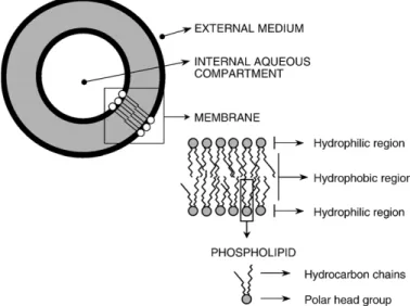
Fig. 1 shows the basic structure and composition of lipo-somes. Liposomes are artificial vesicles, with a size typically in
the nanometer range (diameter within the range of 20–
2000 nm), composed of concentric lipid bilayers, which are separated by internal aqueous compartments. Liposomes are classically made from phosphatidylcholine (PC) and cholesterol (CHOL), but those can be obtained from any bilayer-forming amphiphile. These vesicles are built in such a way that a solute can be encapsulated in the aqueous compartment (polar solutes) or embedded in the lipid bilayers (lipophilic or amphiphilic solutes).
Table 1 summarizes the main requirements for the use of liposomes as local sustained and controlled release systems for short-lived peptides as well as the proposed actions for achieving the desired properties.
2.1. Liposomes as a sustained and controlled release system
As a first requirement, liposomes should effectively retain the encapsulated peptide for a long period of time and release it, preferably, in a controlled manner. The ability of liposomes to act as a sustained-release system after systemic or oral admin-istration has been reported in the case of several short-lived (poly)-peptides including calcitonin, insulin, vasoactive
intes-tinal peptide and muramyl peptides [7–12]. Sustained and
controlled release may be achieved through the manipulation of
membrane fluidity, membrane surface, vesicle size and intravesicular peptide concentration and taking into account our actual knowledge on the factors governing thein vivofate of liposomes. In short, two distinct mechanisms have been shown to contribute to thein vivo release of the encapsulated substance from liposomes: a passive diffusion mechanism consisting of the simple diffusion of the substance across the lipid bilayer, also influenced by the interaction of the liposome membrane with serum components; a cell-mediated mechanism consisting of the endocytosis of liposomes by cells, their degradation by lysosomal phospholipases and the subsequent release of the active substance from cells by exocytosis. In the case of the peptides which act through specific plasma membrane receptors, critical issues to be addressed are whether or not the peptides survive to the lysosomal proteolytic enzymes and are effectively excreted by cells in order to reach their receptor. The answers to these questions will depend on the peptide amino acid sequence, which should determine its rate of permeation across the endosomal membrane and its suscepti-bility to the lysosomal enzymes. Although no direct demon-stration has been provided so far in the case of the RAS peptides, both the preservation of Ang II integrity after endocytosis and its release by endocytic cells are supported by the observation that the intravenous administration of Ang II-containing liposomes, which are rapidly cleared from the circulation by macrophages of the mononuclear phagocyte system, still produced the peptide-specific pressor effect[13].
Membrane fluidity was found to influence the rate of peptide release in both mechanisms, the more rigid membranes being the less permeable [14,15], the more resistant to destabilization by serum components, and the less susceptible to lysosomal degra-dation[16,17]. Certain alterations of the liposome surface can exert a marked influence on the rate of liposome capture by cells and, consequently, on the rate of release of encapsulated substance. For instance, the incorporation into liposome membrane of a phos-pholipid conjugated to ethylene glycol polymer (PEG) markedly reduced the rate of liposome capture by endocytic cells[18,19]. Liposome size was also found to influence the rate of capture of vesiclesin vivo, the smaller liposomes being usually less“visible”
than the larger ones[20,21].
2.2. Liposomes as a site-specific nanoreservoir
As a second requirement, the liposomes should remain localized for a long period of time at the specific site of microinjection and should not extravasate to neighboring areas. Although the factors influencing thein vivofate of liposomes are not available for the brain tissue, previous studies performed with liposomes administered subcutaneously in the dorsal side of the paw of rats have shown that the size of liposomes is a critical parameter that determines their ability to migrate from the injection site[22,23]. It was reported that liposomes with a mean diameter of less than 150 nm readily extravasate from the injection site through the lymphatic path, migrating to the regional lymph nodes and, for a proportion of them, reaching the blood circulation. In the case of liposomes with a mean diameter higher than 150 nm, more than 70% of liposomes were found at the injection site 2 days post-injection. In this context, in order to validate the proposed methodology, relatively large liposomes should be used and a study of liposomes localization should be performed at different times after microinjection.
In order to address this important question, empty liposomes made from distearoylphosphatidylcholine (DSPC), CHOL and distearoylphosphatidylethanolamine-polyethylene glycol 2000 (DSPE-PEG) at a molar ratio of 5:4:0.3 with a mean hydrody-namic diameter of 200 nm, were labelled with the lipophilic fluorescent dye, diI, and their localization was visualized under a fluorescence microscope at different times after microinjec-tion into the RVLM of Wistar rat. Microscopy images showed that the fluorescent marker remained concentrated at the RVLM even after 7 days [24], indicating a long persistence of lipo-somes at the RVLM.
2.3. Preparation and characterization of peptide-containing liposomes
As a third requirement, the liposomes by themselves should be biologically inert and exert minimal interferences on the physiology of the brain area. In this respect, liposomes made from natural lipids, or from lipids derived from them, should be preferred and a control group receiving empty liposomes should be included.
Table 2
Characteristics of the different preparations of angiotensin-containing liposomes Liposomal preparation Membrane compositiona Hydrodyn. diameter (nm) % encaps. Final peptide/ lipid ratio 1. Pegylated/rigid
DSPC/CHOL/DSPE-PEG (5:4:0.3)
200 15 0.004 2. Pegylated/
rigid/conc
DSPC/CHOL/DSPE-PEG (5:4:0.3)
200 15 0.009 3. Pegylated/fluid
DSPC/CHOL/DSPE-PEG (5:4:0.3)
200 15 0.004 4. Conventional/
rigid
DSPC/CHOL (5:4) 200 15 0.004 5. Large/
pegylated/rigid
DSPC/CHOL/DSPE-PEG (5:4:0.3)
100–2000 50 0.013
a
DSPC: distearoylphosphatidylcholine; CHOL: cholesterol; DSPE-PEG: distearoylphosphatidylethanolamine-polyethylene glycol 2000.
Table 1
Desired properties and proposed strategies in the use of liposomes as local sustained and controlled release systems
Desired properties Proposed strategies 1. Sustained release of encapsulated
solute
Use of cholesterol or highTca
phospholipid; use of stealth (PEG) liposomes
2. Long permanence at the injection site (limited extravasation, low cellular uptake)
Use of large liposomes (mean diameterN150 nm) and of stealth (PEG) liposomes
3. Biocompatibility of lipids Use of naturally-occurring lipids or lipids derived from them
4. Preservation of peptide structure Avoid conditions (temperature, solvent, process) that lead to peptide instability 5. Controlled release of encapsulated
solute
Manipulation of size, membrane fluidity and surface, internal peptide concentration
a
As a fourth requirement, the method used to prepare liposomes should preserve the peptide structure and activity.
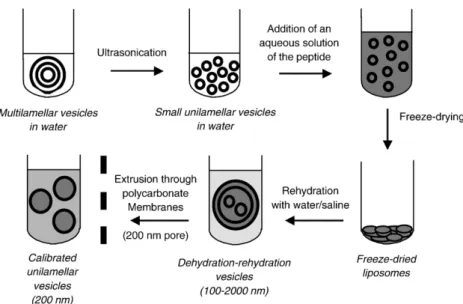
In this context, as shown in Table 2, peptide-containing
liposomes and empty liposomes were made typically from PC and CHOL, in the presence or absence of DSPE-PEG. As illustrated inFig. 2, the dehydration–rehydration method[25]
was used, followed or not by repeated extrusion across
poly-carbonate membrane with 200 nm pore size [26] in order to
obtain a liposome population of relatively homogeneous size distribution (calibrated liposomes). Non-encapsulated peptide was removed by dialysis. In the case of Ang-(1–7) and Ang II, the amount of encapsulated peptide was determined by fluorescence spectroscopy, exploiting the peptide intrinsic
fluorescence, as well as by radioimmunoassay [24]. Both
Fig. 2. Schematic illustration of the method of preparation of peptide-containing liposomes.
assays gave the same results, indicating that the peptide did not undergo any significant alteration in the process of liposome
preparation. Table 2 summarizes the characteristics of the
different liposomal preparations used in our study.
The ability of liposomes to retain encapsulated peptides was very high, as more than 98% of originally encapsulated peptides were found to be retained following a 5-day incubation at 37 °C of the liposome suspension in peptide-free isotonic saline[24].
3. Cardiovascular actions of liposome-encapsulated Ang-(1–7) at the RVLM
3.1. Chronic actions of Ang-(1–7) at the RVLM on blood
pressure and heart rate
Fig. 3shows typical telemetry recordings of mean arterial blood pressure (MAP) and heart rate (HR) obtained in normotensive Wistar rats following unilateral microinjection
into the RVLM of 50 ng of Ang-(1–7) encapsulated in
pegylated rigid calibrated liposomes (Preparation 1) or of
empty liposomes. Ang-(1–7)-containing liposomes produced a
significant increase of MAP for at least 7 days, specifically on day-time. As illustrated inFig. 4, as a consequence of this effect,
a loss of the circadian rhythm of MAP was observed [24].
Moreover, new actions of this peptide on HR were uncovered: a significant decrease of HR on night-time. Therefore, the long-lasting action of this preparation led to unmask a new
physiological role for Ang-(1–7): its modulation of the
circadian rhythms of MAP and HR[24].
3.2. Influence of liposomes characteristics and implications
The potential of the novel method was further investigated, by evaluating the influence of liposome characteristics on the cardiovascular actions of the encapsulated peptide [27]. It is expected that manipulation of vesicle size, membrane surface, membrane fluidity and amount of encapsulated peptide would result in the modulation of the outflux of peptide (see Section 2.1). Ultimately, physiologically-relevant information on the
Fig. 4. Illustration of the cardiovascular actions of Ang-(1–7)-containing liposomes at the RVLM of Wistar rats, involving a pressor effect on day-time and a bradycardia on night-time and resulting in the loss of the circadian variation of MAP and HR.
Table 3
Chronic cardiovascular actionsa elicited by Ang-(1–7) at the RVLM from different liposome preparations
Liposome preparation MAP change HR change
Day-time
Night-time
Day-time
Night-time 1—Pegylated and rigid ↑↑↑↑↑↑↑ ns ns ↓↓↓
2—Pegylated, rigid and conc. ↑ ↑ ns ns 3—Pegylated and fluid ↑ ↑ ns ↓↓↓↓
4—Conventional and rigid ns ns ↑ ns 5—Large, pegylated and rigid ns ↑ ↑↑ ns 6—Empty, calibrated lipos. ns ns ↑ ns 7—Empty, uncalibrated lipos. ns ns ↑↑ ns
↑indicates a significant increase and↓indicates a significant decrease of the parameter.“ns”means no significant difference (PN0.05, one-way ANOVA for repeated measures) (adapted from Ref.[27]).
a
The number of arrows represents the number of days during which a significant difference of the parameter was observed, when compared to its value just before microinjection (control period).
Fig. 5. Effects of Ang II- and Ang-(1–7)-containing liposomes as well as empty liposomes on MAP at the RVLM of Wistar rats. Liposomes were made from DSPC, CHOL and DSPE-PEG at a molar ratio of 5:4:0.3 and contained encapsulated peptide at a peptide/lipid mass ratio of 0.004. Preparations were microinjected unilaterally as 50 ng of peptide in 200 nL of isotonic saline. Changes were determined by calculating the difference between the value of the parameter on each day and the mean of the parameter over the 3 days preceding microinjection. Data represent the means of observed changes ± S.E.⁎Pb0.05 for comparison between the changes induced by peptide-containing liposomes (n= 5) and those induced by empty liposomes (n= 10);#P
relationship between the flux of peptide release and the cardiovascular responses may be obtained.
As illustrated in Table 3, pressor effects and bradycardia were the main actions elicited by the liposome preparations at the RVLM. However, the period, duration and intensity of these effects were found to depend markedly on the liposome charac-teristics. The most intense and prolonged effects were produced by pegylated, rigid and calibrated liposomes (Preparation 1). Less pronounced effects were observed when liposomes were uncalibrated (Preparation 5), contained a higher amount of peptide (Preparation 2) or were presented with a fluid mem-brane (Preparation 3).
The dependence of cardiovascular responses upon liposome characteristics, as evidenced in this study, may be explained by the different release profiles of the liposome preparations. Since
the spontaneous release of Ang-(1–7) was found to be
insignificant on a week period, when these liposome prepara-tions were incubated at 37 °C in peptide-free isotonic saline, cell-mediated release is expected to be the predominant
mechanism of peptide release in vivo. The presence of active
endocytic cells at the microinjection site, presumably microglial cells[28], is also supported by the observation that pegylation, that essentially slows down the uptake of liposomes by cells
[18,29], was required for producing long-lasting responses. Assuming that fluid liposomes are more susceptible to phospholipase degradation than rigid ones [16,17], that large (uncalibrated) liposomes are captured more avidly by endocytic
cells than smaller ones[20,21] and that liposomes containing
higher amount of peptide exhibit a higher flux of peptide release, one expects indeed that pegylated, rigid and calibrated liposomes would behave as the slowest release system.
From the physio-pharmacological point of view, further insights into the neuromodulator actions of Ang-(1–7) at the RVLM and its role in the central control of blood pressure were obtained. This study confirms, in chronic conditions, the pressor
effect of Ang-(1–7) at the RVLM and unmasks a new action of
the peptide on HR. Moreover, it suggests that MAP changes were influenced by HR changes, since bradycardia was usually accompanied by a loss of pressor effect (observed in the case of preparations 1 and 3) and the pressor effect was usually observed in the absence of bradycardia (in the case of Preparation 2). This data also suggests that cardiovascular
responses to Ang-(1–7) strongly depend on the local peptide
concentration, the intensity of the pressor effect and the bradycardia being reduced at high concentrations. Such effect may be related to the presence of Ang-(1–7) receptors in both excitatory (glutamatergic-) and inhibitory neurons at the RVLM, as recently described for Ang II[30].
4. Cardiovascular actions of liposome-encapsulated Ang II at the RVLM
When Ang II-containing liposomes made from distear-oylphosphatidylcholine, cholesterol and DSPE-PEG2000 were microinjected into the RVLM of Wistar rats, a sig-nificant pressor effect was observed on day-time for about 2 days. No significant effect of this peptide was observed on
HR. Fig. 5 compares the changes in MAP elicited by this
preparation to those produced by Ang-(1–7)-containing
liposomes and empty liposomes. The pressor effects of Ang II-containing liposomes showed a significantly lower
inten-sity, when compared to those of Ang-(1–7)-containing
liposomes. This difference may be interpreted by the shorter half-life of the former peptide, as suggested by the shorter duration of its acute action[4]. According to this model, the relation between the flux of peptide release and its rate of degradation may determine the concentration of the peptide available for biological action and the intensity of the resulting biological response.
5. Conclusions and future prospects
In the present review, a novel approach for assessing the chronic actions of short-lived peptides at specific sites of the brain was presented, which has been validated for RAS peptides and the RVLM brain area of rats. This methodology combines the advantages of three different techniques: liposome encap-sulation, site-specific microinjection and telemetry. The dura-tion of the pressor effects of Ang-(1–7) and Ang II at the RVLM was prolonged from a“minute”time scale (for free peptides) to a“day”time scale (for encapsulated peptides). Using the novel
approach, new insights into the effects of Ang-(1–7) on the
circadian variation of blood pressure were achieved. It is felt that this method may be widely applied to a variety of short-lived bioactive substances and several brain areas, encountering numerous applications in the field of neurosciences. Moreover, it may also find applications in protocols of multiple micro-injections in different brain areas.
Although great progresses were achieved towards the validation of the methodology, important questions still re-main to be answered. Although the influence of liposome characteristics on the duration and intensity of peptide chronic actions strongly suggests a modulation of the flux of peptide release, direct experimental evidences supporting this model are required in order to confirm the controlled release properties of this system. Therefore, it would be important to determine the amount of peptide at the micro-injection site as a function of time and liposome character-istics, for instance, by radioimmunoassay. Another important question that remains to be addressed is the identification of the endocytic cells involved in the peptide release. This question may be answered by electron microscopy investi-gation of the microinjection area after administration of li-posomes labelled with electron-dense marker. A third important point that requires confirmation is whether or not the peptide is the final active product and does not suffer metabolic transformation after endocytosis.
Acknowledgments
References
[1] Averill DB, Diz DI. Angiotensin peptides and the baroreflex control of sympathetic outflow: pathways and mechanisms of the medulla oblongata. Brain Res Bull 1999;51:119–28.
[2] Santos RAS, Campagnole-Santos MJ, Andrade SP. Angiotensin-(1–7): an update. Regulatory Pept 2000;91:45–62.
[3] Santos RA, Ferreira AJ, Pinheiro SV, Sampaio WO, Touyz R, Campagnole-Santos MJ. Angiotensin-(1–7) and its receptor as a potential targets for new cardiovascular drugs. Expert Opin Investig Drugs 2005;14:1019–31.
[4] Fontes MAP, Pinge MCM, Naves V, Campagnole-Santos MJ, Lopes OU, Khosla MC, Santos RAS. Cardiovascular effects produced by micro-injection of angiotensins and angiotensin antagonists into the ventrolateral medulla of freely moving rats. Brain Res 1997;750:305–10.
[5] Chappell MC, Pirro NT, Sykes A, Ferrario CM. Metabolism of angiotensin-(1–7) by angiotensin-converting enzyme. Hypertension 1998;31:362–7.
[6] Brockway BP, Mills PA, Azar SH. A new method for continuous measurement and recording of blood pressure, heart rate and activity in rat via radio-telemetry. Clin Exp Hypertens 1991;13:885–95.
[7] Takeuchi H, Matsui Y, Sugihara H, Yamamoto H, Kawashima Y. Effectiveness of submicron-sized, chitosan-coated liposomes in oral administration of peptide drugs. Int J Pharm 2005;303:160–70. [8] Chen D, Li QT, Lee KH. Antinociceptive activity of liposome-entrapped
calcitonin by systemic administration in mice. Brain Res 1993;603:139–42. [9] Kim A, Yun MO, Oh YK, Ahn WS, Kim CK. Pharmacodynamics of insulin
in polyethylene glycol-coated liposomes. Int J Pharm 1999;180:75–81. [10] Woodle MC, Storm G, Newman MS, Jekot JJ, Collins LR, Martin FJ,
Szoka Jr FC. Prolonged systemic delivery of peptide drugs by long-circulating liposomes: illustration with vasopressin in the Brattleboro rat. Pharm Res 1992;9:260–5.
[11] Sethi V, Onyuksel H, Rubinstein I. Liposomal vasoactive intestinal peptide. Methods Enzymol 2005;391:377–95.
[12] Alving CR. Liposomes as carriers of antigens and adjuvants. J Immunol Methods 1991;140:1–13.
[13] Papaioannou S, Yang P-C, Novotney R. Encapsulation of angiotensin II in liposomes: characterization in vitro and in vivo. Clin Exp Hypertens 1978;1:407–22.
[14] Bresseleers GJ, Goderis HL, Tobback PP. Measurement of the glucose permeation rate across phospholipid bilayers using small unilamellar vesicles. Effect of membrane composition and temperature. Biochim Biophys Acta 1984;772:374–82.
[15] Papahadjopoulos D, Nir S, Ohki S. Permeability properties of phospho-lipid membranes: effect of cholesterol and temperature. Biochim Biophys Acta 1971;266:561–83.
[16] Derksen JTP, Baldeschwieler JD, Sherphof GL. In vivo stability of ester-and ether-linked phospholipid-containing liposomes as measured by
perturbed angular correlation spectroscopy. Proc Natl Acad Sci U S A 1988;85:9768–72.
[17] Moghimi SM, Patel HM. Serum-mediated recognition of liposomes by phagocytic cells of the reticuloendothelial system—the concept of tissue specificity. Adv Drug Deliv Rev 1998;32:45–60.
[18] Allen TM, Hansen C, Martin F, Redmann C, Yau-Young A. Liposomes containing synthetic lipid derivatives of poly(ethylene glycol) show prolonged circulation half-lives in vivo. Biochim Biophys Acta 1991;1066:29–36. [19] Oussoren C, Storm G. Role of macrophages in the localization of liposomes
in the lymph nodes after subcutaneous administration. Int J Pharm 1999;183:37–41.
[20] Allen TM, Austin GA, Chonn A, Lin L, Lee KC. Uptake of liposomes by mouse bone marrow macrophages: influence of liposome composition and size. Biochim Biophys Acta 1991;1061:56–64.
[21] Senior J, Crawley JCW, Gregoriadis G. Tissue distribution of liposomes exhibiting long half-lives in the circulation after in vivo injection. Biochim Biophys Acta 1985;839:1–8.
[22] Allen TM, Hansen CB, Guo LSS. Subcutaneous administration of liposomes: a comparison with the intravenous and intraperitoneal routes of injection. Biochim Biophys Acta 1993;1150:9–16.
[23] Oussoren C, Zuidema J, Crommelin DJA, Storm G. Lymphatic uptake and biodistribution of liposomes after subcutaneous injection. II. Influence of liposomal size, lipid composition and lipid dose. Biochim Biophys Acta 1997;1328:261–72.
[24] Silva-Barcellos NM, Frézard F, Caligiorne S, Santos RAS. Long-lasting cardiovascular effects of liposome-entrapped angiotensin-(1–7) at the rostral ventrolateral medulla. Hypertension 2001;38:1266–71.
[25] Kirby C, Gregoriadis G. Dehydration–rehydration vesicles: a simple method for high yield drug entrapment in liposomes. Biotechnology 1984;2:979–84. [26] Naya R, Hope MJ, Cullis PR. Generation of large unilamellar vesicles from long-chain phosphatidylcholines by extrusion technique. Biochim Biophys Acta 1989;986:200–6.
[27] Silva-Barcellos NM, Caligiorne S, Santos RAS, Frézard F. Site-specific microinjection of liposomes into the brain for local infusion of a short-lived peptide. J Control Release 2004;95:301–7.
[28] Magnus T, Chan A, Savill J, Toyka KV, Gold R. Phagocytotic removal of apoptotic inflammatory lymphocytes in the central nervous system by microglia and its functional implications. J Neuroimmunol 2002;130:1–9. [29] Zeisig R, Shimada K, Hirota S, Arndt D. Effect of sterical stabilization on macrophage uptake in vitro and on thickness of the fixed aqueous layer of liposomes made from alkylphosphocholines. Biochim Biophys Acta 1996;1285:237–45.
[30] Hu L, Zhu DN, Yu Z, Wang JQ, Sun ZJ, Yao T. Expression of angiotensin II type 1 (AT1) receptor in the rostral ventrolateral medulla in rats. J Appl