Reactive Balance Control in Response to
Perturbation in Unilateral Stance: Interaction
Effects of Direction, Displacement and
Velocity on Compensatory Neuromuscular
and Kinematic Responses
Kathrin Freyler1*, Albert Gollhofer1, Ralf Colin2, Uli Brüderlin2, Ramona Ritzmann1
1Department of Sport and Sport Science, Albert-Ludwigs-University Freiburg, Freiburg, Germany,
2Department of Mechatronics, University of Applied Science, Esslingen, Germany
*kathrin.freyler@sport.uni-freiburg.de
Abstract
Unexpected sudden perturbations challenge postural equilibrium and require reactive com-pensation. This study aimed to assess interaction effects of the direction, displacement and velocity of perturbations on electromyographic (EMG) activity, centre of pressure (COP) dis-placement and joint kinematics to detect neuromuscular characteristics (phasic and seg-mental) and kinematic strategies of compensatory reactions in an unilateral balance paradigm. In 20 subjects, COP displacement and velocity, ankle, knee and hip joint excur-sions and EMG during short (SLR), medium (MLR) and long latency response (LLR) of four shank and five thigh muscles were analysed during random surface translations varying in direction (anterior-posterior (sagittal plane), medial-lateral (frontal plane)), displacement (2 vs. 3cm) and velocity (0.11 vs. 0.18m/s) of perturbation when balancing on one leg on a movable platform.Phases: SLR and MLR were scaled to increased velocity (P<0.05); LLR was scaled to increased displacement (P<0.05).Segments: phasic interrelationships were accompanied by segmental distinctions: distal muscles were used for fast compensation in SLR (P<0.05) and proximal muscles to stabilise in LLR (P<0.05).Kinematics: ankle joints compensated for both increasing displacement and velocity in all directions (P<0.05), whereas knee joint deflections were particularly sensitive to increasing displacement in the sagittal (P<0.05) and hip joint deflections to increasing velocity in the frontal plane (P<0.05). COP measures increased with increasing perturbation velocity and displacement (P<0.05). Interaction effects indicate that compensatory responses are based on complex processes, including different postural strategies characterised by phasic and segmental specifications, precisely adjusted to the type of balance disturbance. To regain balance after surface trans-lation, muscles of the distal segment govern the quick regain of equilibrium; the muscles of the proximal limb serve as delayed stabilisers after a balance disturbance. Further, a kine-matic distinction regarding the compensation for balance disturbance indicated different
OPEN ACCESS
Citation:Freyler K, Gollhofer A, Colin R, Brüderlin U, Ritzmann R (2015) Reactive Balance Control in Response to Perturbation in Unilateral Stance: Interaction Effects of Direction, Displacement and Velocity on Compensatory Neuromuscular and Kinematic Responses. PLoS ONE 10(12): e0144529. doi:10.1371/journal.pone.0144529
Editor:Andrea Macaluso, University of Rome Foro Italico, ITALY
Received:May 13, 2015
Accepted:November 19, 2015
Published:December 17, 2015
Copyright:© 2015 Freyler et al. This is an open access article distributed under the terms of the
Creative Commons Attribution License, which permits unrestricted use, distribution, and reproduction in any medium, provided the original author and source are credited.
Data Availability Statement:All relevant data are contained within the paper.
plane- and segment-specific sensitivities with respect to the determinants displacement and velocity.
Introduction
In balance research, the setup of a translating platform via externally applied perturbations is used to investigate underlying control mechanisms of compensatory balance responses in stan-dardised laboratory conditions [1–4]. Previous studies examining reactive balance control in response to perturbation have mainly been executed in bipedal paradigms and it is quite well described how postural stability is controlled with double limb support when unidirectional surface translations are induced in predictable experimental settings with constant perturba-tion parameters [2,3,5–8]. However, there is very little knowledge of how individuals recover balance under single leg stance conditions, despite the fact that losses of balance often occur under these conditions [9–11]. Moreover, usually various interdependent variables—i.e., unpredictable magnitude, velocity or direction of stimulus origin—challenge postural equilib-rium simultaneously. Consequently, the composition of the stimulus strongly influences com-pensatory responses [12]. To gain a thorough understanding of the mechanisms contributing to re-stabilisation after perturbation, the complex mechanisms of regaining postural stability in an unilateral stance paradigm must also be part of balance research. Further, the interdepen-dence of various stimulus characteristics should be taken into account, as the interactions of different perturbation variables may have specific effects on the modulation of the postural response [5,13,14].
From studies examining compensatory neuromuscular responses after perturbations in static paradigms, mainly executed in the 1980s and 1990s (see review [15]), it is known that the central nervous system plays a crucial role in governing appropriate muscle forces to prevent falling by relocating the centre of gravity (COG). It has been shown that muscular activation patterns are characterised by phase-specific reflex components indicated as short (SLR), medium (MLR) and long (LLR) latency responses following the onset of perturbation [5,8,16]. Dietz et al. [2,3,7] and Gollhofer [17] demonstrated that functionally relevant muscle activation (>65ms after onset, MLR and LLR) occurs when the COG is shifted away from the vertical. MLR and LLR are supposed to be attributed to spinal, polysynaptic reflexes and have functional significance to induce appropriate active joint moments for the preservation of postural stabil-ity [2,3,8,12,17]. Slight postural disturbances (mainly small rotations around the ankle joint, less visible translation) are compensated by immediate, non-functional monosynaptic stretch responses in the SLR [16,17].
In view of reflexive muscle compensation in response to surface translation, distinction is drawn between perturbation direction, displacement and velocity; experiments executed dur-ing bilateral stance indicate that early, monosynaptic stretch responses are sensitive to pertur-bation velocity [16], whereas the later functional components of muscle activation patterns were demonstrated to compensate for alterations in the displacement of platform translation [5,6]. Further, it is suggested that, by controlling a multi-segment system, neuromuscular con-trol after balance perturbation includes a segmental distribution of compensatory electromyo-graphic (EMG) responses [4,12,18]. Authors speculate that mechanical coupling of sensory inputs at ankle, knee and hip joints induce corresponding activation of distal and proximal limb muscles [4,12], but the functional pattern of interlimb activation during stabilisation is still unclear. Further, researchers showed that the direction of surface translation is also of con-siderable importance for the output of the postural response. Although less examined, there is evidence that muscles play different functions as stabilisers during the postural response, and it
This does not alter the authors' adherence to PLOS ONE policies on sharing data and materials.
has been demonstrated that most muscles primarily act in one direction, independently of measurement condition [19,20]. Apparently, Moore and colleagues [19] discovered that pre-dominantly the distal muscles act in one direction, whereas proximal muscles cover a greater range of trajectories in the horizontal plane.
Based on these aspects, it is assumed that postural responses are modified according to func-tional requirements of a stable equilibrium, and different phasic and segmental strategies are used depending on perturbation characteristics to respond quickly and accurately to the bal-ance disturbbal-ance [5,12,16].
The present study provides a new approach in balance research comprising an unilateral balance design with neuromuscular activity and kinematics expressed as a function of three interrelated perturbation determinants direction, displacement and velocity. In order to achieve a more comprehensive understanding of the neuro-mechanical coupling during unilat-eral balance control, for the first time, random perturbations were applied while balancing in an unstable unilateral stance. To provide unstable balance conditions, the experimental setting comprised a freely swinging platform which was perturbed in each direction in the horizontal plane (Fig 1). Therefore, the purpose of the study was to investigate the interaction effects of three randomly varied determinants (direction, displacement and velocity) and their influence on the phase-specific EMG pattern and segmental regulation of leg muscle activation as well as on the joint deflections and centre of pressure (COP) displacement during perturbed, unilateral stance (Fig 2). We executed the experiments regardless of acceleration and deceleration pro-files. We hypothesised that study would reveal interaction effects for the variables direction, displacement and velocity. We further hypothesised that those determinant-dependent modu-lations would be phase (SLR, MLR and LLR) and segment specific (distal and proximal), and may be associated with differences in the selected balance strategy, accompanied by distinc-tions in kinematic output, depending on the combination of the variables. We expected that the higher the magnitude of displacement and velocity, the higher the neuromuscular and kine-matic postural responses and the more proximal those responses would occur.
Methods
Subjects
Based on the results of a pilot study including five subjects, a power analysis (f = 0.4;
alpha = 0.05; power = 0.9 for ANOVA) revealed that a participation of 20 volunteers is needed in this study. The participants were physically fit students in the department of sports and sports science, with no previous neurological irregularities or injuries to the lower extremities (6 women and 14 men, age 27±3years, weight 73±12kg, height 178±9cm; variables are
expressed as mean±standard deviation). All subjects provided written informed consent for the experiment, which was approved by the ethics committee of the University of Freiburg, and was in accordance with the latest revision of the Declaration of Helsinki.
Experimental design
of randomly applied perturbations. Directions, displacements and velocities were chosen accord-ing to previous experiments executed in bilateral stance conditions [5,20–22] and consequently parameter reliability had been tested in a pilot study including 15 subjects.
Platform construction
Perturbations were generated by means of the Perturmed1(Brüderlin, Germany). The basis of this construction is an already existing device (Posturomed1
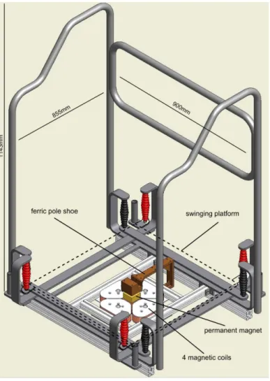
, Haider Bioswing, Germany) with a high test-retest reproducibility, which consists of a platform attached to a solid frame via Fig 1. Hardware construction of the electromagnetically driven perturbation platform (Perturmed1). The basic hardware of the Perturmed1
consists of an already existing device, the Posturomed1
[23–25]. The Perturmed1construction comprises a freely swinging platform (dashed line, 40x40cm) which is fixed with eight steel ropes (red and black): the platform itself is attached to four steel ropes (red), they in turn are attached to another iron frame hanging freely on the other four steel ropes (black). Thus, the freely swinging support surface is in total attached to a solid iron frame via two steel ropes on each corner. The pole shoe with the permanent magnet is fixed beneath the platform; the four magnetic coils are attached below at the bottom of the iron frame. By activation of two opposed interconnected coils via temporal current feed they release attracting and repelling electromagnetic forces, which move the support surface into the respective direction. The safety construction consisting of a solid metallic frame was used to secure the subjects from falling; fall rate within this experiment was below 2%. For the measurements, the electromagnetic forces were used to apply unpredictable horizontal translations of the free-swinging platform, gradually adjustable in direction, displacement and velocity. Platform kinematics were controlled by means of a movable goniometer attached to the platform.
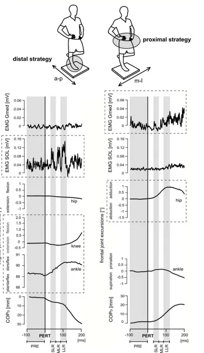
Fig 2. Neuromuscular and kinematic responses of one subject (top) during posterior (left) and lateral (right) perturbation.The top shows the subject standing on his right leg on the support surface (40x40cm), head and eyes directed forward, right knee joint extended and hands on the hips as required for the 30s measurement period. The support surface was a freely swinging platform. Thus, subjects needed to stabilise equilibrium even without perturbation. Below, the time point of perturbation (PERT) is marked as the black line; highlighted with grey/white backgrounds are the different temporal phases of the recorded
measurements (PRE, SLR, MLR, LLR). Perturbations in the sagittal plane cause postural reactions in distal muscles accompanied by ankle and knee joint deflections referring to the ankle strategy using the distal segment for compensation, whereas during perturbations in the frontal plane, mainly hip joint deflections counteract balance disturbance by using proximal muscles, indicating the use of the hip strategy (visualised by the dashed boxes).
two steel ropes on each corner [23–25] (seeFig 1). To move the platform reliably, electromag-netic forces were used to apply unpredictable horizontal translations of the free-swinging plat-form by means of coils affixed beneath the frame and a permanent magnet attached below the platform (Fig 1). Platform trajectories (displacement and velocity) were controlled by means of a movable goniometer attached to the platform and synchronised to the neuromuscular and Fig 3. (A) Platform trajectories for perturbation displacements and velocities and (B) corresponding modulations in neuromuscular responses.(A) Grand means of platform trajectories that illustrate pre-setting for displacement and velocity. (B) Mean changes in neuromuscular activity of all subjects in one representative thigh (a & b) and shank (c & d) muscle in response to increased perturbation displacement (a & c) and velocity (b & d) during the temporal phases before (PRE) and after (SLR, MLR, LLR) the perturbation (separated by dashed line). The phase- and segment-specific interaction effects are elucidated by the dashed boxes: muscles of the shank and thigh are used to compensate for increased perturbation displacement (grey triangle), and the neuromuscular response occurs in LLR (a & c). In contrast, only shank muscles are used for a fast compensation of increased perturbation velocity (grey triangle) during the early reflex phases SLR and MLR (b & d).*indicates a significant difference for pairwise comparisons (p<0.05).
kinematic recordings (grand means are illustrated inFig 3A). The three perturbation determi-nants and their different characteristics were applied stochastically (16 combinations: four directions x two displacements x two velocities). Altogether, 20 perturbations for each combi-nation were collected per subject. To control for the reliability of the subject’s starting position, trials were eliminated when platform trajectories prior to or after perturbation were beyond ±0.2cm (equivalent to 1.33% of the whole platform range).
Test procedure
Prior to measurements, subjects participated in familiarisation sessions for 10 minutes to adapt to the unstable surface and the perturbation mechanism in order to eliminate learning effects within the measurements. During testing, the subjects stood barefoot in an upright position on their right leg, kept hands on the hip and directed their head and eyes forward. They were instructed to stand as still as possible, with the unsupported leg flexed at 45° and not touching the other leg. Foot placement on the platform was controlled by means of a stencil to keep the subjects’feet in the same starting position for all trials. To control for the reliability of the sub-ject’s starting position, the body position of the standing leg was controlled by goniometers and two operators. Perturbations were applied randomly every 2–4 seconds in sets of 10 perturba-tions separated by a minimum of 30 seconds of rest in between for recovery [19,21,26]. Subjects were instructed to stabilise equilibrium as quickly as possible; in case of struggling or falling— defined as attempts where subjects failed to regain postural equilibrium after surface transla-tion and i) touched the safety frame of the Perturmed1
with at least one hand or ii) touched the ground with the unsupported (left) foot to avoid falling—perturbations were repeated.
For normalisation of the EMG data, prior to the measurements subjects performed three isometric maximal voluntary contractions (MVC) for each recorded muscle; we used the trial with the highest EMG for data normalization. The MVCs were executed according to [27] and [28], performed isometrically against resistance and held for three seconds. Between trials and repetitions subjects had recovery pauses of one minute. Body position during MVCs was strictly controlled and standardized by means of supervision by the authors and by goniometric recordings of ankle, knee and hip joint angles. Antagonistic muscle activation was monitored and trials repeated when antagonists were activated.
Dependent variables
The variables EMG data of nine muscles, COP movement, platform trajectories and joint kine-matics were synchronously recorded using a signal (5V, 1ms width) triggered to occur at the instant of platform perturbation. During perturbations, subjects stood on their right leg.
EMG recording. EMG data were obtained by placing bipolar surface electrodes (⊘9mm,
Ag/AgCl, Ambu Blue Sensor P, Ballerup, Denmark) over the m. soleus (SOL), gastrocnemius medialis (GM), tibialis anterior (TA), peroneus longus (PER), rectus femoris (RF), vastus later-alis (VL), biceps femoris (BF), gluteus medius (Gmed) and gluteus maximus (Gmax) of the right leg. Electrodes were placed in line with the direction of the underlying muscle fibres with a centre-to-centre distance of 25mm according to SENIAM guidelines [29]. By shaving and light abrasion of the skin, interelectrode resistance was kept below 2.5kO. Signals were ampli-fied (x1000) and recorded with 1kHz (band-pass filter 10Hz–1kHz).
Postural sway. COP displacement prior to and following perturbation was monitored by
means of a pressure distribution measuring system (pedar1
deformation technology (500g) instead of using a force plate, as the force plate would have enlarged the mass and consequently the inertia of the swinging component of the Perturmed1. To standardize the subject’s starting position and consequently to control for the subject’s for-ward or backfor-ward shifts [31] as well as any shift to the laterals left or right, trials were elimi-nated when COP trajectories prior to perturbation were beyond ±0.2cm.
Joint kinematics. Ankle, knee and hip joint excursions in the sagittal plane in response to anterior and posterior (a-p) perturbations, as well as ankle and hip joint excursions in the fron-tal plane in response to medial and lateral (m-l) perturbations were recorded with electro-goni-ometers (Biometrics1, Gwent, UK) consisting of a centre of rotation and two movable endplates. The endplates have a length of 10cm each and a range of 270°. The centre of rotation was placed over the respective joint centre; each endplate was attached to the prolonged axis of the anatomical structures [32].
Sagittal plane: the centre of rotation was fixed over the lateral malleolus (ankle), over the knee joint cavity (knee) and over the Trochanter major (hip). The two endplates were aligned pointing towards the fifth metatarsal and longitudinal axis of the shank (ankle), towards the lateral malleo-lus and Trochanter major (knee) and towards the longitudinal axis of the femur and thorax (hip). 90° between the fifth metatarsal and the fibula was defined as a 90° ankle angle; plantar flexion was reflected by an angle greater than 90°. The knee and hip flexion angle was set to zero at 0° during an upright stance, and joint flexion was reflected by an angle greater than 0°.
Frontal plane: the centre of rotation was fixed over the heel (ankle) and over the frontal Tro-chanter major (hip); the two endplates were aligned pointing towards the upper Achilles ten-don and the Calcaneus (ankle), and towards the longitudinal axis of the femur and the abdominal wall (hip). The ankle and hip flexion angle was set to zero at 0° during an upright stance; lateral joint flexion was reflected by an angle greater than 0°, medial joint flexion by an angle smaller than 0°. Signals were recorded with 1kHz and filtered (10Hz–1kHz).
Data processing
Each perturbation was analysed in a 500ms interval, comprising 100ms prior to and 400ms after perturbation onset (-100 to 400ms).
EMG during MVC was integrated for each muscle for a time frame of one minute [mVs]; the trial with the highest EMG was used for normalization.
We analysed one perturbation direction for each of the muscles; i.e. the EMG responses of muscles which are mainly used to counteracted surface translation in the respective perturba-tion direcperturba-tion [20]. For each muscle, integrated EMGs (iEMG) were calculated. For data analy-sis, iEMG was divided into four relevant phases: the pre-activation phase 100ms prior onset of perturbation (PRE, -100–0ms) and three compensatory postural responses based on the laten-cies of the reflex phases. Those are defined as follows: the short latency response from 30ms after onset of perturbation until 60ms (SLR, 30–60ms [33]), the medium latency response (MLR, 60–85ms [26]) and the late latency response (LLR, 85–120ms [26]). Subsequently, iEMGs were time normalised [mV/s] for the comparability of iEMGs between phases, then normalised to the respective MVC [%MVC] and averaged for subjects and perturbation conditions.
Ankle, knee and hip joint kinematics were expressed as mean angular displacement [°] for each subject and each perturbation condition, and were calculated as the difference between the peak angle position (defined as the maximum value of the angle excursion within the 400ms window) and the onset position.
COPD[mm] was calculated for each subject and perturbation condition as the difference
COP excursion within the 400ms window) and the onset position. COPVwas calculated
according to [26]: COPV[mm/ms] = COPD/t (with t defined as the time interval from the start
of perturbation to the time point of the peak).
Statistics
To analyse the effects of the three perturbation determinants (direction, displacement and velocity) on the respective neuromuscular and kinematic variables, and to detect interaction effects between the independent variables, within-subject comparisons were performed using a repeated measures analysis of variance (ANOVA). To evaluate the responses of the COP mea-sures (COPDand COPV) and ankle, knee and hip joint excursions, a three-factor ANOVA was
used, respectively [direction (4) x displacement (2) x velocity (2)].
To assess the neuromuscular responses according to varying displacement and velocity, a two-factor ANOVA was calculated for SLR, MLR and LLR, respectively [displacement (2) x velocity (2)]. The level of significance was set at P<0.05. To correct for multiple testing we used Bonferroni correction; each P-value (Pi) for each test was multiplied by the number of
tests (Pi adjusted= Pin, n = number of tests). If Pi adjustedwas<0.05 we considered the
respec-tive testito be of statistical significance. If the assumption of sphericity measured by Mauchly's sphericity test was violated, the Greenhouse-Geisser correction was used. In case of significant main effects, post hoc comparisons (Tukey’s HSD, level of significancep<0.05) were calculated for specification of the direction of the particular differences. Analyses were executed by using SPSS 20.0 (SPSS, Inc., Chicago, IL, USA).
Results
InTable 1, mean values of the COPD, COPVas well as ankle, knee and hip joint excursions are
displayed for the different perturbation conditions. The EMG activity of the nine leg muscles during the four phases (PRE, SLR, MLR, LLR) are shown inTable 2.
Direction of the perturbation
EMG responses were analysed in the direction in which they were maximally active: for poste-rior direction SOL, GM, BF and Gmax; for anteposte-rior direction TA and VL; for medial direction Gmed and RF and for lateral direction PER (Table 2).
The factor direction had a significant main effect on COPD(P<0.001, F = 66.95), COPV
(P<0.001, F = 129.96) as well as on ankle joint deflection in the frontal plane (P = 0.02, F = 18.41) and knee joint deflection in the sagittal plane (P<0.001, F = 39.11). For all condi-tions, COPDshifted contrarily to the perturbation direction, i.e. a forward translation of the
platform caused a backwards shift of the COP.
Ankle and knee joint excursions deflected according to each perturbation direction (in the sagittal plane for a-p, in the frontal plane for m-l perturbations,Fig 2). Perturbation direction had no influence on hip joint excursion.
Displacement of the perturbation
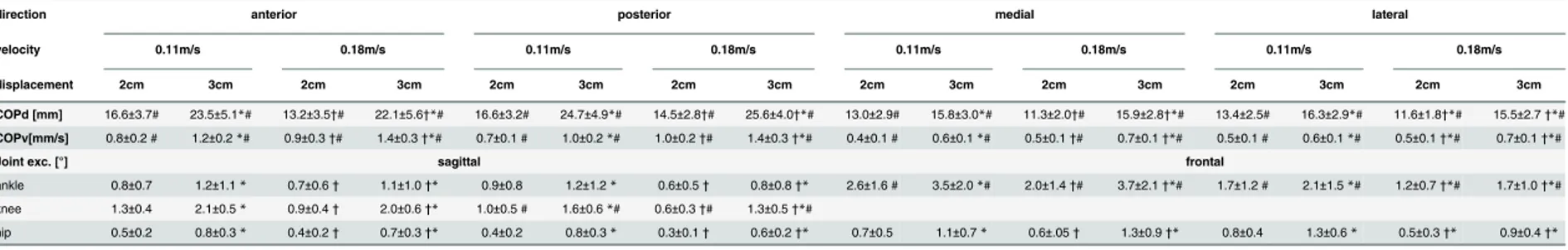
Table 1. Changes of the CODD, COPVand ankle, knee and hip joint deflections with respect to the different permutations of perturbation direction, velocity and displace-ment are illustrated.
direction anterior posterior medial lateral
velocity 0.11m/s 0.18m/s 0.11m/s 0.18m/s 0.11m/s 0.18m/s 0.11m/s 0.18m/s
displacement 2cm 3cm 2cm 3cm 2cm 3cm 2cm 3cm 2cm 3cm 2cm 3cm 2cm 3cm 2cm 3cm
COPd [mm] 16.6±3.7# 23.5±5.1*# 13.2±3.5†# 22.1±5.6†*# 16.6±3.2# 24.7±4.9*# 14.5±2.8†# 25.6±4.0†*# 13.0±2.9# 15.8±3.0*# 11.3±2.0†# 15.9±2.8†*# 13.4±2.5# 16.3±2.9*# 11.6±1.8†*# 15.5±2.7†*#
COPv[mm/s] 0.8±0.2 # 1.2±0.2*# 0.9±0.3†# 1.4±0.3†*# 0.7±0.1 # 1.0±0.2*# 1.0±0.2†# 1.4±0.3†*# 0.4±0.1 # 0.6±0.1*# 0.5±0.1†# 0.7±0.1†*# 0.5±0.1 # 0.6±0.1*# 0.5±0.1†*# 0.7±0.1†*#
Joint exc. [°] sagittal frontal
ankle 0.8±0.7 1.2±1.1* 0.7±0.6† 1.1±1.0†* 0.9±0.8 1.2±1.2* 0.6±0.5† 0.8±0.8†* 2.6±1.6 # 3.5±2.0*# 2.0±1.4†# 3.7±2.1†*# 1.7±1.2 # 2.1±1.5*# 1.2±0.7†*# 1.7±1.0†*# knee 1.3±0.4 2.1±0.5* 0.9±0.4† 2.0±0.6†* 1.0±0.5 # 1.6±0.6*# 0.6±0.3†# 1.3±0.5†*#
hip 0.5±0.2 0.8±0.3* 0.4±0.2† 0.7±0.3†* 0.4±0.2 0.8±0.3* 0.3±0.1† 0.6±0.2†* 0.7±0.5 1.1±0.7* 0.6±.05† 1.3±0.9†* 0.8±0.4 1.3±0.6* 0.5±0.3†* 0.9±0.4†*
Values represent mean values±standard deviations (M±SD). For significant main effects of the ANOVA, a*symbol marks a significant effect of the displacement on the respective parameters measured, a†symbol marks a significant effect of the velocity and a # symbol marks a significant effect of the perturbation direction (P<0.05).
doi:10.1371/journal.pone.0144529.t001
Neuromus
cular
and
Kinematic
Response
s
to
Differe
nt
Perturba
|DOI:10.137
1/journal.p
one.0144529
December
17,
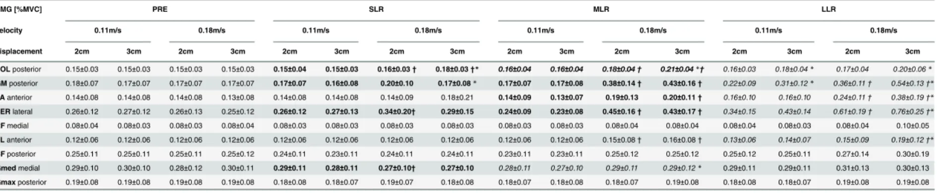
Table 2. Modulations in EMG activity in the time interval before (PRE) and the three reflex phases after perturbation (SLR, MLR, LLR) according to the different perturbation velocities and displacements.
EMG [%MVC] PRE SLR MLR LLR
velocity 0.11m/s 0.18m/s 0.11m/s 0.18m/s 0.11m/s 0.18m/s 0.11m/s 0.18m/s
displacement 2cm 3cm 2cm 3cm 2cm 3cm 2cm 3cm 2cm 3cm 2cm 3cm 2cm 3cm 2cm 3cm
SOLposterior 0.15±0.03 0.15±0.03 0.15±0.03 0.15±0.03 0.15±0.04 0.15±0.03 0.16±0.03† 0.18±0.03†* 0.16±0.04 0.16±0.04 0.18±0.04† 0.21±0.04*† 0.16±0.03 0.18±0.04* 0.17±0.04 0.20±0.06* GMposterior 0.18±0.07 0.17±0.07 0.17±0.07 0.17±0.07 0.17±0.07 0.16±0.08 0.20±0.10 0.17±0.08* 0.17±0.07 0.17±0.08 0.38±0.14† 0.43±0.16† 0.22±0.09 0.31±0.12* 0.36±0.11† 0.54±0.13†* TAanterior 0.14±0.08 0.14±0.08 0.14±0.08 0.13±0.08 0.14±0.08 0.14±0.08 0.14±0.09 0.18±0.21 0.14±0.09 0.13±0.07 0.19±0.13 0.20±0.11† 0.16±0.10 0.16±0.10 0.24±0.11† 0.38±0.19†* PERlateral 0.26±0.12 0.27±0.12 0.26±0.13 0.25±0.12 0.26±0.12 0.27±0.13 0.34±0.20† 0.29±0.15 0.24±0.09 0.23±0.08 0.45±0.16† 0.43±0.17† 0.34±0.15 0.43±0.14 0.61±0.19† 0.76±0.25†* RFmedial 0.08±0.04 0.08±0.03 0.08±0.03 0.08±0.04 0.08±0.03 0.08±0.03 0.08±0.03 0.08±0.03 0.08±0.03 0.08±0.03 0.08±0.04 0.08±0.04 0.08±0.04 0.08±0.03 0.08±0.04 0.10±0.05
VLanterior 0.12±0.06 0.12±0.06 0.12±0.06 0.12±0.06 0.12±0.06 0.12±0.06 0.12±0.06 0.12±0.06 0.12±0.06 0.12±0.06 0.15±0.08† 0.16±0.08† 0.13±0.06 0.14±0.07 0.15±0.09 0.19±0.12†* BFposterior 0.25±0.11 0.25±0.11 0.25±0.11 0.25±0.12 0.24±0.11 0.23±0.11 0.24±0.11 0.24±0.11 0.23±0.11 0.23±0.11 0.25±0.12 0.25±0.12 0.25±0.12 0.25±0.11 0.27±0.14 0.30±0.19
Gmedmedial 0.29±0.10 0.30±0.10 0.28±0.12 0.30±0.11 0.29±0.11 0.28±0.11 0.27±0.10† 0.27±0.10 0.28±0.11 0.27±0.10 0.29±0.11 0.29±0.12* 0.29±0.11 0.29±0.11 0.31±0.13 0.30±0.13
Gmaxposterior 0.19±0.08 0.19±0.08 0.19±0.08 0.19±0.08 0.18±0.08 0.18±0.07 0.19±0.07 0.18±0.08 0.18±0.07 0.18±0.08 0.18±0.07 0.19±0.08 0.18±0.08 0.18±0.07 0.19±0.08 0.19±0.08 Muscles are displayed for the respective perturbation direction, in which they counteracted surface translation. The table elucidates that dominant changes to increased
displacement only occurred within the fast velocity. Moreover, increased perturbation velocity was predominantly compensated in SLR and MLR (bold font), whereas increased displacement was predominantly compensated in LLR (italic font). Values represent mean values±standard deviations (M±SD). For significant main effects of the ANOVA (P<0.05), the lines written in italic font denote a significant effect of the displacement on the specific phase (a*symbol marks significant post hoc comparison, p<0.05) and the lines written in bold font denote a significant effect of the velocity (a†symbol marks significant post hoc comparison, p<0.05). The line written in bold and italic font contains both effects (displacement and velocity) on the specific phase.
doi:10.1371/journal.pone.0144529.t002
Neuromus
cular
and
Kinematic
Response
s
to
Differe
nt
Perturba
|DOI:10.137
1/journal.p
one.0144529
December
17,
COPD(P<0.001; F = 455.66), COPV(P<0.001; F = 335.86) as well as ankle (frontal:
P<0.001; F = 90.19; sagittal: P = 0.03; F = 16.55), knee (sagittal: P<0.001; F = 290.16) and hip (frontal: P<0.001; F = 85.69; sagittal: P<0.001; F = 125.24) joint excursions increased with increasing perturbation displacement.
Velocity of the perturbation
The perturbation velocity affected the early reflex components SLR and MLR, whereas LLR remained unaffected. Muscle activation was scaled to increasing perturbation velocity for the muscles SOL (P<0.001; F = 71.08), GM (P = 0.04; F = 4.81) and PER (P = 0.04; F = 7.08) as well as for Gmed (P = 0.02; F = 16.31) in SLR, and for the shank muscles SOL (P<0.001; F = 126.38), GM (P<0.001; F = 109.25), TA (P = 0.02; F = 17.26) and PER (P<0.001; F = 58.13) also in MLR (Fig 3B).
COPV(P<0.001; F = 82.85) increased with increasing perturbation velocity, whereas COPD
(P<0.001; F = 66.36) and joint excursions in ankle (frontal: P = 0.01; F = 20.14; sagittal: P = 0.04; F = 15.84), knee (sagittal: P<0.001; F = 130.56) and hip (frontal: P = 0.02; F = 10.94; sagittal: P = 0.03; F = 16.39) joints decreased.
Interactions
The ANOVA revealed significant interaction effects(displacement x velocity)for the shank muscles PER (P = 0.02; F = 6.07) in SLR, for GM (P = 0.02; F = 6.44), SOL (P<0.001; F = 17.88) and Gmed (P = 0.04; F = 5.08) in MLR, and for TA (P<0.001; F = 34.87) and GM (P = 0.01; F = 8.65) in LLR; increased perturbation velocity significantly facilitated the effect of an increase in perturbation displacement on EMG activity. For the thigh muscles RF (P = 0.005; F = 10.12), VL (P = 0.009; F = 8.35) and BF (P = 0.03; F = 5.64) significant interaction effects
(displacement x velocity)occurred delayed only in LLR (Table 2,Fig 3B). Accordingly, signifi-cant interaction effects(displacement x velocity)were observed for COPD(P<0.001;
F = 52.18), COPV(P = 0.002; F = 13.66), ankle joint excursions in the frontal (P<0.001;
F = 24.45) and knee joint excursions in the sagittal plane (P = 0.02; F = 6.75), indicating a dis-tinct interrelation in kinematics with increasing velocity in response to increased displacement.
Further, the ANOVA revealed significant interaction effects for COPD(direction x
dis-placement: P<0.001; F = 99.28 anddirection x velocity:P = 0.02; F = 7.46) and for COPV (direction x displacement: P<0.001; F = 70.64 anddirection x velocity:P<0.001; F = 23.48): Increasing perturbation displacement and velocity revealed greater deflections in a-p (sagittal plane) than in m-l (frontal plane) direction.
The significant interaction effects (direction x velocity)for hip joint excursions in the fron-tal (P<0.001; F = 23.91) and for ankle joint excursion in the sagittal plane (P = 0.01; F = 7.51) and(direction x displacement)for ankle joint excursions in the frontal (P<0.001; F = 17.86) and for knee joint excursions in the sagittal plane (P<0.001; F = 25.08) indicating that changes in response to increasing displacement or velocity were differently allocated throughout the limb segments dependent on the direction of perturbation (Fig 2).
The interaction effects between all perturbation determinants(direction x displacement x velocity)were observed with respect to COPD(P = 0.02; F = 3.44) and COPV(P = 0.03;
Discussion
The objective of this study was to assess the interaction effect of three perturbation determi-nants on postural neuromuscular and kinematic responses while balancing in unilateral stance. The study revealed four main results: (1) early reflex components (SLR and MLR) were scaled to increasing velocity, whereas the later component (LLR) was scaled to increasing displace-ment. (2) Moreover, phasic assignments to increasing velocity or displacement of the perturba-tion also revealed segmental preferences to regain balance using distal muscles for fast
compensation in SLR and proximal muscles to stabilise in LLR. (3) Further, kinematic distinc-tions regarding the compensation for balance disturbances indicated plane- and segment-spe-cific dependencies with respect to perturbation displacement and velocity. (4) Velocity is suggested to be the key parameter that significantly facilitates the effect of the other parameters, particularly on neuromuscular activation.
Main effects
There are two aspects elucidated through the main effects:
i. Neuromuscular compensation to changes in velocity of the perturbation occurred predomi-nantly in SLR and MLR. Particularly the short latency compensation of balance disturbance was only present during high velocity perturbations (e.g. SOL, GM, TA, PER, seeTable 2). As known from literature, muscle activity during SLR is commonly not observed during translational perturbations; however, when the velocity is sufficiently high a small SLR is visible. Modulations in muscle activity during SLR are attributed to the spinal input of Ia afferent fibres containing the monosynaptic reflex [5,26,34–37], whereas the functionally relevant MLR [2,3,17] is supposed to be modulated by supraspinal structures via polysynap-tic pathways of group II afferents [2,17,35,36,38–40]. Sensory information transmitted via Ia and II afferent reflex circuits are related to the velocity-sensitive muscle spindle to imme-diately counteract increased perturbation velocity [3,5]. There is evidence that the ability to detect stimulus velocity instantaneously [5,38] is attributed to the high conductibility of the Ia afferent fibres, which enables muscle spindle receptors to deliver fast information needed for corrective responses [37,41]. In contrast to velocity-induced changes, the effect of increasing perturbation displacement revealed phase-specific compensation only in LLR, which is supposed to involve direct corticospinal pathways [5,26,36,38,42,43]. As it is reported that the SLR is not sensitive to changes in displacement and hence was only pres-ent after the fastest perturbations in some muscles, the late componpres-ent LLR may be neces-sary to compensate for substantial balance disturbances [3,5,43].
Functional consequences of the phase-specific differences in displacement- and velocity-induced neuromuscular responses are reflected by modulated joint kinematics and COP dis-placement; fast compensation in SLR may provide an appropriate torque in the ankle joint to counteract the perturbation and regain balance at an early stage [44]. Thus, although velocity was enhanced, joint excursions and COP displacement remained unchanged or even decreased. Delayed compensation on the neuromuscular level, however,–as it is observed in LLR for enhanced perturbation displacement–caused increased kinematic reactions, i.e. aug-mented joint deflections and enhanced COP displacement and velocity, associated with signifi-cant difficulties to regain postural equilibrium [45], for instance also observed after knee surgery [46].
For compensation of augmented perturbation velocity, the shank muscles are of consider-able importance to regain equilibrium, while proximal muscles are barely involved (Fig 3B). According to literature, quickly delivered reflex activations in distal muscle groups are linked to stretch velocity and, thus, provide appropriate and fast isolated distal joint torques to restore balance [21]. Hence, ankle joint stiffness is increased, leading to less joint excursions and COP displacement. In contrast, our study revealed that both shank and thigh muscles were activated to regain equilibrium when perturbation displacement was increased (Fig 3B). It is suggested that the late reaction in LLR required a recruitment of the proximal muscles in addition to the distal muscles [3,5,21]. Consequently, an increase in displacement caused an overall increase in kinematics involving the proximal segments reflected in larger COP displacements and knee and hip joint deflections.
Interaction effects
There are three aspects elucidated through the interaction effects:
1. Reflex phases and limb segments: Our findings suggest that the timing of the neuromuscular response to both increasing displacement and velocity was different for the distal and proxi-mal limb. Compensatory muscle activity of the shank muscles occurred in SLR, whereas the majority of thigh muscles contributed to balance recovery only in LLR (Fig 3B). This obser-vation seems to be largely determined by the anatomical properties and function of the tar-get muscles—distal muscles acting on joints near to and proximal muscles stabilising joints far from the postural disturbance [3,19]. According to Moore et al. [19], the distal limb seg-ment is related to platform velocity and thus, muscles may serve as“prime movers”to pro-duce fast corrective responses around the ankle joint. Conversely, containing delayed EMG bursts in LLR, the proximal muscles are supposed to act as“stabilisers”to compensate for the resulting torque transferred between limb segments after distal muscle activity [3,19]. In view of the latter aspect, our results detected different phase- and segment-specific neuro-muscular strategies between the distal and proximal limb, finely attuned to the augmented balance disturbance.
throughout increasing velocity [8]. Interlinked with the EMG data, we suggest that, by con-trolling a multi-segment system, kinematic control after balance perturbation includes a segmental distribution of compensatory responses (Fig 3B). As the base of support does not restrict the subjects freedom of movement, hardware constrains are excluded to be responsi-ble for the observed differences.
3. Key parameter velocity: Interactions elucidate perturbation velocity to be the target parame-ter, which predominantly facilitates the effect of the other determinants. As changes in velocity considerably influenced early reflexive muscle activation detected in the SLR and MLR [16], our results indicate that displacement-induced adaptations, mainly visible in the LLR, are influenced by the extent of the velocity as well. As a major consequence, neuromus-cular compensation due to increased displacement showed gradually elevated activation, however, and most importantly, those effects were only detected during the fast velocity condition, whereas displacement-induced changes during the slow velocity condition remained mostly unaffected. Aforementioned aspects indicate a significant increase in pos-tural demand associated with considerably elevated pospos-tural reactions during increased velocity.
Limitation
Although acceleration and deceleration profiles are coded in the displacement and velocity of the perturbation, they may have an additional effect on the output of postural responses [13]. It is assumed that the higher the displacement and velocity of the perturbation, the higher the acceleration and deceleration profile of the platform movement and the bigger the postural response. However, the amount of 320 perturbations needed for an assessment of the three parameters direction, displacement and velocity with their characteristics did not make it pos-sible to control for two more parameters. Consequently, there is a need for further investiga-tions to clearly assess effects and interacinvestiga-tions of acceleration and deceleration profiles with other variables.
Conclusion
This study provided new insights on the neurophysiological and kinematic regulation of pos-tural responses during unilateral balancing by applying random, unexpected perturbations. This is considered to be a challenging postural task which requires appropriate neuromuscular control to regain equilibrium after surface translation [1], as it is needed during slip-like condi-tions. For the first time, reactive balance control was examined during unilateral balance tasks assessing how the different perturbation determinants interact and how these interactions are represented in the postural response. Main and interaction effects indicate that compensatory postural responses are based on complex processes that include different postural strategies characterised by phasic and segmental specifications, precisely adjusted to the respective type of balance disturbance.
Author Contributions
References
1. Horak FB, Henry SM, Shumway-Cook A. Postural perturbations: new insights for treatment of balance disorders. Phys Ther. 1997; 77(5): 517–533. PMID:9149762
2. Dietz V, Horstmann G, Berger W. Involvement of different receptors in the regulation of human posture. Neurosci. Lett. 1988; 94(1–2): 82–87. PMID:3241675
3. Dietz V, Horstmann GA, Berger W. Interlimb coordination of leg-muscle activation during perturbation of stance in humans. J. Neurophysiol. 1989; 62(3): 680–693. PMID:2769353
4. Hughes MA, Schenkman ML, Chandler JM, Studenski SA. Postural responses to platform perturbation: kinematics and electromyography. Clin Biomech (Bristol, Avon). 1995; 10(6): 318–322.
5. Diener HC, Horak FB, Nashner LM. Influence of stimulus parameters on human postural responses. J. Neurophysiol. 1988; 59(6): 1888–1905. PMID:3404210
6. Horak FB, Diener HC. Cerebellar control of postural scaling and central set in stance. J. Neurophysiol. 1994; 72(2): 479–493. PMID:7983513
7. Dietz V, Trippel M, Discher M, Horstmann GA. Compensation of human stance perturbations: selection of the appropriate electromyographic pattern. Neurosci. Lett. 1991; 126(1): 71–74. PMID:1866087 8. Horak FB, Nashner LM. Central programming of postural movements: adaptation to altered
support-surface configurations. J. Neurophysiol. 1986; 55(6): 1369–1381. PMID:3734861
9. Richardson JK, Ashton-Miller JA, Lee SG, Jacobs K. Moderate peripheral neuropathy impairs weight transfer and unipedal balance in the elderly. Archives of Physical Medicine and Rehabilitation. 1996; 77(11): 1152–1156. PMID:8931527
10. Blake AJ, Morgan K, Bendall MJ, Dallosso H, Ebrahim SB, Arie TH, et al. Falls by elderly people at home: prevalence and associated factors. Age Ageing. 1988; 17(6): 365–372. PMID:3266440 11. Chomiak T, Pereira FV, Hu B. The Single-Leg-Stance Test in Parkinson’s Disease. J Clin Med Res.
2015; 7(3): 182–185. doi:10.14740/jocmr1878wPMID:25584104
12. Nashner LM. Fixed patterns of rapid postural responses among leg muscles during stance. Exp Brain Res. 1977; 30(1): 13–24. PMID:590411
13. Brown LA, Jensen JL, Korff T, Woollacott MH. The translating platform paradigm: perturbation displace-ment waveform alters the postural response. Gait Posture. 2001; 14(3): 256–263. PMID:11600329 14. Murnaghan CD, Robinovitch SN. The effects of initial movement dynamics on human responses to
pos-tural perturbations. Human Movement Science. 2013; 32(4): 857–865. doi:10.1016/j.humov.2013.07. 002PMID:23958475
15. Dietz V. Human neuronal control of automatic functional movements: interaction between central pro-grams and afferent input. Physiol. Rev. 1992; 72(1): 33–69. PMID:1731372
16. Gollhofer A, Rapp W. Recovery of stretch reflex responses following mechanical stimulation. Eur J Appl Physiol Occup Physiol. 1993; 66(5): 415–420. PMID:8330609
17. Gollhofer A, Horstmann GA, Berger W, Dietz V. Compensation of translational and rotational perturba-tions in human posture: stabilization of the centre of gravity. Neurosci. Lett. 1989; 105(1–2): 73–78. PMID:2485889
18. Winter DA. Human balance and posture control during standing and walking. Gait Posture. 1995(3: ): 193–214.
19. Moore S, Rushmer D, Windus S, Nashner LM. Human automatic postural responses: responses to hor-izontal perturbations of stance in multiple directions. Exp Brain Res. 1988(73: ): 648–658.
20. Henry SM, Fung J, Horak FB. EMG responses to maintain stance during multidirectional surface trans-lations. J. Neurophysiol. 1998; 80(4): 1939–1950. PMID:9772251
21. Nardone A, CorràT, Schieppati M. Different activations of the soleus and gastrocnemii muscles in
response to various types of stance perturbation in man. Exp Brain Res. 1990; 80(2): 323–332. PMID:
2358046
22. Runge CF, Shupert CL, Horak FB, Zajac FE. Ankle and hip postural strategies defined by joint torques. Gait Posture. 1999(10: ): 161–170.
23. Giboin L, Gruber M, Kramer A. Task-specificity of balance training. Human Movement Science. 2015; 4422–31.
25. Boeer J, Mueller O, Krauss I, Haupt G, Axmann D, Horstmann T. Effects of a sensory-motor exercise program for older adults with osteoarthritis or prosthesis of the hip using measurements made by the Posturomed oscillatory platform. J Geriatr Phys Ther. 2010; 33(1): 10–15. PMID:20503728
26. Taube W, Schubert M, Gruber M, Beck S, Faist M, Gollhofer A. Direct corticospinal pathways contribute to neuromuscular control of perturbed stance. J. Appl. Physiol. 2006; 101(2): 420–429. PMID:
16601305
27. Roelants M, Verschueren SMP, Delecluse C, Levin O, Stijnen V. Whole-body-vibration-induced increase in leg muscle activity during different squat exercises. Journal of Strength and Conditioning Research. 2006; 20(1): 124–129. PMID:16503671
28. Wiley EM, Damiano DL. Lower-extremity strenght profiles in spastic cerebral palsy. Developmental Medicine & Child Neurology. 1998; 40:100–107.
29. Hermens HJ, Freriks B, Disselhorst-Klug C, Rau G. Development of recommendations for SEMG sen-sors and sensor placement procedures. J Electromyogr Kinesiol. 2000; 10(5): 361–374. PMID:
11018445
30. Lobo da Costa PH, Azevedo Nora FG, Vieira MF, Bosch K, Rosenbaum D. Single leg balancing in bal-let: Effects of shoe conditions and poses. Gait & Posture. 2013; 37(3): 419–423.
31. Tokuno CD, Carpenter MG, Thorstensson A, Cresswell AG. The influence of natural body sway on neu-romuscular responses to an unpredictable surface translation. Exp Brain Res. 2006; 174(1): 19–28. PMID:16544134
32. Ritzmann R, Freyler K, Weltin E, Krause A, Gollhofer A, McCrory JL. Load Dependency of Postural Control—Kinematic and Neuromuscular Changes in Response to over and under Load Conditions. PLoS ONE. 2015; 10(6): e0128400. doi:10.1371/journal.pone.0128400PMID:26053055
33. Rinalduzzi S, Trompetto C, Marinelli L, Alibardi A, Missori P, Fattapposta F, et al. Balance Dysfunction in Parkinson’s Disease. BioMed Research International. 2015; 2015(3): 1–10.
34. Gottlieb GL, Agarwal GC. Response to sudden torques about ankle in man: myotatic reflex. J. Neuro-physiol. 1979; 42(1 Pt 1): 91–106. PMID:430116
35. Dietz V, Quintern J, Berger W, Schenck E. Cerebral potentials and leg muscle e.m.g. responses associ-ated with stance perturbation. Exp Brain Res. 1985(57: ): 348–354.
36. Diener HC, Ackermann H, Dichgans J, Guschlbauer B. Medium- and long-latency responses to dis-placements of the ankle joint in patients with spinal and central lesions. Electromyography and clinical Neurophysiology. 1985(60: ): 407–416.
37. Horak FB, Diener HC, Nashner LM. Influence of central set on human postural responses. J. Neurophy-siol. 1989; 62(4): 841–853. PMID:2809706
38. Diener HC, Dichgans J, Bootz F, Bacher M. Early stabilization of human posture after a sudden distur-bance: influence of rate and amplitude of displacement. Exp Brain Res. 1984(56: ): 126–134.
39. Gottlieb GL, Agarwal GC, Jaeger R. Response to sudden torques about the ankle in man. IV. A Func-tional Role of a-y Linkage. J. Neurophysiol. 1981; 46(1): 179–190. PMID:7264709
40. Grey M, Ladouceur M, Anderson J, Nielsen J, Sinkjaer T. Group II muscle afferents probably contribute to the medium latency soleus stretch reflex during walking in humans. Journal of Physiology. 2001; 3 (534): 925–933.
41. Honeycutt CF, Nardelli P, Cope TC, Nichols TR. Muscle spindle responses to horizontal support sur-face perturbation in the anesthetized cat: insights into the role of autogenic feedback in whole body pos-tural control. Journal of Neurophysiology. 2012; 108(5): 1253–1261. doi:10.1152/jn.00929.2011
PMID:22673334
42. Jacobs JV, Horak FB. Cortical control of postural responses. J Neural Transm. 2007; 114(10): 1339–
1348. PMID:17393068
43. d'Avella A, Fernandez L, Portone A, Lacquaniti F. Modulation of Phasic and Tonic Muscle Synergies With Reaching Direction and Speed. Journal of Neurophysiology. 2008; 100(3): 1433–1454. doi:10. 1152/jn.01377.2007PMID:18596190
44. Mergner T, Rosemeier T. Interaction of vestibular, somatosensory and visual signals for postural con-trol and motion perception under terrestrial and microgravity conditions—a conceptual model. Brain Res. Brain Res. Rev. 1998; 28(1–2): 118–135. PMID:9795180
45. Strang AJ, Haworth J, Hieronymus M, Walsh M, Smart LJ. Structural changes in postural sway lend insight into effects of balance training, vision, and support surface on postural control in a healthy popu-lation. Eur J Appl Physiol. 2011; 111(7): 1485–1495. doi:10.1007/s00421-010-1770-6PMID:
21165641
47. Oude Nijhuis LB, Bloem BR, Carpenter MG, Allum JHJ. Incorporating voluntary knee flexion into non-anticipatory balance corrections. J. Neurophysiol. 2007; 98(5): 3047–3059. PMID:17898137 48. Di Giulio I, Baltzopoulos V, Maganaris CN, Loram ID. Human standing: does the control strategy
pre-program a rigid knee? J. Appl. Physiol. 2013; 114(12): 1717–1729. doi:10.1152/japplphysiol.01299. 2012PMID:23620493