Research Article
Inhibition of Macrophage Functions by the C-Terminus of
Murine S100A9 Is Dependent on B-1 Cells
Rosana Lima Pagano,
1,2Natassja Foizer Moraes,
1Beatriz Helena De Lorenzo,
3,4Sandra
Coccuzzo Sampaio,
1Mario Mariano,
3,5and Renata Giorgi
11Laboratory of Pathophysiology, Butantan Institute, Avenida Vital Brazil 1500, Butant˜a, 05503-000 S˜ao Paulo, SP, Brazil
2Laboratory of Neuromodulation and Experimental Pain, Hospital S´ırio-Libanˆes, Rua Coronel Nicolau dos Santos 69, Bela Vista,
01308-060 S˜ao Paulo, SP, Brazil
3Discipline of Immunology, Department of Microbiology, Immunology and Parasitology, Federal University of S˜ao Paulo,
Vila Clementino, 04023-900 S˜ao Paulo, SP, Brazil
4Discipline of Immunology, Centro Universit´ario S˜ao Camilo, Ipiranga, 04263-200 S˜ao Paulo, SP, Brazil
5Discipline of Immunology, Universidade Paulista, Vila Clementino, 04026-002 S˜ao Paulo, SP, Brazil
Correspondence should be addressed to Renata Giorgi; [email protected]
Received 5 June 2014; Revised 16 August 2014; Accepted 18 August 2014; Published 2 September 2014
Academic Editor: Helen C. Steel
Copyright © 2014 Rosana Lima Pagano et al. his is an open access article distributed under the Creative Commons Attribution License, which permits unrestricted use, distribution, and reproduction in any medium, provided the original work is properly cited.
he protein S100A9 plays a key role in the control of inlammatory response. he C-terminus of the murine S100A9 protein (mS100A9p) downregulates the spreading and phagocytic activity of adherent peritoneal cells. Murine peritoneal cells are constituted by macrophages and B-1 cells, and the latter exert an inhibitory efect on macrophage functions by secreting interleukin-(IL-) 10. Here, we investigated the inluence of B-1 cells on the inhibitory efect evoked by mS100A9p on macrophages. mS100A9p did not alter spreading and phagocytosis either by peritoneal macrophages obtained from mice deprived of B-1 cells or by bone marrow-derived macrophages (BMDM�). Nevertheless, when BMDM�were cocultivated by direct or indirect contact with B-1 cells treated with mS100A9p, the phagocytosis by BMDM�was decreased, showing that the efect of mS100A9p on macrophages was modulated by B-1 cells and/or their secretory compounds. Furthermore, the inhibitory action of mS100A9p on phagocytosis by adherent peritoneal cells was abolished in cells obtained from IL-10 knockout mice. Taken together, the results show that mS100A9p has no direct inhibitory efect on macrophages; however, mS100A9p modulates B-1 cells, which in turn downregulates macrophages, at least in part, via IL-10. hese data contribute to the characterization of S100A9 functions involving B-1 cells in the regulation of the inlammatory process.
1. Introduction
Phagocytes that express S100A8 and S100A9 proteins belong to the irst group of cells that iniltrate in inlammatory sites and play a pivotal role in innate immune responses [1,2]. hese proteins have attracted a special interest due to their high cytosolic concentration in phagocytes and their high intracellular calcium-binding capacity [3,4]. Increased plasma levels of S100A8/A9 have been found in patients sufering from a number of inlammatory disorders, includ-ing rheumatoid arthritis, inlammatory bowel disease, cystic ibrosis, psoriasis, diabetes, systemic lupus erythematosus,
multiple sclerosis, and atherosclerosis, making this complex a very useful biomarker of inlammatory diseases [5–8].
Extracellular S100A9 induces neutrophil chemotaxis and adhesion [9–11], macrophage chemotaxis [12], degranula-tion, and activation of neutrophils [13–15] and enhances proinlammatory cytokine production by macrophages and peripheral blood mononuclear cells [16,17]. S100A9 regulates myeloid cell function by binding to Toll-like receptors- (TLR-) 4 [13] and the receptor for advanced glycation end products (RAGE) [18] and by modulating microtubule reorganiza-tion during transendothelial migrareorganiza-tion [19], resulting in proinlammatory efects. However, S100A9 expression also
has anti-inlammatory efects, by deactivation of activated peritoneal macrophages [20] and suppression of macrophage activation following phagocytosis of apoptotic neutrophils [21]. Our group has previously demonstrated that human S100A9 and the synthetic peptide corresponding to the C-terminal portion of the murine S100A9 protein (mS100A9p) have antinociceptive activity in inlammatory pain models [22–26]. Further, we showed that mS100A9p inhibits the spreading and phagocytic activity of adherent peritoneal cells stimulated or not with proteinase-activated receptor-1 [27,28]. hus, S100A9 has both proinlammatory and anti-inlammatory activities, emphasizing the need to further study its dual roles.
he peritoneal cavity is a unique compartment within which a variety of immune cells reside, such as diferent macrophages subsets [29] and B-1 cells [30]. B-1 cells rep-resent the main B lymphocyte population in the peritoneal and pleural cavities of mice [30]. B-1 cells express high levels of IgM, low levels of IgD, and CD11b on their cell surface and can be subdivided into B-1a (CD5+) and B-1b (CD5−) cells, which develop from distinct progenitor cells [31, 32]. B-1b cells proliferate spontaneously in stationary cultures of adherent mouse peritoneal cells and diferentiate into a novel type of mononuclear phagocytes [33]. B-1a cells are able to diferentiate into phagocytes when cocultivated with ibrob-lasts [34]. Furthermore, B-1b cells leave peritoneal cavity and migrate to inlammatory sites, where they are transformed into a novel type of mononuclear phagocytes, which perform the functions of adhesion, spreading and phagocytosis [33]. he egress from the peritoneal cavity occurs by direct signals through Toll-like receptors, resulting in downregulation of integrins and CD9 expression on B-1 cells, which are essential for their mobilization and participation in immune responses [35].
B-1 cells can also inluence the inlammatory milieu once they are pivotal for giant cell formation [36], wound-healing process via IL-10 [37], and inhibition of macrophage activi-ties, also mediated by IL-10 [38]. Additionally, data support the hypothesis that B-1 cells downregulate the macrophage inlammatory response to eliminate parasites [39], exert a tolerogenic function in a model of allergic reaction [40], and modulate the innate immune system in the early phase of endotoxemia [41,42].
Considering that peritoneal cavity is constituted by macrophages and B-1 cells, which have phagocytic activ-ity, and that mS100A9p inhibits the activities of adher-ent peritoneal cells, the aim of the presadher-ent study was to investigate whether the inhibitory efect induced by the C-terminus of S100A9 protein is dependent on B-1 cells and/or macrophages.
2. Material and Methods
2.1. Animals. Male BALB/c and BALB/xid mice (18–22 g) and C57BL/6 and C57BL/6 IL-10 knockout (KO) mice (25–30 g) were provided by Institutional Animal Facilities of Federal University of S˜ao Paulo (CEDEME). Male Swiss mice (18– 22 g) were provided by the Central Animal House of Butantan
Institute. Five animals were housed per cage, with wood shaving, at a constant ambient room with controlled temper-ature (22∘C±2∘C) and light/dark cycle (12/12 hours), with free access to water and mice chow pellets, at least two days before the experiments. Experimental proceedings were in accordance with the guidelines for animal experimentation, and the practices were approved by the Institutional Animal Care Committee at the Butantan Institute (CEUAIB, protocol number 072/2002).
2.2. Synthesis and Treatments with Peptide of S100A9.
he peptide H-E-K-L-H-E-N-N-P-R-G-H-G-H-S-H-G-K-G (H92-G110; MW 2128.55 Da), which is identical to the C-terminus of mS100A9p, was synthesized based on a previ-ously reported sequence [43]. his peptide was synthesized in solid phase by FMOC technique. he characterization and puriication of the peptide were made by HPLC, and its mass was evaluated by MALDI-TOF spectrometry. he peptide was diluted in DMSO solution (15%) in Milli-Q water and stored at−20∘C until the experiments. he peptide was used in the concentrations of 0.59, 1.17, and 2.35�M (0.12, 0.25, and 0.5�g, resp.), based on previously published results [27].
2.3. Peritoneal Cell Preparation. BALB/c, BALB/xid, C57BL/6, and C57BL/6 IL-10 KO mice were euthanized in a CO2 chamber and their peritoneal cavity was washed with 5 mL of cold phosphate-bufered saline (PBS), pH 7.4. Ater a gentle massage of the abdominal wall, the peritoneal luid, containing resident cells, was collected. Cell viability was assessed by the Trypan blue exclusion test (>95%). Total peritoneal cells were counted in a Neubauer’s chamber, and diferential counts were carried out in smears stained with a panchromatic dye [44]. For all measurements, samples of individual animals were used. he assays were always performed in duplicates.
2.4. Bone Marrow-Derived Macrophages Culture (BMDM�).
2.5. B-1 Cell Culture. B-1 cells were obtained as previously described [33,38]. Briely, peritoneal cells were collected from the abdominal cavity of Swiss mice by repeated lavage with 2 mL of RPMI-1640 medium. Cell viability was evaluated using the Trypan blue dye exclusion method. Cells (2 × 10⁵/mL) were dispensed on round glass coverslips (13 mm) in 24-well plates (Costar, Tokyo, Japan) and cultures incubated at 37∘C in 5% CO2for 40 min; ater incubation, nonadherent cells were discarded. Adherent monolayers were rinsed with RPMI, and subsequently R-10 medium (RPMI-1640 con-taining 10% heat-inactivated fetal bovine serum) was added. Cultures were then maintained at 37∘C in 5% CO2for 5 days, without changing the medium, and at that time loating B-1 cells were in large numbers. Nonadherent B-B-1 cells were collected and submitted to cell function assays. mS100A9p was incubated for 24 h with adherent B-1 cells, during cell spreading.
2.6. Coculture of BMDM�and B-1 Cells. BMDM� (1×10⁵ cells) were dispensed on round glass coverslips in 24-well plates for 15 min. Ater this period, B-1 cells (1×10⁵cells) were added directly over adherent macrophages. In another assay, B-1 cells were added to the upper compartments of transwell chambers (Costar). In both assays, in either the absence or presence of transwell chambers, mS100A9p was incubated concomitantly with B-1 cells for 1 h.
2.7. Spreading Assay. he spreading ability of peritoneal cells obtained from BALB/c and BALB/xid mice, BMDM�, or B-1 cells was estimated according to a previously described method [47]. Briely, 100�L of cell suspensions in PBS (1×10⁵cells) was placed onto glass coverslips and let to adhere for 15 min at room temperature. For B-1 cells, the adherence was for 3 h. Coverslips were washed with PBS and incubated in RPMI medium at 37∘C for 1 h, in the presence of mS100A9p, during cell spreading. For B-1 cells, R-10 medium was used for 24 h, incubated with mS100A9p. Cells were ixed in a 2.5% glutaraldehyde solution and the index of cell spreading was determined by phase contrast microscopy. his index was deined as percentage of spread cells in 100 cells counted. Spread cells were deined as adherent cells which changed their rounded shape to a lattened shape, showing a lower refractilebody and a higher diameter compared to unspread cells [47].
2.8. Phagocytic Activity. Coverslips containing adherent and spread cells of the peritoneal cavity of BALB/c, BALB/xid, C57BL/6, and C57BL/6 IL-10 KO mice or BMDM�or B-1 cells were incubated with 1 mL of RPMI medium containing nonopsonizedCandida albicansin an atmosphere contain-ing 5% CO2 for 1 h at 37∘C. he percentage of cells that phagocytosed more than three particles was determined in smears stained with a panchromatic dye [44], by examination under light microscopy.Candida albicans(ATCC Y-537) was cultured in 8% Sabouraud’s dextrose broth (Microbiology and Mycology Laboratories, Department of Clinical Analyses, College of Pharmaceutics Science, University of S˜ao Paulo) at 30∘C, for one day. Fungi were suspended in 3 mL of
Dulbecco’s PBS for determiningCandida albicans count in Neubauer’s chamber, and then the particles were suspended in RPMI medium for the phagocytosis assay.Candida albi-cansviability was determined by exclusion of 0.01% methy-lene blue (>98%). he particle count was approximately 1× 10⁶per coverslip.
2.9. Statistical Analysis. Results are expressed as means ± standard deviation (SD). Comparisons between experimental and control groups were initially tested by analysis of variance (ANOVA). he alpha level (signiicance level related to the probability of rejecting a true hypothesis) was set at� ≤0.05. Signiicant diferences were then compared using Tukey’s test.
3. Results
3.1. mS100A9p Did Not Alter the Spreading and the Phagocytic Activity of Macrophages Obtained from BALB/xid Mice. In order to evaluate the involvement of B-1 cells in mS100A9p-induced inhibitory efect on macrophage spreading, we used peritoneal cells obtained from BALB/xid mice, which are deicient in B-1 cells [48]. For comparison, peritoneal cells obtained from isogenic BALB/c mice were used. mS100A9p downregulated spreading of peritoneal cells obtained from BALB/c mice in the concentrations of 1.17 and 2.35�M (23 and 19% of inhibition, resp.), when compared with control cells, incubated only with culture medium (Figure 1(a)). On the other hand, mS100A9p, in both concentrations, did not change spreading of peritoneal cells obtained from BALB/xid mice (Figure 1(a)). In relation to the phagocytic activity of
Candida albicans particles, mS100A9p did not change the phagocytosis by peritoneal cells obtained from BALB/xid mice either (Figure 1(b)). mS100A9p inhibited the phagocy-tosis by peritoneal cells obtained from BALB/c mice, in the concentrations of 1.17 and 2.35�M (44 and 28% of inhibition, resp.) (Figure 1(b)).
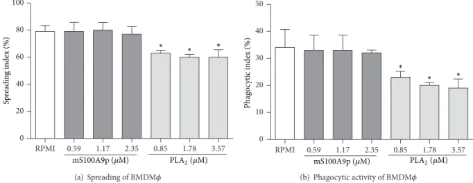
3.2. mS100A9p Did Not Change the Spreading and the Phagocytic Activity of BMDM�. To conirm that mS100A9p was unable to downregulate macrophage functions in the absence of B-1 cells, mS100A9p was incubated with BMDM� during the spreading assay. mS100A9p, in concentrations of 0.59, 1.17, and 2.35�M, did not alter BMDM� spread-ing (Figure 2(a)). PLA2, the positive control of inhibition of macrophage spreading and phagocytosis, downregulated spreading of BMDM�(20% to 0.85�M, 24% to 1.78�M, and 24% to 3.57�M) (Figure 2(a)). Diferent concentrations of mS100A9p incubated for 1 h with BMDM�during spreading did not downregulate phagocytosis of Candida albicans
particles (Figure 2(b)). On the other hand, PLA2 inhibited phagocytosis by BMDM�in all concentrations used (32% to 0.85�M, 41% to 1.78�M, and 44% to 3.57�M) (Figure 2(b)).
RPMI 1.17 2.35 RPMI 1.17 2.35 0
15 30 45 60 75
BALB/c BALB/xid
RPMI
mS100A9p (�M)
Sp
re
adin
g index (%)
∗ ∗
(a) Spreading of peritoneal cells
RPMI 1.17 2.35 RPMI 1.17 2.35
0 5 10 15 20 25
BALB/c BALB/xid
Phag
o
cytic index (%)
RPMI
mS100A9p (�M) ∗
∗
(b) Phagocytic activity of peritoneal cells
Figure 1: Spreading and phagocytosis assays by peritoneal cells obtained from BALB/c and BALB/xid mice ater incubation with mS100A9p. (a) Peritoneal cells were adhered to coverslips (1×10⁵cells/well) and incubated with mS100A9p (1.17 or 2.35�M in RPMI-1640 medium), for 1 h at 37∘C in 5% CO2. Control adherent cells were incubated only with culture medium. (b) Ater spreading, cells were incubated with 1× 10⁶Candida albicansparticles for 1 h. Results are expressed as means±SD, using six animals per group.∗� ≤0.05, when compared to the respective control group (RPMI).
RPMI 0.59 1.17 2.35 0.85 1.78 3.57 0
20 40 60 80 100
∗ ∗
∗
Sp
re
adin
g index (%)
PLA2(�M)
mS100A9p (�M)
(a) Spreading of BMDM�
RPMI 0.59 1.17 2.35 0.85 1.78 3.57 0
10 20 30 40 50
Phag
o
cytic index (%)
∗
∗ ∗
PLA2(�M)
mS100A9p (�M)
(b) Phagocytic activity of BMDM�
Figure 2: Spreading and phagocytosis assays by bone marrow-derived macrophages (BMDM�) ater incubation with mS100A9p. (a) BMDM� adhered to coverslips (1×10⁵cells/well) were incubated with mS100A9p (0.59; 1.17; or 2.35�M in RPMI-1640 medium), for 1 h at 37∘C in 5% CO2. Phospholipase A2(PLA2: 0.85; 1.78; 3.57�M), in the same conditions of peptide, was used as positive control. BMDM�controls were incubated only with culture medium. (b) Ater spreading, BMDM�were incubated with 1×10⁶Candida albicansparticles for 1 h. Results are expressed as means±SD, using ive samples per group.∗� ≤0.05, when compared to the control group (RPMI).
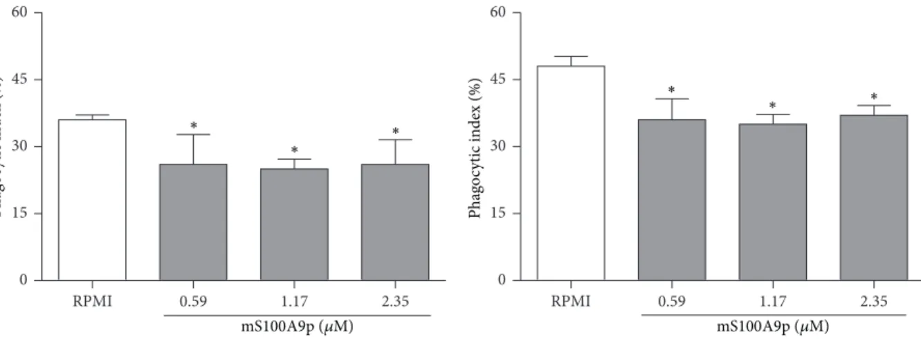
directly with B-1 cells. In the presence of the B-1 cells, phago-cytosis ofCandida albicansparticles by BMDM�was inhib-ited 28, 31, and 28%, which corresponded to concentrations of 0.59, 1.17, and 2.35�M mS100A9p, respectively (Figure 3(a)). To verify whether the contact between these two cell types was necessary to the inhibitory efect of mS100A9p, cocul-tures of BMDM� and B-1, in the presence of mS100A9p, were performed using transwell chambers. he absence of direct contact between BMDM� and B-1 cells maintained the downregulation of the macrophage phagocytic activity in
response to mS100A9p. he fall in phagocytosis was 25, 27, and 23%, corresponding to concentrations of 0.59, 1.17, and 2.35�M mS100A9p, respectively (Figure 3(b)).
RPMI 0.59 1.17 2.35 0
15 30 45 60
Phag
o
cytic index (%)
∗ ∗
∗
mS100A9p (�M)
(a) Phagocytic activity of BMDM�cocultivated with B-1 cells, with
direct contact
RPMI 0.59 1.17 2.35
0 15 30 45 60
∗
Phag
o
cytic index (%)
∗ ∗
mS100A9p (�M)
(b) Phagocytic activity of BMDM�cocultivated with B-1 cells, without
direct contact
Figure 3: Phagocytosis assays by bone marrow-derived macrophages (BMDM�), cocultivated with B-1 cells, treated with mS100A9p. BMDM�adhered to coverslips (1×10⁵cells/well) were incubated in the presence of B-1 cells (1×10⁵cells/well) concomitant with mS100A9p (0.59; 1.17; or 2.35�M in RPMI-1640 medium), for 1 h at 37∘C in 5% CO2. Coculture assays were evaluated with (a) or without (b) direct contact of these two cell populations, using transwell chambers. Experimental controls were cocultures of BMDM�and B-1 cells incubated only with culture medium. Ater 1 h, both cocultures were incubated with 1×10⁶Candida albicansparticles for 1 h. Results are expressed as means±SD, using ive samples per group.∗� ≤0.05, when compared to the control group (RPMI).
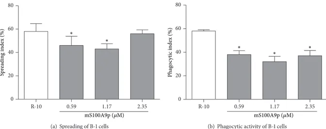
of inhibition, resp.) (Figure 4(a)). In addition, mS100A9p inhibited phagocytosis by B-1 cells, at the concentrations of 0.59, 1.17, and 2.25�M (34, 45, and 36%, resp.) (Figure 4(b)).
3.5. mS100A9p Did Not Alter Phagocytosis by Adherent Peri-toneal Cells Obtained from IL-10 KO Mice. Since B-1 cells secrete IL-10 [49], which inhibits murine macrophage phago-cytosisin vitro[38], we evaluated the involvement of IL-10 in mS100A9p-induced inhibitory efect on phagocytosis by peritoneal cells obtained from IL-10 KO mice. Peritoneal cells obtained from wild-type mice (C57BL/6) were used as con-trol. mS100A9p downregulated phagocytosis by peritoneal cells obtained from wild-type C57BL/6, in the concentration of 2.35�M (25% of inhibition), when compared with control cells incubated only with culture medium (Figure 5). he peptide, in both concentrations (1.17 and 2.35�M), did not modify phagocytosis by peritoneal cells from C57BL/6 IL-10 KO mice, when compared with control cells (Figure 5).
4. Discussion
S100A8/A9 are important proteins expressed by phago-cytes during the inlammatory response [5]. he expres-sion of these proteins is restricted to cells of the mono-cytic/granulocytic lineage, but under certain conditions they may also be expressed by keratinocytes [50,51]. It was also demonstrated that newly arrived inlammatory macrophages, but not the resident ones, express S100A8/A9 at inlamma-tory sites for a short time [50, 52]. Both proinlammatory and anti-inlammatory functions have been reported for S100A8/A9, which may depend on several factors, including their concentration, receptors involved in their recognition, their posttranslational modiications, cell types studied, and
mediators of the local milieu [53, 54]. Previous studies of our group demonstrated an inhibitory efect of either S100A9 or its C-terminal portion on an inlammatory nociception model [22, 24, 26], suggesting that mS100A9p induces a similar efect to that evoked by the complete protein. his evidence supports the hypothesis that this peptide has a crucial role in inlammatory events. In addition, mS100A9p downregulated the spreading and phagocytosis by adherent peritoneal cellsin vivoandex vivo[27,28]. Considering the inhibitory efect of mS100A9p on the activities of peritoneal cells and that B-1 cells represent the main B cell population in normal peritoneal cavity of mice [55], in this study it was investigated whether the inhibitory efect induced by C-terminus of S100A9 protein is related to an action on macrophages and/or B-1.
Initially, we investigated the efect of mS100A9p on spreading and phagocytosis by peritoneal cells from BALB/xid mice, which are deicient in B-1 cells [48]. Our data showed that mS100A9p did not inhibit the functions of cells obtained from BALB/xid mice, suggesting an important role of B-1 cells in mS100A9p-induced inhibitory efect on adherent peritoneal cells. hese data were supported by other studies showing that absence of B-1 cells in BALB/xid mice interfered with the inlammatory response, including the activity and proliferation of peritoneal macrophages [38,42], control of infection [39, 56], and wound-healing process [37].
R-10 0.59 1.17 2.35 0
20 40 60 80
Sp
re
adin
g index (%)
∗ ∗
mS100A9p (�M)
(a) Spreading of B-1 cells
R-10 0.59 1.17 2.35
0 20 40 60 80
Phag
o
cytic index (%)
∗ ∗
∗
mS100A9p (�M)
(b) Phagocytic activity of B-1 cells
Figure 4: Spreading and phagocytosis assays by B-1 cells ater incubation with mS100A9p. (a) B-1 cells, isolated by stationary culture of peritoneal cells obtained from Swiss mice, were adhered per 3 h to coverslips (1×10⁵cells/well) and incubated with mS100A9p (0.59; 1.17; or 2.35�M in R-10, RPMI-1640 medium containing 10% of heat-inactivated fetal bovine serum, R-10), for 24 h at 37∘C in 5% CO2. Control adherent cells were incubated only with R-10. (b) Ater spreading, cells were incubated with 1×10⁶Candida albicansparticles for 1 h. Results are expressed as means±SD, using six animals per group.∗� ≤0.05, when compared to the control group (R-10).
RPMI 1.17 2.35 RPMI 1.17 2.35
0 20 40 60 80
WT IL-10KO
Phag
o
cytic index (%)
∗
RPMI
mS100A9p (�M)
Figure 5: Phagocytic activity by peritoneal cells obtained from C57BL/6 wild-type (WT) and C57BL/6 IL-10 KO (IL-10 KO) mice ater incubation with mS100A9p. Peritoneal cells obtained were adhered to coverslips (1×10⁵cells/well) and incubated with mS100A9p (1.17 or 2.35�M in RPMI-1640 medium) for 1 h at 37∘C in 5% CO2. Control adherent cells were incubated only with culture medium. Ater this time, cells were incubated with 1×10⁶Candida albicansparticles for 1 h. Results are expressed as means±SD, using six animals per group.∗� ≤ 0.05, when compared to the control group (RPMI).
described elsewhere [46]. hese results demonstrated that macrophages used in our assays were functioning normally. Despite the indings obtained here, suggesting that the inhibitory efect of mS100A9p is not due to a direct action on macrophages, it has been showed that mS100A9p down-regulates the phagocytic activity of apoptotic neutrophils by macrophages [21]. Nevertheless, isolated macrophages have not been used, and the mS100A9p efect was observed on adherent peritoneal cells, which comprise macrophages and B-1 cells. his fact could explain the discrepancy between the results presented here and those reported by De Lorenzo et al. [21].
was performed in the absence or presence of transwell inserts. Once a downregulatory efect of mS100A9p was observed in both assays, we can suggest that the inhibitory efect of mS100A9p on the adherent peritoneal cells, as previously observed [27], is mediated by B-1 cells. hese results also showed that the direct contact between macrophages and B-1 cells is not essential to mS100A9p activity but is related to the secretory activity of B-1 cells.
Considering that B-1 cells diferentiate into a macro-phage-like cells, exhibiting the ability to phagocytose either in vitro orin vivo[33,59], we decided to evaluate whether mS100A9p had an efect on B-1 cell functions. he data presented here show that mS100A9p downregulated the spreading and phagocytosis by B-1 cells, suggesting that the C-terminus of S100A9 not only modulates the action of B-1 cells, which in turn inhibits the phagocytic activity of macrophages, but also has a direct efect on B-1 cells, probably by a communication via soluble autocrine and paracrine signals. Also, we did not observe a concentration-dependent efect of mS100A9p on spreading and phagocytosis assays carried out.
Many factors have a regulatory role in macrophage activ-ity, acting as key orchestrators of the inlammatory response. In this sense, IL-10 has been found to downregulate a number of diferent macrophage functions, including cytokine pro-duction and the respiratory burst [60,61], acting thereby as an important negative regulator of cell-mediated immunity [62]. Peritoneal B-1 cells are the major producers of IL-10 and they use it to regulate their own development and/or the function of other immunocompetent cells [49, 63]. As mentioned earlier, IL-10 secreted by B-1 cells downregulates the phagocytic activity of macrophages [38] and modulates the kinetics of wound-healing process [37]. Further, B-1 cells from IL-10 KO mice were not able to inhibit macrophage functions [38].
To investigate the IL-10 participation in mS100A9p-induced inhibitory efect, we evaluated whether mS100A9p altered the phagocytic activity of peritoneal cells obtained from IL-10 KO mice. Considering that mS100A9p did not change the activity of these cells, it is plausible to suggest that mS100A9p induces the secretion of IL-10 by B-1 cells and consequently inhibits its own functions and the activities of peritoneal macrophages. In this regard, it was demonstrated,
in vitro and in vivo, that B-1 cells induce macrophage polarization to an M2-like phenotype, which is anti-inlammatory and immunosuppressive in nature, evidenc-ing that IL-10 is crucial to modulate macrophage function [64].
he mechanism by which mS100A9p regulates IL-10 secretion by B-1 cells is not known. However, both B-1 cell activation and IL-10 release are regulated by TLR-4 [65–67], and S100A9 protein acts as an endogenous TLR-4 ligand, regulating thereby myeloid cell function [13, 68,
69]. In this sense, it is plausible to hypothesize that the C-terminus of S100A9 stimulates IL-10 secretion by B-1 cells via TLR-4, suppressing thereby macrophage activities. hese data emphasize the importance of S100A9 and B-1 cells to orchestrate inlammatory response and its resolution.
5. Conclusion
he downregulatory efects of mS100A9p on macrophages are mediated by B-1 cells, possibly via IL-10 secretion. Taken together, our data showed that mS100A9p is a good research tool to understand S100A9 function on the development and maintenance of the inlammatory process and demonstrated a pivotal role of B-1 cells mediating the action of the C-terminus of S100A9 on control of innate immunity.
Conflict of Interests
he authors declare that there is no conlict of interests regarding the publication of this paper.
Authors’ Contribution
Rosana Lima Pagano, Sandra Coccuzzo Sampaio, and Renata Giorgi conceived and designed the experiments. Rosana Lima Pagano, Natassja Foizer Moraes, Beatriz Helena De Lorenzo, and Sandra Coccuzzo Sampaio performed the experiments. Rosana Lima Pagano, Sandra Coccuzzo Sam-paio, and Renata Giorgi analyzed the data. Rosana Lima Pagano, Sandra Coccuzzo Sampaio, and Renata Giorgi con-tributed reagents, materials, and analysis tools. Rosana Lima Pagano, Sandra Coccuzzo Sampaio, Natassja Foizer Moraes, Mario Mariano, and Renata Giorgi wrote the paper.
Acknowledgments
he authors thank Drs. Maria Aparecida Juliano and Luis Juliano at the Department of Biophysics, Pharmacology Institute, Federal University of S˜ao Paulo, Brazil, for synthesis, characterization, and puriication of mS100A9p. his work was supported by Fundac¸˜ao de Amparo `a Pesquisa do Estado de S˜ao Paulo (FAPESP-2002/08277-7) and Fundac¸˜ao Butantan.
References
[1] K. Odink, N. Cerletti, J. Bruggen et al., “Two calcium-binding proteins in iniltrate macrophages of rheumatoid arthritis,”
Nature, vol. 330, no. 6143, pp. 80–82, 1987.
[2] W. Nacken, J. Roth, C. Sorg, and C. Kerkhof, “S100A9/S100A8: myeloid representatives of the S100 protein family as prominent players in innate immunity,” Microscopy Research and Tech-nique, vol. 60, no. 6, pp. 569–580, 2003.
[3] J. Edgeworth, M. Gorman, R. Bennett, P. Freemont, and N. Hogg, “Identiication of p8,14 as a highly abundant het-erodimeric calcium binding protein complex of myeloid cells,”
he Journal of Biological Chemistry, vol. 266, no. 12, pp. 7706– 7713, 1991.
[4] J. Roth, F. Burwinkel, C. Van den Bos, M. Goebeler, E. Vollmer, and C. Sorg, “MRP8 and MRP14, S-100-like proteins associ-ated with myeloid diferentiation, are translocassoci-ated to plasma membrane and intermediate ilaments in a calcium-dependent manner,”Blood, vol. 82, no. 6, pp. 1875–1883, 1993.
molecules,”Trends in Immunology, vol. 24, no. 4, pp. 155–158, 2003.
[6] D. Foell, M. Frosch, C. Sorg, and J. Roth, “Phagocyte-speciic calcium-binding S100 proteins as clinical laboratory markers of inlammation,”Clinica Chimica Acta, vol. 344, no. 1-2, pp. 37–51, 2004.
[7] C. Gebhardt, J. N´emeth, P. Angel, and J. Hess, “S100A8 and S100A9 in inlammation and cancer,”Biochemical Pharmacol-ogy, vol. 72, no. 11, pp. 1622–1631, 2006.
[8] D. Foell, H. Wittkowski, T. Vogl, and J. Roth, “S100 proteins expressed in phagocytes: a novel group of damage-associated molecular pattern molecules,”Journal of Leukocyte Biology, vol. 81, no. 1, pp. 28–37, 2007.
[9] R. A. Newton and N. Hogg, “he human S100 protein MRP-14 is a novel activator of the�2 integrin Mac-1 on neutrophils,”
Journal of Immunology, vol. 160, no. 3, pp. 1427–1435, 1998. [10] C. Ryckman, K. Vandal, P. Rouleau, M. Talbot, and P. A. Tessier,
“Proinlammatory activities of S100: proteins S100A8, S100A9, and S100A8/A9 induce neutrophil chemotaxis and adhesion,”
Journal of Immunology, vol. 170, no. 6, pp. 3233–3242, 2003.
[11] K. Vandal, P. Rouleau, A. Boivin, C. Ryckman, M. Talbot, and P. A. Tessier, “Blockade of S100A8 and S100A9 suppresses neutrophil migration in response to lipopolysaccharide,”he Journal of Immunology, vol. 171, no. 5, pp. 2602–2609, 2003. [12] M. A. Raquil, N. Anceriz, P. Rouleau, and P. A. Tessier,
“Block-ade of antimicrobial proteins S100A8 and S100A9 inhibits phagocyte migration to the alveoli in streptococcal pneumonia,”
Journal of Immunology, vol. 180, no. 5, pp. 3366–3374, 2008. [13] T. Vogl, K. Tenbrock, S. Ludwig et al., “Mrp8 and Mrp14 are
endogenous activators of Toll-like receptor 4, promoting lethal, endotoxin-induced shock,”Nature Medicine, vol. 13, no. 9, pp. 1042–1049, 2007.
[14] J.-C. Simard, D. Girard, and P. A. Tessier, “Induction of neutrophil degranulation by S100A9 via a MAPK-dependent mechanism,”Journal of Leukocyte Biology, vol. 87, no. 5, pp. 905– 914, 2010.
[15] J.-C. Simard, M.-M. Simon, P. A. Tessier, and D. Girard, “Damage-associated molecular pattern S100A9 increases bac-tericidal activity of human neutrophils by enhancing phagocy-tosis,”he Journal of Immunology, vol. 186, no. 6, pp. 3622–3631, 2011.
[16] K. Sunahori, M. Yamamura, J. Yamana et al., “he S100A8/A9 heterodimer ampliies proinlammatory cytokine production by macrophages via activation of nuclear factor kappa B and p38 mitogen-activated protein kinase in rheumatoid arthritis,”
Arthritis Research and herapy, vol. 8, no. 3, article R69, 2006.
[17] J. C. Simard, A. Cesaro, J. Chapeton-Montes et al., “S100A8 and S100A9 induce cytokine expression and regulate the NLRP3 inlammasome via ROS-dependent activation of NF-�B(1.),”
PLoS ONE, vol. 8, no. 8, Article ID e72138, 2013.
[18] J. H. Boyd, B. Kan, H. Roberts, Y. Wang, and K. R. Walley, “S100A8 and S100A9 mediate endotoxin-induced cardiomy-ocyte dysfunction via the receptor for advanced glycation end products,”Circulation Research, vol. 102, no. 10, pp. 1239–1246, 2008.
[19] T. Vogl, S. Ludwig, M. Goebeler et al., “MRP8 and MRP14 control microtubule reorganization during transendothelial migration of phagocytes,”Blood, vol. 104, no. 13, pp. 4260–4268, 2004.
[20] T. Aguiar-Passeti, E. Postol, C. Sorg, and M. Mariano, “Epithe-lioid cells from foreign-body granuloma selectively express the calcium-binding protein MRP-14, a novel down-regulatory molecule of macrophage activation,”Journal of Leukocyte Biol-ogy, vol. 62, no. 6, pp. 852–858, 1997.
[21] B. H. P. De Lorenzo, L. C. Godoy, R. R. Novaes e Brito et al., “Macrophage suppression following phagocytosis of apoptotic neutrophils is mediated by the S100A9 calcium-binding pro-tein,”Immunobiology, vol. 215, no. 5, pp. 341–347, 2010.
[22] R. Giorgi, R. L. Pagano, M. A. Amorim Dias, T. Aguiar-Passeti, C. Sorg, and M. Mariano, “Antinociceptive efect of the calcium-binding protein MRP-14 and the role played by neutrophils on the control of inlammatory pain,”Journal of Leukocyte Biology, vol. 64, no. 2, pp. 214–220, 1998.
[23] R. L. Pagano, M. A. A. Dias, C. S. Dale, and R. Giorgi, “Neutrophils and the calcium-binding protein MRP-14 mediate carrageenan-induced antinociception in mice,” Mediators of Inlammation, vol. 11, no. 4, pp. 203–210, 2002.
[24] R. L. Pagano, M. Mariano, and R. Giorgi, “Neutrophilic cell-free exudate induces antinociception mediate by the protein S100A9,”Mediators of Inlammation, vol. 2006, Article ID 36765, 6 pages, 2006.
[25] C. S. Dale, N. Cenac, L. R. G. Britto et al., “he C-terminus of murine S100A9 protein inhibits hyperalgesia induced by the agonist peptide of protease-activated receptor 2 (PAR 2),”British Journal of Pharmacology, vol. 149, no. 4, pp. 374–384, 2006.
[26] C. S. Dale, R. D. L. Pagano, C. C. Paccola et al., “Efect of the C-terminus of murine S100A9 protein on experimental nociception,”Peptides, vol. 27, no. 11, pp. 2794–2802, 2006.
[27] R. L. Pagano, S. C. Sampaio, L. Juliano, M. A. Juliano, and R. Giorgi, “he C-terminus of murine S100A9 inhibits spreading and phagocytic activity of adherent peritoneal cells,” Inlamma-tion Research, vol. 54, no. 5, pp. 204–210, 2005.
[28] R. L. Pagano, S. C. Sampaio, M. A. Juliano, L. Juliano, and R. Giorgi, “Involvement of proteinase-activated receptors 1 and 2 in spreading and phagocytosis by murine adherent peritoneal cells: modulation by the C-terminal of S100A9 protein,” Euro-pean Journal of Pharmacology, vol. 628, no. 1–3, pp. 240–246, 2010.
[29] E. E. Bou Ghosn, A. A. Cassado, G. R. Govoni et al., “Two phys-ically, functionally, and developmentally distinct peritoneal macrophage subsets,”Proceedings of the National Academy of Sciences of the United States of America, vol. 107, no. 6, pp. 2568– 2573, 2010.
[30] L. A. Herzenberg and A. B. Kantor, “B-cell lineages exist in the mouse,”Immunology Today, vol. 14, no. 2, pp. 79–83, 1993.
[31] A. B. Kantor and L. A. Herzenberg, “Origin of murine B cell lineages,”Annual Review of Immunology, vol. 11, pp. 501–538, 1993.
[32] J. D. Bouaziz, K. Yanaba, and T. F. Tedder, “Regulatory B cells as inhibitors of immune responses and inlammation,”
Immunological Reviews, vol. 224, no. 1, pp. 201–214, 2008.
[33] S. R. Almeida, L. S. Aroeira, E. Frymuller et al., “Mouse B-1 cell-derived mononuclear phagocyte, a novel cellular compo-nent of acute non-speciic inlammatory exudate,”International Immunology, vol. 13, no. 9, pp. 1193–1201, 2001.
macrophage characteristics,”Journal of Immunology, vol. 155, no. 9, pp. 4155–4161, 1995.
[35] S. A. Ha, M. Tsuji, K. Suzuki et al., “Regulation of B1 cell migration by signals through Toll-like receptors,”he Journal of Experimental Medicine, vol. 203, no. 11, pp. 2541–2550, 2006.
[36] C. S. B. Bogsan, R. R. Novaes E Brito, M. da Cruz Palos et al., “B-1 cells are pivotal for in vivo inlammatory giant cell formation,”
International Journal of Experimental Pathology, vol. 86, no. 4, pp. 257–265, 2005.
[37] H. C. Oliveira, A. F. Popi, A. L. L. Bachi, S. Nonogaki, J. D. Lopes, and M. Mariano, “B-1 cells modulate the kinetics of wound-healing process in mice,”Immunobiology, vol. 215, no. 3, pp. 215– 222, 2010.
[38] A. F. Popi, J. D. Lopes, and M. Mariano, “Interleukin-10 secreted by B-1 cells modulates the phagocytic activity of murine macrophages in vitro,”Immunology, vol. 113, no. 3, pp. 348–354, 2004.
[39] A. F. Popi, L. C. Godoy, P. Xander, J. D. Lopes, and M. Mariano, “B-1 cells facilitate Paracoccidioides brasiliensis infection in mice via IL-10 secretion,”Microbes and Infection, vol. 10, no. 7, pp. 817–824, 2008.
[40] B. H. P. De Lorenzo, R. R. N. Brito, L. C. Godoy, J. D. Lopes, and M. Mariano, “Tolerogenic property of B-1b cells in a model of allergic reaction,”Immunology Letters, vol. 114, no. 2, pp. 110–118, 2007.
[41] D. F. Barbeiro, H. V. Barbeiro, J. Faintuch et al., “B-1 cells temper endotoxemic inlammatory responses,”Immunobiology, vol. 216, no. 3, pp. 302–308, 2011.
[42] A. F. Popi, L. Osugui, K. R. Perez, I. M. Longo-Maug´eri, and M. Mariano, “Could a B-1 cell derived phagocyte “be one” of the peritoneal macrophages during LPS-driven inlammation?”
PLoS ONE, vol. 7, no. 3, Article ID e34570, 2012.
[43] M. J. Ratery, C. A. Harrison, P. Alewood, A. Jones, and C. L. Geczy, “Isolation of the murine S100 protein MRP14 (14 kDa migration-inhibitory-factor-related protein) from acti-vated spleen cells: characterization of post-translational modii-cations and zinc binding,”he Biochemical Journal, vol. 316, no. 1, pp. 285–293, 1996.
[44] G. Rosenfeld, “M´etodo r´apido de colorac¸˜ao de esfregac¸os de sangue,” inNoc¸˜oes pr´aticas sobre corantes pancrˆomicos e estudos de diversos fatores, pp. 315–328, 1947.
[45] G. Boltz-Nitulescu, C. Wiltschke, C. Holzinger et al., “Difer-entiation of rat bone marrow cells into macrophages under the inluence of mouse L929 cell supernatant,”Journal of Leukocyte Biology, vol. 41, no. 1, pp. 83–91, 1987.
[46] S. C. Sampaio, A. C. Rangel-Santos, C. M. Peres, R. Curi, and Y. Cury, “Inhibitory efect of phospholipase A2 isolated from Crotalus durissus terriicus venom on macrophage function,”
Toxicon, vol. 45, no. 5, pp. 671–676, 2005.
[47] M. Rabinovitch and M. J. DeStefano, “Macrophage spreading in vitro. I. Inducers of spreading,”Experimental Cell Research, vol. 77, no. 1-2, pp. 323–334, 1973.
[48] A. Narendran, D. Ramsden, A. Cumano, T. Tanaka, G. E. Wu, and C. J. Paige, “B cell developmental defects in X-linked immunodeiciency,”International Immunology, vol. 5, no. 2, pp. 139–144, 1993.
[49] A. O'Garra, R. Chang, N. Go, R. Hastings, G. Haughton, and M. Howard, “Ly-1 B (B-1) cells are the main source of B cell-derived
interleukin 10,”European Journal of Immunology, vol. 22, no. 3, pp. 711–717, 1992.
[50] N. Hogg, C. Allen, and J. Edgeworth, “Monoclonal antibody 5.5 reacts with p8,14, a myeloid molecule associated with some vascular endothelium,”European Journal of Immunology, vol. 19, no. 6, pp. 1053–1061, 1989.
[51] M. M. Wilkinson, A. Busuttil, C. Hayward, D. J. Brock, J. R. Dorin, and V. van Heyningen, “Expression pattern of two related cystic ibrosis-associated calcium-binding proteins in normal and abnormal tissues,”Journal of Cell Science, vol. 91, no. 2, pp. 221–230, 1988.
[52] G. Zwadlo, J. Bruggen, G. Gerhards, R. Schlegel, and C. Sorg, “Two calcium-binding proteins associated with speciic stages of myeloid cell diferentiation are expressed by subsets of macrophages in inlammatory tissues,”Clinical and Experimen-tal Immunology, vol. 72, no. 3, pp. 510–515, 1988.
[53] J. Goyette and C. L. Geczy, “Inlammation-associated S100 proteins: new mechanisms that regulate function,”Amino Acids, vol. 41, no. 4, pp. 821–842, 2011.
[54] R. Donato, B. R. Cannon, G. Sorci et al., “Functions of S100 proteins,”Current Molecular Medicine, vol. 13, no. 1, pp. 24–57, 2013.
[55] B. A. Graf, D. A. Nazarenko, M. A. Borrello et al., “Biphenotypic B/macrophage cells express COX-1 and up-regulate COX-2 expression and prostaglandin E(2) production in response to pro-inlammatory signals,”European Journal of Immunology, vol. 29, no. 11, pp. 3793–3803, 1999.
[56] W. N. Khan, F. W. Alt, R. M. Gerstein et al., “Defective B cell development and function in Btk-deicient mice,”Immunity, vol. 3, no. 3, pp. 283–299, 1995.
[57] J. Hendriks, J. Riesle, and C. A. van Blitterswijk, “Co-culture in cartilage tissue engineering,”Journal of Tissue Engineering and Regenerative Medicine, vol. 1, no. 3, pp. 170–178, 2007.
[58] D. J. Holt, L. M. Chamberlain, and D. W. Grainger, “Cell-cell signaling in co-cultures of macrophages and ibroblasts,”
Biomaterials, vol. 31, no. 36, pp. 9382–9394, 2010.
[59] R. R. Novaes e Brito, B. A. Cortez, G. M. Machado-Santelli et al., “In vitroandin vivophagocytic ability of mouse B-1 cells,”
Immunology and Immunogenetics Insights, vol. 2, no. 1, pp. 31–39, 2010.
[60] D. F. Fiorentino, A. Zlotnik, T. R. Mosmann, M. Howard, and A. O’Garra, “IL-10 inhibits cytokine production by activated macrophages,”he Journal of Immunology, vol. 147, no. 11, pp. 3815–3822, 1991.
[61] C. Bogdan, Y. Vodovotz, and C. Nathan, “Macrophage deacti-vation by interleukin 10,”he Journal of Experimental Medicine, vol. 174, no. 6, pp. 1549–1555, 1991.
[62] K. W. Moore, A. O’Garra, R. de Waal Malefyt, P. Vieira, and T. R. Mosmann, “Interleukin-10,”Annual Review of Immunology, vol. 11, pp. 165–190, 1993.
[63] V. Sindhava, M. E. Woodman, B. Stevenson, and S. Bondada, “Interleukin-10 mediated autoregulation of murine B-1 B-cells and its role in Borrelia hermsii infection,”PLoS ONE, vol. 5, no. 7, Article ID e11445, 2010.
[65] S.-A. Ha, M. Tsuji, K. Suzuki et al., “Regulation of B1 cell migration by signals through Toll-like receptors,”he Journal of Experimental Medicine, vol. 203, no. 11, pp. 2541–2550, 2006.
[66] T. A. Barr, S. Brown, G. Ryan, J. Zhao, and D. Gray, “TLR-mediated stimulation of APC: distinct cytokine responses of B cells and dendritic cells,”European Journal of Immunology, vol. 37, no. 11, pp. 3040–3053, 2007.
[67] G. Tumurkhuu, N. Koide, J. Dagvadorj et al., “B1 cells produce nitric oxide in response to a series of toll-like receptor ligands,”
Cellular Immunology, vol. 261, no. 2, pp. 122–127, 2010.
[68] J. M. Ehrchen, C. Sunderk¨otter, D. Foell, T. Vogl, and J. Roth, “he endogenous Toll-like receptor 4 agonist S100A8/S100A9 (calprotectin) as innate ampliier of infection, autoimmunity, and cancer,”Journal of Leukocyte Biology, vol. 86, no. 3, pp. 557– 566, 2009.
Submit your manuscripts at
http://www.hindawi.com
Stem Cells
International
Hindawi Publishing Corporationhttp://www.hindawi.com Volume 2014
Hindawi Publishing Corporation
http://www.hindawi.com Volume 2014 INFLAMMATION
Hindawi Publishing Corporation
http://www.hindawi.com Volume 2014
Behavioural
Neurology
Endocrinology
International Journal of Hindawi Publishing Corporationhttp://www.hindawi.com Volume 2014
Hindawi Publishing Corporation
http://www.hindawi.com Volume 2014
Disease Markers
Hindawi Publishing Corporation
http://www.hindawi.com Volume 2014
BioMed
Research International
Oncology
Journal ofHindawi Publishing Corporation
http://www.hindawi.com Volume 2014
Hindawi Publishing Corporation
http://www.hindawi.com Volume 2014
Oxidative Medicine and Cellular Longevity
Hindawi Publishing Corporation
http://www.hindawi.com Volume 2014
PPAR Research
The Scientiic
World Journal
Hindawi Publishing Corporationhttp://www.hindawi.com Volume 2014
Immunology Research
Hindawi Publishing Corporation
http://www.hindawi.com Volume 2014
Journal of
Obesity
Journal ofHindawi Publishing Corporation
http://www.hindawi.com Volume 2014
Hindawi Publishing Corporation
http://www.hindawi.com Volume 2014 Computational and Mathematical Methods in Medicine
Ophthalmology
Journal ofHindawi Publishing Corporation
http://www.hindawi.com Volume 2014
Diabetes Research
Journal ofHindawi Publishing Corporation
http://www.hindawi.com Volume 2014
Hindawi Publishing Corporation
http://www.hindawi.com Volume 2014 Research and Treatment
AIDS
Hindawi Publishing Corporation
http://www.hindawi.com Volume 2014
Gastroenterology Research and Practice
Hindawi Publishing Corporation
http://www.hindawi.com Volume 2014
Parkinson’s
Disease
Evidence-Based Complementary and Alternative Medicine
Volume 2014 Hindawi Publishing Corporation