Ethyl 2-cyanoacrylate tissue adhesive
in partial lobectomy in rats*
,**
Adesivo cirúrgico de etil-2-cianoacrilato em lobectomia parcial em ratos
Ariani Cavazzani Szkudlarek, Paula Sincero, Renato Silva de Sousa, Rosalvo Tadeu Hochmuller Fogaça
Abstract
Objective: To determine the efficacy of ethyl 2-cyanoacrylate adhesive in repairing the lung parenchyma after partial lobectomy in rats, in terms of hemostasis/aerostasis, scarring, and surgical time. Methods: The study involved 30 Wistar rats, randomly divided into five groups (one control group and four study groups). In the study groups, the lung parenchyma was repaired with either cyanoacrylate adhesive or surgical suture following resection of a small or large fragment (25% or 50%, respectively) of the left caudal lung lobe. Results: Surgical time and hemostasis time were shorter in the two groups treated with the adhesive than in the two submitted to suture. There were no significant differences among the groups regarding specific lung compliance. Adherences and inflammatory reactions were more severe in the groups submitted to suture. Conclusions: In this study, the use of cyanoacrylate adhesive helped reduce the surgical time and the intensity of inflammatory reactions, as well as preserving lung compliance. Cyanoacrylate adhesives should be considered an option for lung parenchyma repair, decreasing the risk of complications after partial lobectomy in humans.
Keywords: Lung; Pneumonectomy; Cyanoacrylates.
Resumo
Objetivo: Determinar experimentalmente a eficácia do adesivo cirúrgico de etil-2-cianoacrilato na reparação do parênquima pulmonar após lobectomias parciais em ratos em relação a hemostasia/aerostasia, cicatrização e tempo cirúrgico. Métodos: O estudo envolveu 30 ratos Wistar, divididos aleatoriamente em cinco grupos (grupo controle e quatro grupos de estudo. Nos grupos de estudo, o reparo do parênquima pulmonar foi realizado ou com o adesivo ou por sutura após lobectomia parcial de um fragmento pequeno ou grande (25% ou 50%, respectivamente) do lobo inferior caudal esquerdo. Resultados: O tempo cirúrgico e o tempo de hemostasia foram menores nos grupos submetidos ao uso do adesivo. Não houve diferenças significativas na complacência pulmonar específica entre os grupos. Aderências e reações inflamatórias foram mais severas nos grupos submetidos a sutura.
Conclusões: Neste estudo, o uso de adesivo de cianoacrilato ajudou a reduzir o tempo cirúrgico e a intensidade de reações inflamatórias, assim como preservou a complacência pulmonar. Adesivos de cianoacrilato devem ser considerados como uma opção no reparo do parênquima pulmonar, diminuindo o risco de complicações após lobectomia parcial em humanos.
Descritores: Pulmão; Pneumonectomia; Cianoacrilatos.
* Study carried out at the Federal University of Paraná, Curitiba, Brazil.
Correspondence to: Ariani C Szkudlarek. Rua Abílio Peixoto, 298. apto. 34, CEP 80035-260, Curitiba, PR, Brasil. E-mail: [email protected]
Financial support: This study received financial support from Fundação Araucária. Submitted: 14 June 2011. Accepted, after review: 19 September 2011.
have been experimentally and clinically used as topical hemostatic agents in parenchymal tissues, as well as in oral surgery, incisions, and skin grafts.(4,5,11). Therefore, the use of
ethyl 2-cyanoacrylate tissue adhesive might be an alternative method to repair the lung parenchyma after partial lobectomy.
The objective of the present study was to determine the efficacy of ethyl 2-cyanoacrylate adhesive in repairing the lung parenchyma after partial lobectomy and providing adequate hemostasis/aerostasis and appropriate scarring, as well as in reducing anesthesia time, so that post-surgical complications are minimized. We evaluated parenchymal and chest wall scarring after the use of either conventional suture or ethyl 2-cyanoacrylate tissue adhesive in rats, by morphological/histopathological analysis and functional analysis of lung compliance, using pressure-volume curves. In addition, we evaluated the surgical time after the application of either method.
Methods
The Animal Care Committee of the Federal University of Paraná approved the study. All animals received humane care in compliance with the European Convention on Animal Care.
Our sample comprised 30 male adult Wistar rats weighing 190-210 g. The animals were housed in a temperature- and humidity-controlled environment (22 ± 1°C; 65-70% relative humidity), on a 12/12-h light/dark cycle, with ad libitum access to standard pellet rat chow.
Experimental procedures included resection of a small or large fragment (25% or 50%, respectively) of the left caudal lung lobe and repair of lung parenchyma with either ethyl 2-cyanoacrylate adhesive or surgical absorbable suture (Vicryl 6-0). The animals were randomly divided into five groups of 6 animals each: sham-operated (SO), in which the chest was opened with no pulmonary resection, a thin layer of adhesive was applied on the surface of the lung parenchyma, and absorbable suture was used to close the chest wall; sutured, small fragment (SuSF), in which the lung parenchyma was repaired with absorbable suture; sutured, large fragment (SuLF); adhesive, small fragment (AdSF), in which the lung parenchyma was repaired with cyanoacrylate adhesive; and
Introduction
Partial lobectomy is a useful therapeutic procedure to remove small lesions, abscesses, and tumors, as well as to surgically treat post-traumatic intrapulmonary hematomas and cystic fibrosis.(1,2) The techniques commonly used
to surgically repair the lung parenchyma are conventional suture with absorbable suture or the use of metallic staples. After the resection of the lung fragment, the greatest difficulty lies in achieving hemostasis and aerostasis. This difficulty results from the fragility of the lung tissue, which can become even more fragile when there is edema or contusion, tending to lacerate when sutured. In addition, there are risks to patients because of prolonged anesthesia, which commonly occurs with conventional suture techniques.
Cyanoacrylates have been extensively used as surgical tissue adhesives,(2-5) and they
have been shown to be safe.(6-8) Their use as a
surgical adhesive has been recommended due to their characteristics: fast polymerization after application and contact with fluids, such as water and blood, or with moist tissues. However, in order to obtain better gluing results, cyanoacrylate adhesives should be applied to surfaces that are as dry as possible.
(6) The first cyanoacrylates developed for surgical
procedures were short-chain adhesives, and this kind of adhesive was extensively studied and consequentially rejected for surgical use because of the intense local inflammation and foreign body reactions that they caused.(8) That type
of cyanoacrylate adhesive is rapidly degraded, producing toxic substances, such as cyanide gas and formaldehyde.(8-10) Long-chain cyanoacrylate
After surgery, the animals were kept in proper boxes, and the wounds were inspected daily to verify suture integrity and the presence of an infectious process. Following the inspection, asepsis was performed with iodide polyvinyl pyrrolidone. Seven days after the procedure, the suture was removed.
On post-operative day 30, the rats were killed, after which the chest wall and the lung parenchyma were inspected for adherences. Adherences were classified from 1 to 4, using the criteria devised by Knightly et al.(12) For
the histopathological analysis, small pieces of the surgical areas of the chest and of the lung parenchyma, perpendicular to the wound surface and including normal lung tissue, were removed and fixed using a solution consisting of 10% buffered formaldehyde and 1% agar at 42°C for 2 h. Following dehydration in graded ethanol solutions, the samples were embedded in paraffin blocks, cut into 4-µm sections, mounted on glass slides, deparaffinized with xylene, and counterstained with H&E.(13) Specific
lung compliance was measured under static conditions, and the slope of the volume-pressure curve was calculated for each animal. The curves were obtained from functional residual capacity up to TLC.
Statistical analyses were performed using the Mann-Whitney rank sum test for inflammatory reaction and adherences. For multiple comparisons, we used one-way ANOVA, followed by pairwise comparison between the means adhesive, large fragment (AdLF). In all groups,
anesthesia was induced using diethyl ether and maintained during the surgical procedures with a combination of ketamine and xylazine (i.m., 90 mg/kg and 5 mg/kg, respectively).
Initially, the surgical area was cleansed. After the trachea had been checked for obstructions, each rat was intubated with a 16-G endotracheal cannula (Vasofix; B. Braun Meslungen AG., Meslungen, Germany). Tidal volume was adjusted to 10 mL/kg. A rodent ventilator (Ugo Basile Biological Research Apparatus, Comerio, Italy) was used for artificial respiration.
The intervention was carried out as follows: an incision was made in the sixth intercostal space; capillary hemostasis was achieved by compression and ligation; the scalene and serratus posterior muscles were sectioned in order to expose the external intercostal muscles; after an exhalation, the external and internal intercostal muscles, as well as the parietal pleura, were sectioned, and the incision was extended dorsoventrally; the edges of the incision were protected using a saline-moistened gauze and separated to expose the left caudal pulmonary lobe; the distal edge of the lobe was secured with hemostats, adapted at the level of the resection; and the distal fragment was removed.
After the removal of the fragment, the following procedures were performed: in the SuSF and SuLF groups, the repair was made with an absorbable synthetic suture and everting sutures, followed by continuous simple sutures with the same material; in the AdSF and AdLF groups, the repair was made with the application of a thin layer of ethyl 2-cyanoacrylate tissue adhesive. The hemostats were removed 60 s after adhesive application.
Following these procedures, the lobe was replaced into the thoracic cavity, and the thorax was filled with saline solution until the lung was submerged, in order to identify air or blood leakage. After that, we closed the thoracic cavity, respecting the anatomic layers.
To restore the negative intrathoracic pressure, a drain was surgically inserted at two caudal intercostal spaces from the incision line. Aspiration was carried out until the animal began to breathe spontaneously. After a forced inhalation, the drain was rapidly removed and, at the same time, the orifice in the subcutaneous tissue was closed. The surgical time was noted.
to control, 10 min). In terms of specific lung compliance, there were no significant differences among the SO, SuSF, and AdSF groups (0.92 ± 0.39 mL/cmH2O, 1.15 ± 0.56 mL/cmH2O, and 0.89 ± 0.68 mL/cmH2O, respectively), nor between the SuLF and AdLF groups (0.64 ± 0.09 mL/cmH2O vs. 0.69 ± 0.22 mL/cmH2O).
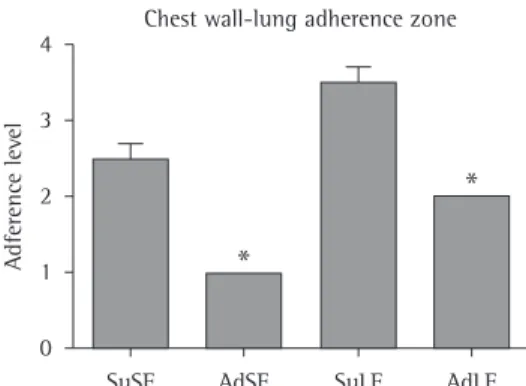
Upon macroscopic examination, adherence zones in the chest wall and in the lung were identified in all of the animals. However, adherences were more severe in the SuSF group than in the AdSF group (level 2 in 50% and level 3 in 50% of the animals vs. level 1 in 100% of the animals), as were those in the SuLF group when compared with those seen in the AdLF group (level 3 in 50% and level 4 in 50% vs. level 2 in 100%; Figure 2). The adherence zones found in the SO group were all classified as level 1.
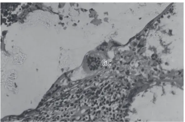
The histopathological examination showed that the inflammatory reaction was more severe in the SuSF group than in the AdSF group (moderate in all animals vs. absent in 50% and moderate in 50%, p = 0.39), as well as being more severe in the SuLF group than in the AdLF group (moderate in 50% and severe in 50% vs. absent in all animals; p = 0.01). In the animals with no inflammatory reaction, pleural thickness was found to be greater. The inflammatory reaction in the SO group did not significantly differ from that observed in the AdSF and AdLF groups (p = 0.17 and p = 0.69, respectively), although it did differ from that observed in the SuSF and SuLF groups (p = 0.01 and p = 0.008, respectively). Figures 3, 4, and 5 show the histopathological findings in the lung tissue of animals in the SO, SuLF, and AdLF groups, respectively.
Discussion
Ethyl 2-cyanoacrylate adhesive can be used as an alternative to the classical suture technique because of its capacity to promote satisfactory hemostasis/aerostasis and to significantly reduce the time under anesthesia, which always presents a risk to the patients. The mechanism of sealing the tissue is carried out by the chemical connection of tissue proteins with polyisocyanate.(11)
In humans and in animals, it should be borne in mind that, except in the case of lung lobe donors, lung tissue is excised for a reason—to using Tukey’s test. The level of significance was
set at p < 0.05.
Results
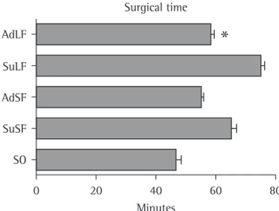
The surgical time in the SO group (48.0 ± 4.0 min) was significantly shorter than in all of the other groups. Surgical time was also significantly shorter in the SuSF group than in the SuLF group (65.0 ± 6.2 min vs. 75.0 ± 4.78 min; Figure 1). However, no such difference was found between the AdSF and AdLF groups (55.0 ± 3.2 min and 58.0 ± 4.0 min, respectively).
After the partial lobectomy, all of the animals showed minor hemorrhage. However, the bleeding was controlled faster in the AdSF and AdLF groups (mean time to control, 4 min) than in the SuSF and SuLF groups (mean time
Figure 2 - Classification of adherence zones in the chest wall/lung in the groups studied. SO: sham-operated; SuSF: suture, small fragment; AdSF: adhesive, small fragment; SuLF: suture, large fragment; and AdLF: adhesive, large fragment. *AdSF group was significantly different from SuSF, and AdLF was significantly different from SuLF.
The lung compliance test is one of the most relevant tests to confirm the appropriate functioning of the lung. Therefore, this test has been used to monitor patients with acute respiratory failure, acute lung injury, or ARDS.(22) The compliance test showed no
significant differences among our groups. To our knowledge, this is the first time that ethyl 2-cyanoacrylate adhesive has been shown to neither impair the scarring process in the lung nor significantly interfere with lung compliance.
Adherence zones in the chest wall were identified in all of the animals and were considered a normal consequence of the hypoxia caused by the incision and the procedures employed in order to achieve hemostasis. The adherences were more severe in the SuSF and SuLF groups than in the AdSF and AdLF groups. In the two former groups, the adherences were attributed to ischemic injury of the tissue caused by the surgical procedure and to the presence of the absorbable suture. In the latter two groups, the adherences might be related to the toxicity of cyanoacrylate degradation by-products.
(11,23) Although the presence of toxic residues
14 days after surgery has been previously reported,(21) no adhesive residues were found
30 days after surgery in the present study. The inflammatory reactions observed during the histopathological examination of the pulmonary fragments were more intense and numerous in the animals submitted to conventional suture with absorbable suture. Although we also found inflammatory reactions in the animals treated with the tissue adhesive, they were less intense. The inflammatory reactions might have occurred due to the toxicity of cyanoacrylate degradation by-products and to the intense heating during the polymerization process.
(10,11,21) In addition, the amount of vascular
tissue in the lung parenchyma should be taken into consideration, because the inflammatory response is proportional to that.(20)
In conclusion, in the present study, the use of cyanoacrylate adhesive helped reduce the surgical time and the intensity of inflammatory reactions, as well as preserving lung compliance, in rats. Therefore, the use of this type of adhesive should be considered an option for lung parenchyma repair, decreasing the risk of complications after partial lobectomy in humans. resect bronchial carcinoma, for example—
and there are certain aspects, such as age, smoking status, and diabetes, that might lead to complications after lobectomy.(14) Therefore,
alternative methods to repair lung tissue can help reduce postoperative complications. Unlike other studies, showing a significant incidence of mild and moderate complications after pulmonary lobectomy(15) or segmentectomy in
humans,(16) our study shows that, in most of the
animals, the pulmonary reserve allowed them to tolerate and adapt to the loss of large segments of the lobe, as has been shown previously.
(17) Considering the application of the tissue
adhesive technique itself, it should be noted that this technique is simple and rapid, which can be life-saving.(12,18-21)
Figure 4 - Typical histopathological sample of lung tissue in the suture, large fragment (SuLF) group. Notice the thicker pleura, the presence of giant cells (GC), and inflammatory reaction (H&E; magnification, ×40).
13. Kandemir O, Buyukates M, Kandemir NO, Aktunc E, Gul AE, Gul S, et al. Demonstration of the histopathological and immunohistochemical effects of a novel hemostatic agent, Ankaferd Blood Stopper, on vascular tissue in a rat aortic bleeding model. J Cardiothorac Surg. 2010;5:110.
14. Camargo SM, Camargo Jde J, Schio SM, Sánchez LB, Felicetti JC, Moreira Jda S, et al. Complications related to lobectomy in living lobar lung transplant donors. J Bras Pneumol. 2008;34(5):256-63.
15. Miyasaka Y, Oh S, Takahashi N, Takamochi K, Suzuki K. Postoperative complications and respiratory function following segmentectomy of the lung - comparison of the methods of making an inter-segmental plane. Interact Cardiovasc Thorac Surg. 2011;12(3):426-9. 16. Sánchez PG, Vendrame GS, Madke GR, Pilla ES, Camargo
Jde J, Andrade CF, et al. Lobectomy for treating bronchial carcinoma: analysis of comorbidities and their impact on postoperative morbidity and mortality. J Bras Pneumol. 2006;32(6):495-504.
17. Matsumoto I, Oda M, Tsunezuka Y, Tamura M, Kawakami K, Watanabe G. Experimental study of extracorporeal lung resection in dogs: ex situ sleeve resection and autotransplantation of the pulmonary lobe after extended pneumonectomy for central lung cancer. J Thorac Cardiovasc Surg. 2004;127(5):1343-9. 18. Quinn J, Maw J, Ramotar K, Wenckebach G, Wells
G. Octylcyanoacrylate tissue adhesive versus suture wound repair in a contaminated wound model. Surgery. 1997;122(1):69-72.
19. Mizrahi S, Bickel A, Ben-Layish E. Use of tissue adhesives in the repair of lacerations in children. J Pediatr Surg. 1988;23(4):312-3.
20. Trott AT. Cyanoacrylate tissue adhesives. An advance in wound care. JAMA. 1997;277(19):1559-60.
21. Petter-Puchner AH, Simunek M, Redl H, Puchner KU, Van Griensven M. A comparison of a cyanoacrylate [corrected] glue (Glubran) vs. fibrin sealant (Tisseel) in experimental models of partial pulmonary resection and lung incision [corrected] in rabbits. J Invest Surg. 2010;23(1):40-7.
22. Vieira SR. Curvas de complacência ou curvas pressão-volume na insuficiência respiratória aguda. J Pneumol. 1999;25(6):335-9.
23. Kaplan M, Bozkurt S, Kut MS, Kullu S, Demirtas MM. Histopathological effects of ethyl 2-cyanoacrylate tissue adhesive following surgical application: an experimental study. Eur J Cardiothorac Surg. 2004;25(2):167-72.
Acknowledgments
We would like to thank Dr. Peter Reinach for reviewing the manuscript.
References
1. Camargos P, Le Bourgeois M, Revillon Y, Tatsuo E, Sermet-Gaudelus I, Scheinmann P, et al. Lung resection in cystic fibrosis: a survival analysis. Pediatr Pulmonol. 2008;43(1):72-6.
2. Silver IA. Tissue adhesives. Vet Rec. 1976;98(20):405-6. 3. Cagirici U, Cetin Y, Cakan A, Samancilar O, Veral A,
Askar FZ. Experimental use of N-butyl cyanoacrylate tissue adhesive on lung parenchyma after pulmonary resection. Thorac Cardiovasc Surg. 2007;55(3):180-1. 4. Obretenov E, Petrov D, Kalaĭdzhiev G, Plochev M.
Surgical treatment of post-traumatic intrapulmonary haematomas [Article in Bulgarian]. Khirurgiia (Sofiia). 2002;58(1):24-7.
5. Karmy-Jones R, Jurkovich GJ, Shatz DV, Brundage S, Wall MJ Jr, Engelhardt S, et al. Management of traumatic lung injury: a Western Trauma Association Multicenter review. J Trauma. 2001;51(6):1049-53. 6. Beam JW. Tissue adhesives for simple traumatic
lacerations. J Athl Train. 2008;43(2):222-4.
7. Quinn J, Wells G, Sutcliffe T, Jarmuske M, Maw J, Stiell I, et al. A randomized trial comparing octylcyanoacrylate tissue adhesive and sutures in the management of lacerations. JAMA. 1997;277(19):1527-30.
8. Toriumi DM, Raslan WF, Friedman M, Tardy ME. Histotoxicity of cyanoacrylate tissue adhesives. A comparative study. Arch Otolaryngol Head Neck Surg. 1990;116(5):546-50.
9. Farion K, Osmond MH, Hartling L, Russell K, Klassen T, Crumley E, et al. Tissue adhesives for traumatic lacerations in children and adults. Cochrane Database Syst Rev. 2002;(3):CD003326.
10. Toriumi DM, O’Grady K, Desai D, Bagal A. Use of octyl-2-cyanoacrylate for skin closure in facial plastic surgery. Plast Reconstr Surg. 1998;102(6):2209-19.
11. Kodama K, Doi O, Higashiyama M, Yokouchi H. Pneumostatic effect of gelatin-resorcinol formaldehyde-glutaraldehyde glue on thermal injury of the lung: an experimental study on rats. Eur J Cardiothorac Surg. 1997;11(2):333-7.
12. Knightly JJ, Agostino D, Cliffton EE. The effect of fibrinolysin and heparin on the formation of peritoneal adhesions. Surgery. 1962;52:250-8.
About the authors
Ariani Cavazzani Szkudlarek
Professor. Dom Bosco University, Curitiba, Brazil.
Paula Sincero
Veterinarian. Federal University of Paraná, Curitiba, Brazil.
Renato Silva de Sousa
Researcher. Federal University of Paraná, Curitiba, Brazil.
Rosalvo Tadeu Hochmuller Fogaça