Intramammary infusion of
Weissella confusa
affects somatic
cell counts and milk differential leukocyte count
Infusão intramamária de Weissella confusa afeta a contagem de células somáticas e a contagem diferencial de leucócitos no leite
Cruz Elena Enríquez-ValenciaI* Liliana Serna-CockII Rómulo Campos-GaonaI ISSN 0103-8478
ABSTRACT
The use of lactic acid bacteria (LAB) as an alternative to antimicrobial usage has been proposed for the control of bovine mastitis. However, before its application, the in vivo effects of the LAB on mammary gland must be carefully evaluated. The objective of this study was to determine whether the intramammary infusion of Weissella confusa and its metabolites in cows affects the somatic cell counts (SCCs) and milk differential leukocyte count. Twenty-four mammary quarters of six Hartón del Valle cows were selected for the investigation. A 5mL aliquot of an aqueous solution of W. confusa cells (W) (concentration of 109cfu mL-1), 5mL of W. confusa
cells with its metabolites (W+W10b) or 5mL of metabolites (W10b) was randomly applied to three mammary quarters of each cow. The remaining teat on each udder represented the experimental control units (C). At 1, 2, 3, 5, 7, 9, 13 and 15 days post-infusion (PI), the SCCs, pH, titrable acidity and milk differential leukocyte count were evaluated. The application of the three biological
substances produced significant increases in SCCs, pH and polymorphonuclear neutrophil counts. No significant differences
between W and W+W10b infusion were found. W10b resulted in a lesser alteration on the variables evaluated. The results suggest that the intramammary infusion of W. confusa and its metabolites affect the SCCs and milk differential leukocyte count in cows.
Key words: lactic acid bacteria, leukocytes,mastitis.
RESUMO
A utilização de bactérias do ácido láctico (LAB) como uma alternativa ao uso de antimicrobianos tem sido proposta para o controle da mastite bovina. No entanto, antes dessas aplicações, o efeito in vivo das LAB sobre a glândula deve ser cuidadosamente avaliado. O objetivo deste estudo foi determinar se a infusão intramamária de Weissella confusa e seus metabólitos em vacas afeta a contagem de células somáticas (CCS) e a contagem diferencial de leucócitos no leite. Vinte e quatro tetos de seis vacas Hartón del Valle foram selecionados para a pesquisa. Uma
alíquota de 5mL de uma solução aquosa de células de W. confusa
(W) (concentração 109u.f.c. mL-1), 5mL de células de W. confusa
com seus metabólitos (W + W10b) ou 5mL de metabólitos (W10b) foi aplicada ao acaso em três tetos de cada vaca. O teto restante em cada úbere representou a unidade experimental de controle (C). Nos dias 1, 2, 3, 5, 7, 9, 13 e 15 após infusão (PI), a CCS, o pH, a acidez titulável e a contagem diferencial de leucócitos no leite foram avaliadas. A aplicação das três substâncias biológicas
produziu aumentos significativos na CCS, no pH e na contagem de neutrófilos polimorfonucleares. Não foram encontradas diferenças significativas entre W e W+W10b. O W10b mostrou a menor
alteração sobre as variáveis avaliadas. Os resultados sugerem que a aplicação de W. confusa e seus metabólitos afeta a CCS e a contagem diferencial de leucócitos no leite.
Palavras-chave: bactérias lácticas, leucócitos, mastite.
INTRODUCTION
Bovine mastitis is an inflammation of the udder that is usually a result of microbial infection
(KULKARNI & KALIWAL, 2013). It is considered
to be one of the costliest diseases in dairy cows due to the decrease in milk quality and production by the infected animals and associated costs of antimicrobial
treatment (HOGEVEEN et al., 2011). In order to
reduce the problem of mastitis in dairy cows, several strategies have been established for its treatment and prevention. These measures include antimicrobial therapy and studies of the potentials of molecules with bactericidal and/or bacteriostatic activities (BEECHER et al., 2009). The demand for dairy products without antimicrobials has continued to
IFacultad de Ciencias Agropecuarias, Universidad Nacional de Colombia, Sede Palmira, Valle del Cauca, Colombia. E-mail: [email protected]. *Corresponding Author.
IIFacultad de Ingeniería y Administración, Universidad Nacional de Colombia, Sede Palmira, Valle del Cauca, Colombia.
increase in recent years. For this reason, new mastitis control methods continue to be studied. The use of live lactic acid bacterial cultures as therapeutic agents against bovine mastitis is currently a topic of research
(BEECHER et al., 2009; SERNA-COCK et al., 2013).
Weissella spp. is a lactic acid bacteria that
exhibit antimicrobial activity due to the production of lactic acid, hydrogen peroxide, diacetyl, and compounds that act as bacteriocins (MATAMOROS et al., 2009). There is scientific evidence that the
genus Weissella has activity against pathogens
causing bovine mastitis. SERNA-COCK et al.
(2010) reported that W. confusa isolated from
bovine rumen fluid has in vitro antimicrobial activity
against Staphylococcus aureus and Streptococcus agalactiae. Based on the findings from earlier
investigations, the present study was carried out to
determine whether the intramammary infusion of W.
confusa and its metabolites into cows’ udders affects
the somatic cell counts (SCCs) and milk differential leukocyte count. This is to assess the suitability of W. confusaas intramammary probiotic for the control of
mastitis in cattle.
MATERIALS AND METHODS
The experiment was conducted in a farm at the National University of Colombia in Palmira, Valle del Cauca, Colombia. Twenty-four mammary quarters from six healthy cows of the Hartón del Valle
(Bos taurus Colombian creole breed) were employed
as experimental units. According to SHARIF &
MUHAMMAD (2008), the SCCs of milk from non-mastitic udder of an apparently healthy cow is usually
less than 200,000 cells mL-1, thus the cows and experimental units were selected based on their SCCs
(<200,000 cells mL-1). The cows were routinely hand milked once a day with the presence of calf. During the day, cows were released to graze in open pasture of
Cynodon plectostachyus and were locked in the barn
at night. The animals were under the supervision of a veterinarian during the entire period of the experiment according to the recommendation of Experimentation Ethics Committee of the Universidad Nacional de Colombia, Palmira. The milk from cows selected for investigation was discarded.
Growth of W. confusa was carried out
in Man Rogosa Sharpe (MRS) broth (Scharlau, Scharlab, Sentmenat, Spain). Fermentation was performed in two Erlenmeyer flasks each with a capacity of 1,000mL (700mL of effective volume). The Erlenmeyer flasks were maintained with continuous agitation (100rpm) on an orbital agitator
(model 5000I, VWR, USA) at 33°C for four
hours. The initial inoculum of W. confusa used for
fermentation was 10% of the volume fermented, and the fermentation pH was adjusted to 6 using 4M NaOH. After fermentation, three types of biological substances were separated as described by SERNA-COCK et al. (2012). The separated biological
substances were W. confusa cells (W), W. confusa
cells together with its metabolites (W+W10b), and metabolites alone (W10b). After separation, 5mL of each of the three biological substances were aseptically placed into different sterile tubes of 15mL capacity. These samples were then lyophilized under freezing conditions of -20°C, vacuum pressure of 0.120mB and a condenser temperature of -50°C (LabConco, England) for later use. Before infusion of the substances into bovine mammary glands, each lyophilized substance was reconstituted in sterile distilled water and drawn into a sterile syringe. To determine the final concentration of W. confusa, a
sample (1mL) per treatment was collected, serially diluted in peptone water and pour plated in agar petri plates. Triplicate plates per dilution were incubated at 32ºC and colony forming units (cfu) counted after 24
hours (ICMSF, 2000). The final concentration of W.
confusacells present in W and W+W10b treatments
was 1x10⁹cfu mL-1. This concentration of W. confusa was infused because in previous experiments it showed the highest antimicrobial activity against
S. aureus and S. agalactiae (SERNA-COCK et al., 2010; SERNA-COCK et al., 2012).
Prior to inoculation, the cows were milked in the morning and the mammary quarters washed with water, dried with disposable towels and disinfected using 70% alcohol soaked in cotton swabs. Three of the quarters were randomly chosen to receive an application of one of the three treatments. Essentially,
a 5mL aqueous solution of each of W (109cfu mL-1),
W+W10b (109cfu mL-1) and W10b was infused into
different quarters of the mammary gland. Treatments were applied inside the teat duct at a depth of 50mm. The remaining teat on each udder was not infused and
represented the experimental control units (C).
flasks on days 1, 2, 3, 5, 7, 9, 13 and 15 post-infusion (PI) and the SCCs and milk differential leukocyte count (polymorphonuclear neutrophils PMNs, macrophages and lymphocytes) were evaluated. The SCCs was measured using a De Laval Direct Cell Counter (DCC 2828, Stockholm, Sweden) and milk differential leukocyte count was determined
via microscopy of stained milk smears. For this
evaluation, it was used the methodology proposed by LINDMARK-MANSSON et al. (2006), and each leukocyte cell in the smear was differentiated based on the description of GARGOURI et al. (2008), adapted for cow’s milk and expressed in percentages.
In addition, the effect of W. confusa and
its metabolites on milk quality was evaluated. For this, pH of the milk and the titrable acidity (TA) (% lactic acid) were determined. The pH of milk was recorded using a pH meter. Titrable acidity was
determined according to the volumetric method
based on Colombian technical standard (ICNTC, 1999). The samples were titrated with N/9 NaOH solution using titration kit with phenolphthalein as an indicator. The volume of alkali used was noted, and calculation was made using following formula:
TA (%) = . Thus,
TA is the amount of NaOH needed to increase the pH of a given amount of milk to pH 8.4 at which phenolphtalein changes color from colorless to pink.
To test the effects of the treatments on each of the response variables, it was used an analysis of variance. Minimal significant difference test was used to compare means. Six repetitions, three biological substances (W, W+W10b and W10b) and the control treatment (C) were performed. In the case of SCCs values, it was
carried out a logarithmic transformation (Log10)
prior to conducting the analysis of variance. The analysis was done using SAS version 9.13 (SAS
Institute, Inc., Cary, NC).
RESULTS AND DISCUSSION
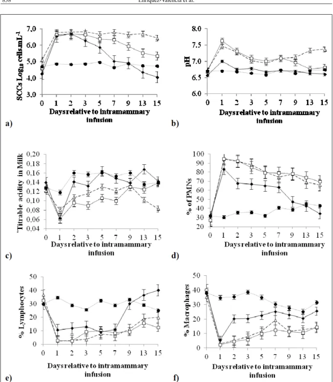
Twenty-four hours PI of the three biological substances, clinical signs of inflammation were observed, such as severe induration of udder with skin temperature changes and asymmetry of the teat. Clots were visible in the milk samples with the exception of milk from C quarters. Similarly, the SCCs increased significantly in milk samples from the W, W+W10b and W10b quarters (Figure 1a). The milk from the quarters inoculated with W+W10b
did not show a significant decrease in SCCs during the evaluation period (Figure 1a). The SCCs of milk from W+W10b quarters initially exhibited an
average value of Log10 6.8 cells mL
-1 at 24 hours PI. This value remained within the same order of
magnitude after 15 days (Log10 6.4 cells mL
-1). Milk from the quarters inoculated with W showed a significant decrease (P<0.001) in the SCCs from day 9 PI of the bacteria, finally reaching an average
of Log10 5.34 cells mL
-1 on day 15 (Figure 1a). Milk from the quarters inoculated with W10b also showed a significant decrease (P<0.001) in SCCs beginning
72 hours PI reaching a value of Log105.03 cells mL
-1
on the seventh day. Hence these quarters could be considered healthy and free from inflammation at this time. These results suggested that W10b was the treatment that had the least effect on the SCCs (Figure 1a). In milk samples from the C quarters, it was not detected significant increases in SCCs
throughout the entire evaluation period (Figure 1a).
These findings showed the initial innate immune response PI of W and W+W10b substances given that innate defense mechanisms that are activated quickly upon exposure to bacteria (AITKEN et al., 2011). The resident leukocytes together with epithelial cells initiate the inflammatory response via release of chemoattractants for the rapid recruitment of PMNs to the site of infection and consequently
the SCC increases (SOUZA et al., 2012).
Similar results to those found in this study were reported by BEECHER et al. (2009), who evaluated in detail the immune response following the infusion of live cultures of Lactococcus lactis
DPC 3147 into the mammary glands of cows. These authors noted that during the first 72 hours PI,
quarters where L. lactis was applied showed higher
levels of SCC than control quarters. According to these investigators, teats treated with L. lactis were
considered to be infection free by the seventh day PI, as
the average SCCs was Log10 5.3 cells mL
PI, the TA values increased significantly in milk samples from the W, W+W10b and W10b quarters and no significant differences were found between the TA of milk samples from quarters inoculated with W and milk samples from quarters inoculated with W+W10b. After 24 hours PI, there were also
no significant differences observed when the TA of milk samples from quarters inoculated with W10b was compared with the TA of milk from C quarters (Figure 1c). In both cases (pH and TA), the results showed that the biological substance W10b caused a different effect on the milk composition than the
Figure 1 - a) Somatic cell counts (SCCs Log10 cells mL
-1); b) Milk pH; c)*Milk titrable acidity (% lactic acid); d) percentage of polymorphonuclear
neutrophils (PMNs); e) percentage of lymphocytes and f) percentage of macrophages in milk following the application of (□) W. confusa, (Δ) W. confusa + metabolites, (♦) metabolites alone and in (●) control. Day zero corresponds to the analysis prior to the
application of the treatments. The bars indicate standard deviation of means. *Milk titrable acidity is the amount of NaOH needed to
biological substances W and W+W10b, with W10b being observed to induce an effect of shorter duration (up to 24 hours) on pH and TA when compared with W and W+W10b (Figure 1b and c).
The changes observed with respect to the pH and TA of milk caused by the infusion of W and W+W10b were associated with increases in SCCs. Milk composition is correlated with changes in the cellular response, possibly because PMNs exhibit different enzyme profiles than macrophages or perhaps because of a cellular metabolism reorientation (LE ROUX et al., 2003). The sequence of events that occurs in the udder during the inflammatory process involves an exchange of components between the blood and the milk, such as the migration of PMNs from the blood to the milk. On the other hand, as shown in the Figure 1b, it was found pHs above 7 PI of W and W+W10b. The pH of bovine milk is commonly stated to be between 6.6 and 6.8 at room
temperature (WALSTRA et al., 2006) and the pH
of mastitic milk is higher, up to pH 7.5 (SINGH et al., 1997). This increase in pH is presumably due to increased permeability of the mammary cells, with an
increase in [Na+] and [Cl-] and a reduction in lactose
content (SINGH et al., 1997).
As would be expected, the changes observed with respect to TA of milk could be influenced by the lactic acid production of W. confusa. According to SERNA-COCK et al. (2010) W. confusa grows
adequately in milk-based substrates producing up
13.12g L-1 of lactic acid. However, the lowest values of TA in milk were found at 24 hours PI of the three biological substances (Figure 1c). Given that, TA is the amount of NaOH needed to increase the pH of a given amount of milk to pH 8.4; these lowest values of TA are associated with highest values of pH (pHs>6.5) caused by changes in the cellular response as a consequence of the infusion of W. confusa and its metabolites.
The percentages of PMNs, lymphocytes and macrophages evaluated in the milk are shown in Figures 1d, 1e and 1f. At 24 hours PI of the three biological substances, significant increases in the percentage of PMNs (P<0.001) were found. In milk obtained following W10b treatment, the initially high levels of PMNs decreased significantly (P<0.001)
throughout the evaluation time (Figure 1d). With
respect to PMNs in milk from quarters inoculated with W+W10b and W, no significant differences (P>0.001) were observed during the evaluation period (Figure 1d). No significant increase of PMNs was observed in the milk samples of C quarters. It was observed the highest percentages of PMNs with lowest percentage of lymphocytes and macrophages in milk samples from the W, W+W10b and W10b quarters.
The augment in SCCs and PMNs 24 hours PI of W and W+W10b can be due to recognition of Pattern Recognition Receptors (PRRs). The PRRs recognize specific patterns of microbial components that are conserved among pathogens known as Pathogen-Associated Molecular Patterns (PAMPs) (SOUZA et al., 2012). The PRRs, such as Toll-Like Receptors (TLRs) recognize a wide range of PAMPs and initiate an inflammatory response with a rapid recruitment of PMNs to the mammary gland and consequently the SCC increases. This fact may also explain the lower effect on the SCCs in W10b than W and W+W10b treatment, once the SCC changes depend of bacterial recognition and infection
development (AITKEN et al., 2011).
Weissella (W) treatment showed in milk
a significant decrease in SCCs from day 9 PI of the bacteria; however, the percentage of PMNs did not show a significant decrease during the evaluation
time. According to MEHRZAD et al. (2005) and
MERLE et al. (2007), SCCs do not seem to describe completely the defense activities within the udder, which require the determination of functional parameters such as viability and activity of PMNs.
No significant variations in the percentage of PMNs, lymphocytes and macrophages were observed in the C treatment (Figures 1d, 1e and 1f). In the present study, each quarter of the mammary gland was used as an experimental unit based on the concept that the four quarters of the bovine mammary gland act as independent units because of the anatomical structure of the mammary gland. Nevertheless, there are indications that substantial interdependence exists
among quarters (BERRY & MEANEY, 2006) and
infection of one udder quarter possibly influences the cell activity of neighboring quarters (MERLE et al., 2007). Since it was not evaluated the cell activity, the interdependence was not entirely clear. Therefore, different cows should be used for each treatment in future experiments. In addition, the effect of W. confusa and its metabolites on the viability and
activity of PMNs should be evaluated.
CONCLUSION
The application of W. confusa and
its metabolites to the bovine udder resulted in elevated values of SCC in milk accompanied by high percentages of PMNs. More research on the mechanism of response and activation of PMNs in
the udder following application of W. confusaand its
desirable to inoculate bacteria that increase the SCCs.
The use of W. confusa would only be interesting
for dairy industry if this modulates mammary gland immune mechanisms like PMNs function without a substantive increase of PMNs that would impair milk quality. Thus, future studies should assess the viability and activity of PMNs PI of bacteria. In addition, it is also important to elucidate the ability of
W. confusato adhere to epithelial cells of bovine teat
canal, the capacity of the strain to persist in the udder and histopathological changes of the canal and cistern of the udder PI of W. confusa. All these are necessary
in order to find a possible use of W. confusa in a
nonantimicrobial formulation for the prevention and treatment of bovine mastitis.
ACKNOWLEDGEMENTS
The authors would like to express their appreciation to Banco Santander and the “VIRGINIA GUTIERREZ DE PINEDA” program for young researchers and innovators of Colciencias by financing this project.
REFERENCES
AITKEN, S.L. et al. Immunopathology of mastitis: Insights into
disease recognition and resolution. Journal of Mammary Gland Biology and Neoplasia, v.16, n.3, p.291-304, 2011. Available
from: <http://link.springer.com/article/10.1007/s10911-011-9230-4>. Accessed: Jun. 06, 2014. doi: 10.1007/s10911-011-9230-4.
BEECHER, C. et al. Administration of a live culture of
Lactococcus lactis DPC 3147 into the bovine mammary gland
stimulates the local host immune response, particularly IL-1β and
IL-8 gene expression. Journal of Dairy Research, v.76, n.3,
p.340-348, 2009. Available from: <http://dx.doi.org/10.1017/ S0022029909004154>. Accessed: Oct. 24, 2013. doi:10.1017/
S0022029909004154.
BERRY, D.P.; MEANEY, W.J. Interdependence and distribution of subclinical mastitis and intramammary infection among udder
quarters in dairy cattle. Preventive Veterinary Medicine, v.75,
n.1-2, p.81-91, 2006. Avaliable from: <http://www.sciencedirect. com/science/article/pii/S0167587706000420>. Accessed: Apr. 1, 2014. doi:10.1016/j.prevetmed.2006.02.001.
GARGOURI, A. et al. Total and differential bulk cow milk somatic cell counts and their relation with lipolysis. Livestock Science,
v.113, n.2-3, p.274-279, 2008. Available from: <http://www. sciencedirect.com/science/article/pii/S1871141307003502#>. Accessed: Oct. 24, 2013. doi: 10.1016/j.livsci.2007.05.007.
HOGEVEEN, H. et al. Economic aspects of mastitis: New
developments. New Zealand Veterinary Journal, v.59, n.1,
p.16-23, 2011. Available from: <http://www.tandfonline.com/doi/pdf/1 0.1080/00480169.2011.547165>. Accessed: Oct. 24, 2013. doi:10
.1080/00480169.2011.547165.
INSTITUTO COLOMBIANO DE NORMAS TÉCNICAS Y CERTIFICACIÓN. Productos de frutas y verduras:
Determinación de la acidez titulable (NTC 4623). Bogotá:
ICONTEC, 1999. 6p.
INTERNATIONAL COMMISSION ON MICROBIOLOGICAL
SPECIFICATIONS FOR FOODS (ICMSF). Microorganismos de los alimentos 1: su significado y métodos de enumeración.
2.ed. España: Acribia, 2000. 439p.
KULKARNI, A.G.; KALIWAL, B.B. Bovine mastitis: a review.
International Journal of Recent Scientific Research, v.4, n.5, p.543-548, 2013. Available from: <http://www.recentscientific.com/
sites/default/files/Download_492.pdf>. Accessed: Oct. 24, 2013.
LE ROUX, Y. et al. Polymorphonuclear proteolytic activity and
milk composition change. Veterinary Research, v.34, n.5,
p.629-645, 2003. Available from: <http://www.vetres.org/articles/vetres/ pdf/2003/05/V3502.pdf>. Accessed: Oct. 24, 2013. doi: 10.1051/
vetres:2003021.
LINDMARK-MǺNSSON, H. et al. Relationship between somatic
cell count, individual leukocyte populations and milk components
in bovine udder quarter milk. International Dairy Journal, v.16,
n.7, p.717-727, 2006. Available from: <http://www.sciencedirect. com/science/article/pii/S0958694605001433#>. Accessed: Oct. 24, 2013. doi:10.1016/j.idairyj.2005.07.003.
MATAMOROS, S. et al. Selection and evaluation of seafood-borne psychrotrophic lactic acid bacteria as inhibitors of pathogenic and spoilage bacteria. Food Microbiology, v.26, n.6, p.638-644, 2009.
Available: <http://www.sciencedirect.com/science/article/pii/ S074000200900104X>. Accessed: Oct. 24, 2013. doi:10.1016/j. fm.2009.04.011.
MERLE, R. et al. Cell function in the bovine mammary gland: a preliminary study on interdependence of healthy and infected
udder quarters. Journal of Dairy Research, v.74, n.2, p.174-179,
2007. Available from: <http://journals.cambridge.org/action/displ ayAbstract?fromPage=online&aid=1022180>. Accessed: Apr. 1,
2014. doi:10.1017/S002202990600238X F.
MEHRZAD, J. et al. High milk neutrophil chemiluminescence limits the severity of bovine coliform mastitis. Veterinary Research, v.36, n.1, p.101-116, 2005. Avaliable from: <http://
www.vetres.org/articles/vetres/pdf/2005/01/V4042.pdf>.
Accessed: Oct. 24, 2013. doi: 10.1051/vetres:2004055.
SHARIF, A.; MUHAMMAD, G. Somatic cell count as an indicator
of udder health status under modern dairy production. Pakistan Veterinary Journal, v.28, n.4, p.194-200, 2008. Available from:
<http://www.pvj.com.pk/pdf-files/28_4/194-200.pdf>. Accessed: Apr. 15, 2014. doi: pvj.com.pk/pdf-files/28_4/194-200.
SERNA-COCK, L. et al. Cellular response of the bovine mammary gland after Weissella confusainfusion to control Streptococcus agalactiae. Revista Colombiana de Ciencias Pecuarias, v.26,
n.4, p.280-287, 2013. Available from: <http://rccp.udea.edu.co/ index.php/ojs/article/view/907/1029>. Accessed: Apr. 08, 2014.
SERNA-COCK, L. et al. Effects of fermentation substrates and conservation methods on the viability and antimicrobial activity of Weissella confusaand its metabolites. Electronic Journal of Biotechnology,v.15, n.3, p.1-8, 2012. Available from: <http://
www.scielo.cl/pdf/ejb/v15n3/a06.pdf>. Accessed: Oct. 24, 2013. doi:10.2225/vol15-issue3-fulltext-9.
aureus y Streptococcus agalactiae. Revista Facultad de Ingeniería Universidad de Antioquia, v.55, p.55-65, 2010.
Available from: <http://www.scielo.org.co/pdf/rfiua/n55/n55a06. pdf>. Accessed: Oct. 24, 2013.
SINGH, H. et al. Physico-chemical properties of milk. In: Fox, P.F.
Advanced dairy chemistry volume 3Lactose, water, salts and vitamins. 2.ed. London: Chapman & Hall, 1997. Chap. 11, p.469-511.
SOUZA, F.N.D. et al. The innate immunity in bovine mastitis: the role of pattern-recognition receptors. American Journal of Immunology, v.8, n.4, p.166-178, 2012. Available from: <http://
thescipub.com/abstract/10.3844/ajisp.2012.166.178>. Accessed: Jun. 20, 2014. doi:10.3844/ajisp.2012.166.178.