Arq Bras Oftalmol. 2005;68(2):270-2
Tratamento de infecção corneana causado pelo Fonsecaea pedrosoi - Relato de caso
1Professora Titular do Departamento de Oftalmologia da Universidade Federal de São Paulo - UNIFESP. 2Professor de Microbiologia, Facultat de Medicina I
Ciencies de la Salut, Universitat Rovira I Virgili 43201-Reus and Institut d’Estudis Avançats - Chile. 3Livre Docente e Afiliada do Departamento de
Oftalmo-logia da UNIFESP. São Paulo (SP).
4Pós-graduando do Departamento de Microbiologia e Parasitologia da UNIFESP. São Paulo (SP). 5Professor de Microbiologia, Facultat de Medicina I
Ciencies de la Salut, Universitat Rovira I Virgili 43201-Reus and Institut d’Estudis Avançats - Chile. 6Doutora do Departamento de Oftalmologia da UNIFESP.
São Paulo (SP).
7Microbiology Unitat Clinic, University Austral de Chile - Chile.
8Médico Residente do Departamento de Oftalmologia da UNIFESP. São Paulo (SP).
Endereço para Correspondência: Ana Luisa Höfling-Lima - Av. Ibijaú, 331 - 17th andar - São Paulo (SP) CEP 04524-020
E-mail: anahofling@uol.com.br Recebido para publicação em 12.12.2003 Versão revisada recebida em 21.07.2004 Aprovação em 23.07.2004
Ana Luisa Höfling-Lima1 Josep Guarro2
Denise de Freitas3 Patricio Godoy4 Josepa Gené5
Luciene Barbosa de Souza6 Luis Zaror7
Andre C. Romano8
Clinical treatment of corneal infection due to Fonsecaea
pedrosoi - Case report
To report an unusual case of fungus keratitis due to Fonsecaea pedrosoi
that developed after corneal trauma. Case report: A 18-year-old male presented with a corneal ulcer in the right eye, 28 days after a trauma with glass fragments. Corneal scrapings were collected for smears and culture. Dematiaceous hyphae were seen on wet mounts of the scrapings and dark pigmented colonies grew repetitively on the culture media; all colonies were identical, and were subsequently identified as Fonsecaea pedrosoi. Treatment was initiated with topical natamycin at one hour intervals, 200 mg oral ketoconazole per day and later changed to a combination of 200 mg ketoconazole and amphotericin B. In humid tropical regions
Fonsecaea pedrosoi is one of the primary causes of human chronic cutaneous mycosis, chromoblastomycosis. Combination of systemic and topical antifungal medications may provide the best option for cure in corneal chromoblastomycosis.
ABSTRACT
Keywords: Corneal diseases/microbiology; Keratitis/microbiology; Eye injuries/complications; Eye infections, fungal; Antifungal agents/therapeutic use; Mitosporic fungi; Case report
INTRODUCTION
Fonsecaea pedrosoi is the commonest causative agent of chromoblas-tomycosis(1), a chronic mycotic cutaneous and subcutaneous skin
infec-tion, which primarily occurs in humid tropical regions.
Treatment of the mycosis caused by this agent is unrewarding not only because of the scarcity of effective antifungals but also due to the need for prolonged periods of treatment, which in some reports has required prolon-ged therapeutic regimens of up to 2 years to obtain a mycologic cure(2).
Several studies have indicated, by minimal inhibitory concentration (MIC) values, that itraconazole presents no resistance to Fonsecaea pedrosoi(3).
In the ophthalmic literature, only one previous case of corneal chromo-blastomycosis caused by Fonsecaea pedrosoi was reported(4). In the
mana-gement of that case, two penetrating keratoplasties (PK) were performed, and itraconazole was introduced as the first line option after the first PK and continued for 6 months after second PK when the eye became quiet and the infection controlled. This result correlates with the clinical experience in medical reports, which show that 10% of chromoblastomycosis patients are not cured in spite of extended courses (20 months) of itraconazole therapy(2).
Arq Bras Oftalmol. 2005;68(2):270-2 Clinical treatment of corneal infection due to Fonsecaea pedrosoi - Case report 271
tival flap. The second case (Phialofora verrucosa) was trea-ted with keratectomy, conjunctival flap, topical natamycin and a penetrating keratoplasty led to a final visual acuity of 20/60. The third case (Phialofora bubakii) was treated with topical 1% fluocytosine.
CASE REPORT
An 18-year-old male was referred to the corneal service in August 2001 with a corneal ulcer of the right eye 28 days after glass trauma to the eye. The patient had been treated with neomycin, ciprofloxacin, and 0.1% dexamethasone for a week without improvement. His distance visual acuity was 20/400 in the right eye and 20/30 in the left at presentation. Intraocular pressure measured by bimanual examination was normal. There was a 3 x 2 mm diameter central epithelial defect with feathery borders. The anterior segment showed a mild inflam-matory reaction with a few keratic precipitates under the corneal infiltrate. The posterior segment was quiet.
Corneal scrapings were collected for fungus and bacteriolo-gic microscopic examination and culture. Potassium hydroxide mounts and smears with Gram and Giemsa stains were made. Scrapings were inoculated directly onto Sabouraud glucose agar (DIFCO) with chloramphenicol (200 mg/L) and blood agar. Gram staining showed filamentous structures on the next day. Neomycin, ciprofloxacin, and 1% dexamethasone were dis-continued and topical natamycin (5% pimaricin) was initiated hourly along with 200 mg per day oral systemic ketoconazole. A good reepithelialization of the ulcer was observed 10 days later and natamycin treatment was decreased to application every 2 hours. No rebound inflammatory reaction was noted after dexamethasone was suspended. Two days later, recurren-ce of the infection was noted and natamycin was restored to every 2 hours. On the next day, dematiaceous hyphae were seen on wet mounts of the scrapings and dark pigmented colonies grew on the culture media; all colonies were identical and were subsequently identified as Fonsecaea pedrosoi.
Natamycin therapy was discontinued and treatment with topical amphotericin B (0.5%) was initiated every hour along with the oral ketoconazole. After 100 days the treatment was discontinued because clinical healing occurred. A small cen-tral stromal leukoma remained and the final distance visual acuity was 20/200. No relapse of infection was observed three months after the cessation of therapy.
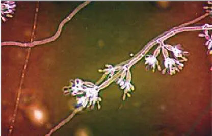
The fungus was cultured on oatmeal agar and showed the typical features of Fonsecaea pedrosoi, i.e. spreading, lanose and olivaceous-green colonies with a blackish reverse. Cultures also showed the two typical morphologic forms(1): i) light brown
conidiophores, profusely branched with cylindrical, intercalary or terminal conidiogenous cells with clusters of prominent den-ticles and ii) the Phialophora synanamorph, characterized by ampulliform, darker phialides with conspicuous funnel-shaped collarettes. The conidia were smooth-walled, clavate, pale oliva-ceous and measured 3.5 - 5 x 1.5 - 2 µm (Figure 1).
COMMENTARY
A combination of two antimycotic agents was prescribed to treat this reported case: ketoconazole and amphotericin B. This treatment have led to a quiet state of the infection and apparently cessation of fungal penetration and no relapse of this infection was eminent after termination of therapy. This result might be explained by the MIC levels being lower for amphotericin B than for natamycin in the afore mentioned case report, and ketoconazole being less resistant in our sensitivity testing (see Table 1).
Although in vitro susceptibility testing does not reliably predict in vivo efficacy for fungi, and MICs can not be used as strict guidelines for therapy, the possibility of performing in vitro tests should be considered. In that way, one may avoid ineffective, costly and a prolonged course of treatment. In view of the lack of susceptibility of this agent to the current antimycotic drugs, it may be wiser to seriously consider the possibility of combining the best available drugs in order to obtain a synergistic effect.
The authors open the possibility of clinical treatment for
Fonsecaea pedrosoi corneal infection and discuss the impor-tance of determination of MIC levels for antifungal agents.
Table 1. Common antifungal medication sensitivities to Fonsecaea pedrosoi ocular isolate
Antifungal drug MIC (µg/ml)
Amphotericin B 10.0
Flucitosine 80.0
Itraconazole 0.25
Ketoconazole 0.03
Voriconazole 0.03
Sensitivity testing was performed according to the guidelines of the National
Committee for Clinical Laboratory Standards (NCCLS) for molds5, using RPMI
1640 medium buffered to pH7 with 0.165 M morpholinepropanesulfonic acid
(MOPS), an inoculum of 1.5x10⁶ CFU/ml, an incubation temperature of 30°C,
an incubation time of 72h, and an additive drug dilutions regimen. MIC: minimal inhibitory concentration
Arq Bras Oftalmol. 2005;68(2):270-2
272Clinical treatment of corneal infection due to Fonsecaea pedrosoi - Case report
Further studies should be performed to determine the value of combined topical and systemic clinical treatment and also additional surgical treatment.
ACKNOWLEDGEMENTS
The authors thank Maria Cecilia Zorat Yu for her help in the laboratory investigation.
RESUMO
Relato de um caso atípico de infecção fúngica da córnea causada pelo microrganismo Fonsecaea pedrosoi após trau-ma ocular. Paciente, trau-masculino, estudante de 18 anos, apre-sentou-se ao Setor de Doenças Externas Oculares do Departa-mento de Oftalmologia da UNIFESP com úlcera de córnea paracentral de 3,5 x 3,5 mm e aspecto branco-acinzentado com bordas infiltradas, 28 dias após trauma em ocular por vidro. Foi realizado raspado da córnea e o material enviado para análise microbiológica. Foi observado crescimento de colô-nias em meio de cultura e posteriormente colocadas em solu-ção de lactofenol-azul de algodão. Verificou-se a presença de hifas dermáceas de pigmento escuro, identificado como Fon-secaea pedrosoi. Tratamento foi iniciado com natamicina 5%
tópica a cada hora e cetoconazol 200 mg por dia. Subseqüente-mente foi substituído pela combinação cetoconazol e anfoteri-cina B. Fonsecaea pedrosoi é uma das principais causas em humanos de micose crônica cutânea, cromoblastomicose, em regiões úmidas tropicais. A combinação de antimicóticos sis-têmicos e tópicos pode ser a melhor opção para pacientes no tratamento de cromoblastomicose da córnea.
Descritores: Doenças da córnea/microbiologia; Ceratite/mi-crobiologia; Traumatismos oculares/complicações; Infecções oculares fúngicas; Antimicóticos/uso terapêutico; Fungos mitospóricos; Relato de caso
REFERENCES
1. Hoog GS, Guarro J. Gené J, Figueras MJ. Atlas of clinical of fungi. Baarn, The Netherlands; Centraalbureau Voor Schimmelultures - Universitat, Rovira i Virgili, 2000.
2. Esterre P, Andriantsimhavandy A, Ramarcel ER, Pecarrere JL. Forty years of chromoblastomycosis in Madagascar: a review. Am J Trop Med Hyg. 1996; 55(1):45-7.
3. Bedout C de, Gomez BL, Restrepo A. In vitro susceptibility testing of Fon-secaea pedrosoi to antifungals. Rev Inst Med Trop Sao Paulo. 1997;39(3):145-8. 4. Barton K, Miller D, Pflugfelder SC. Corneal chromoblastomycosis. Cornea.
1997;16(2):235-9.