Correspondence to: Dominik Faust, Asklepios Hospital, Department of Medicine, Bad Muender, Germany.
C A S E R E P O R T S UDC: 616.36-008.6-08
Ursodeoxycholic acid for treatment of cholestasis in patients with
hepatic amyloidosis
Ursodeoksiholna kiselina za le
č
enje holestaze kod bolesnika
sa amiloidozom jetre
Dominik Faust*, Bora Akoglu†, Gordana Ristic†, Vladan Milovic†
Asklepios Hospital, *Department of Medicine, Langen, Germany; MediClin Deister Weser Hospital, †Department of Gastroenterology and Oncology, Bad Muender, Germany
Abstract
Background. Amyloidosis represents a group of different dis-eases characterized by extracellular accumulation of pathologic fibrillar proteins in various tissues and organs. Severe amyloid deposition in the liver parenchyma has extrahepatic involve-ment predominantly in the kidney or heart. We evaluated the effect of ursodeoxycholic acid, in four patients with severe he-patic amyloidosis of different etiologies, who presented with increased alkaline phosphatase and γ-glutamyl transferase. Case report. The study included four patients who presented with amyloidosis-associated intrahepatic cholestasis. Three of them had renal amyloidosis which developed 1–3 years before chole-stasis occurred, the remaining one having intrahepatic cholesta-sis as the primary sign of the disease. Amyloidocholesta-sis was identi-fied from liver biopsies in all patients by its specific binding to Congo red and green birefringence in polarized light. The bio-chemical nature and the class of amyloid deposits were identi-fied immunohistochemically. In addition to their regular treat-ment, the patients received 750 mg ursodeoxycholic acid per day. After 2–4 weeks all patients had a significant decrease of serum alkaline phosphatase and γ-glutamyl transferase, and their general status significantly improved. Conclusion. Treatment with ursodeoxycholic acid may be beneficial in pa-tients with hepatic amyloidosis, and do extend indications for the use of ursodeoxycholic acid in amyloidotic cholestatic liver disease.
Key words:
amyloidosis; cholestasis; biopsy;
immunohistochemistry; deoxycholic acid.
Apstrakt
Uvod. Amiloidoza predstavlja grupu različitih oboljenja koju karakteriše vanćelijsko nakupljanje patoloških fibrilnih proteina u različtim tkivima i organima. Značajno nakuplja-nje depozita u parenhimu jetre prati ekstrahepatična
zahva-ćenost pre svega bubrega ili srca. Ispitivan je efekat ursode-oksiholne kiseline kod četiri bolesnika sa amiloidozom jetre različite etiologije koji su primljeni zbog povišenja alkalne fosfataze i gama glutamil transferaze. Prikaz bolesnika. Opisali smo četiri bolesnika sa intrahepatičkom holestazom udruženom sa amiloidozom. Tri od četiri bolesnika imala su amioloidozu bubrega koja se razvila 1–3 godine pre pojave holestaze, a jedan bolesnik holestazu kao prvi znak bolesti. Amiloidoza je dokazana iz biopsija jetre nakon specifičnog bojenja Kongo crvenim. Biološka priroda i klasa amiloida ispitana je imunohistohemijski. Zajedno sa njihovom
uobi-čajenom terapijom, bolesnici su lečeni ursodeoksiholnom kiselinom, 750 mg dnevno. Posle 2–4 nedelje kod svih bole-snika dokazan je značajni pad alkalne fosfataze i gama glu-tamil transferaze, i njihovo opšte stanje značajno se popra-vilo. Zaključak. Terapija ursodeoksiholnom kiselinom us-pešna je kod bolesnika sa amiloidozom jetre, čime se proši-ruje indikacija za davanje ursodeoksiholne kiseline kod bole-snika sa holestazom koja nastaje kao posledica jetrene ami-loidoze.
Ključne reči:
amiloidoza; holestaza; biopsija; imunohistohemija; dezoksiholna kiselina.
Introduction
Amyloidosis represents a group of different diseases characterized by extracellular accumulation of pathologic fibrillar proteins (called amyloid on the basis of special tinctorial and optical properties) in various tissues and
involvement predominantly in the kidney (47%) or heart (42%) 2. Regarding poor median survival rate of patients with severe hepatic amyloidosis and the failure of existing treatment options to improve survival and relieve cholestasis, novel therapeutical approaches are obviously needed.
When used to dissolve gallstones in patients with chronic active hepatitis, the dihydroxylated bile acid, urso-deoxycholic acid, improved both serum transaminases and cholestasis-indicating enzymes 3. Ursodeoxycholic acid has also been used in patients with cystic fibrosis, and is the treatment of choice in primary biliary cirrhosis 4–6. In the present study, we administered ursodeoxycholic acid to four patients with cholestasis due to amyloidosis with hepatic in-volvement. Rapid improvement of cholestasis after the treatment was initiated, recurrence of cholestasis after urso-deoxycholic acid was temporarily discontinued and repeated improvement after the drug was reintroduced, suggest that ursodeoxycholic acid may be an efficient therapy in patients with cholestasis due to hepatic amyloidosis.
Case report
The study included four patients who presented with amyloidosis-associated intrahepatic cholestasis (Table 1). Three of the four patients had renal amyloidosis which de-veloped 1–3 years before cholestasis occurred. The patient 1 was a 44-year-old Caucasian female with immunoglobu-lin-light-chain-λ-related (ALλ) amyloidosis, while two males, patient 2 (65-year-old Caucasian) and patient 3 (40-year-old African), had amyloid protein A (AA) amyloido-sis. In contrast, the patient 4 (70-years-old Caucasian male)
had no overt renal amyloidosis, and intrahepatic cholestasis was the primary sign of the disease. Familial Mediterranean fever was the cause of amyloidosis in one of two patients with AA amyloidosis (patient 2). In patients 1, 3 and 4 no cause could initially be identified, and, in particular, chronic inflammatory bowel disease and Mediterranean fe-ver were ruled out.
Two out of four patients (patients 1 and 2) complained to pruritus as the only cholestasis-related symptom. Pa-tients 1 and 2 were in the end-stage renal disease and un-dergoing hemodialysis. They were both receiving ACE in-hibitors and calcium antagonists to treat renal hypertension. The patient 3 had moderately impaired renal function with-out indications for hemodialysis, and was receiving no medication. The patient 4 had coronary heart disease and, as regular therapy, was receiving acetylsalicylic acid and metoprolol daily.
Laboratory data are summarized in Tables 1 and 2. Ex-trahepatic cholestasis was excluded in all patients by ultra-sound. Magnetic resonance cholangiopancreatography was additionally done in Patient 3, confirming normal morphol-ogy of intra- and extrahepatic bile ducts.
Liver biopsies were performed in all patients and evalu-ated separately by two pathologists. They revealed severe capillary amyloid deposits along the sinusoids and in the walls of hepatic arteries, and were diagnostic for amyloido-sis, according to the criteria described before 7. Amyloid was identified at light microscopy by its specific binding to Congo red and its green birefringence in polarized light. The biochemical nature and the class of amyloid deposits were identified immunohistochemically, as previously described 8.
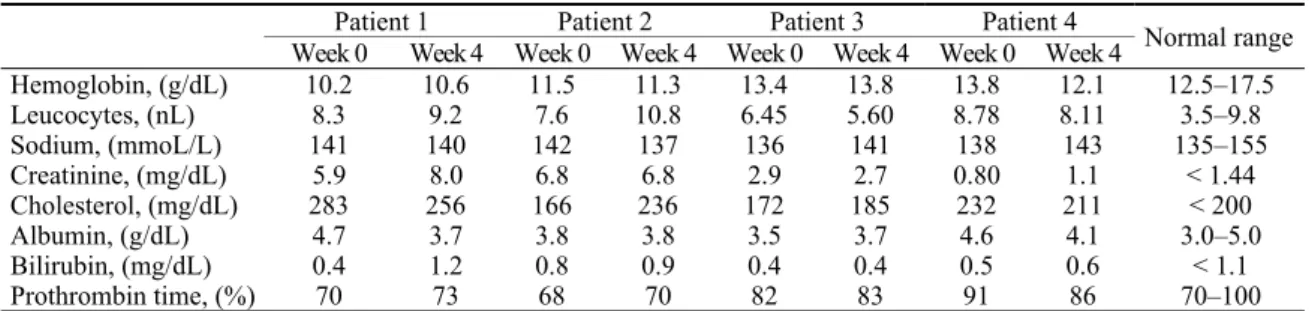
Table 1 Survey of blood cell count, clinical chemistry and clotting tests before and after four weeks of treatment
with ursodeoxycholic acid (750 mg per day)
Patient 1 Patient 2 Patient 3 Patient 4
Week 0 Week 4 Week 0 Week 4 Week 0 Week 4 Week 0 Week 4 Normal range
Hemoglobin, (g/dL) 10.2 10.6 11.5 11.3 13.4 13.8 13.8 12.1 12.5–17.5
Leucocytes, (nL) 8.3 9.2 7.6 10.8 6.45 5.60 8.78 8.11 3.5–9.8
Sodium, (mmoL/L) 141 140 142 137 136 141 138 143 135–155
Creatinine, (mg/dL) 5.9 8.0 6.8 6.8 2.9 2.7 0.80 1.1 < 1.44
Cholesterol, (mg/dL) 283 256 166 236 172 185 232 211 < 200
Albumin, (g/dL) 4.7 3.7 3.8 3.8 3.5 3.7 4.6 4.1 3.0–5.0
Bilirubin, (mg/dL) 0.4 1.2 0.8 0.9 0.4 0.4 0.5 0.6 < 1.1
Prothrombin time, (%) 70 73 68 70 82 83 91 86 70–100
Patient 1: 44-year-old Caucasian female with AL-lambda amyloidosis; patient 2: 65-year-old Caucasian male with AA amyloidosis and familial Mediterranean fever; patient 3: 40-year-old African male with AA amyloidosis but without clinical features of chronic inflammatory disease; patient 4: 70-years-old Caucasian male, with hereditary ApoAI-amyloidosis
Table 2 Immunological markers of patients studied
Patient 1 Patient 2 Patient 3 Patient 4 Normal values
ANA negative negative negative negative negative
AMA negative negative negative negative negative
ANCA (screen) negative negative negative negative negative
IgG (g/dL) 0.85 1.32 1.07 1.48 0.8–1.8
IgA (mg/dL) 0.22 0.35 0.29 0.31 0.1–0.45
IgM (mg/dL) 0.01 0.12 0.20 0.19 0.06–0.26
Liver biopsies of all four patients revealed amyloid deposits which were positive to Congo red staining (Figure 1). Fur-ther molecular characterization of the type of amyloidosis showed that the patient 4 suffered from hereditary ApoAI-amyloidosis, a rare disorder characterized by a mutation in the gene for apolipoprotein AI 9.
Fig. 1– Liver biopsy of patient 3. The other two patients exhibited identical findings. In the vicinity of a portal tract, extensive extracellular amyloid deposits are seen in all
sinu-soids (arrow), with concomitant finding of atrophic hepatocytes (Congo red staining, magnification 400).
Prior to the initiation of the treatment, informed consent was obtained from each patient and the study protocol (con-forming to the ethical guidelines of the 1975 Declaration of Helsinki) was approved by the Ethics Committee of the Jo-hann Wolfgang Goethe University School of Medicine in Frankfurt.
Independent from body weight, 750 mg ursodeoxy-cholic acid per day (Ursofalk®, Dr Falk Pharma GmbH, Freiburg, Germany) divided into three daily doses were ad-ministered to all patients.
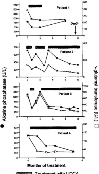
After 2 to 4 weeks of treatment, a significant decrease in serum alkaline phosphatase and gamma-glutamyl transfer-ase levels in comparison to pretreatment values was observed in all patients, while other liver function tests remained un-changed (Figure 2). Patients 1 and 2 also reported a signifi-cant improvement of their pruritus. There was no change in the liver size on follow up (ultrasound) and renal function tests remained unchanged. Four weeks after the treatment with ursodeoxycholic acid was started the patients were dis-charged from our outpatients clinic and were treated by their general practitioners, with regular (1–3 months) follow-up visits to us. Three of the patients (patients 1–3), however, interrupted ursodeoxycholic acid treatment only a few weeks after being discharged, since their family physicians decided, due to high costs, not to prescribe the drug any longer. In the patient 4 alkaline phosphatase decreased to normal (100 U/L), and in patients 2 and 4 gamma-glytamyl transferase fell to one third of the pretreatment values.
The patients 1–3 were seen again in our clinic after ur-sodeoxycholic acid treatment was discontinued; in all three,
cholestatic parameters again markedly increased (Figure 2). Reintroducing ursodeoxycholic acid (750 mg per day) again resulted in a decrease in serum alkaline phosphatase and gamma-glutamyl transferase levels as early as one week after the treatment was reintroduced. Again, there was no change in other liver function tests.
Fig. 2 – Decrease in serum alkaline phosphatase and gamma-glutamyl transferase during treatment with ursodeoxycholic acid (UDCA; 750 mg per day) and their reversal after the treatment was
temporarily discontinued. Patient 1: 44-year-old Caucasian female with AL-lambda amyloidosis; patient 2: 65-year-old Caucasian male with AA amyloidosis and familial Mediterranean fever;
pati-ent 3: 40-year-old African male with AA amyloidosis but without clinical features of chronic inflammatory disease; patient 4: 70-years-old Caucasian male, with hereditary ApoAI-amyloidosis.
(normal range: AP < 180 U/L, γ-GT < 28 U/L).
again interrupted ursodeoxycholic acid therapy after a treat-ment period of six weeks; alkaline phosphatase and gamma-glutamyl transferase again increased, and returned to previ-ous values after the treatment was reintroduced. The patient 4 was taking ursodeoxycholic acid without any interruption for almost six months, and, on the last follow-up, had nearly normalized parameters of cholestasis (Figure 2).
Discussion
Here we describe four patients with hepatic amylodosis and cholestasis, who were successfully treated with ursode-oxycholic acid and stabilized their laboratory parameters of cholestasis within weeks after the initiation of therapy. The effect of ursodeoxycholic acid was specific, since temporary discontinuation of the drug resulted in a recurrent increase in alkaline phosphatase and gamma-glutamyl transferase, which again returned to nearly normal values after ursodeoxycholic acid was reintroduced. Among our four patients, however, one patient died during the course of treatment due to heart failure caused by amyloidosis of the heart.
A relatively small series of patients with liver amyloi-dosis available for this study at present does not allow any conclusion whether treatment with ursodeoxycholic acid can influence survival. If any analogy between the two chole-static diseases can be helpful, in patients with primary biliary cirrhosis it has been shown that ursodeoxycholic acid delays the need for transplantation, while the posttransplantation outcome of ursodeoxycholic acid-treated patients is not dif-ferent from those who were administered placebo 10, 11.
Chronic cholestatic liver diseases are characterized by impaired bile flow, caused by different mechanisms and cell structures, like the bile salt dependent and independent bile flow, different export pumps, ATP, glutathione and the
cyto-skeleton 12–14. Amyloidosis with liver involvement is rela-tively rare in comparison to amyloidotic disease of the kid-neys, lung and heart, is characterized by the accumulation of amyloid fibrilis in the liver parenchyma and ultimately may result in chronic intrahepatic cholestasis 15. Amyloid is de-posited in the parenchyma and in the wall of blood vessels in the liver, as well as around the bile canaliculi.
Experimental evidence suggests that, in principle, ur-sodeoxycholic acid acts at least on two major levels in re-lieving cholestasis in man: it protects cholangiocytes against cytotoxic effects of hydrophobic bile acids and bile acid–induced apoptosis, and it stimulates hepatobiliary se-cretion 16. On cellular level, ursodeoxycholic acid stimu-lates ATP secretion in the liver, mobilizes intracellular cal-cium and activates phospholipase A, induces a pleiotropic metabolic response in the hepatocyte by activating protein kinase C, inserts bile acid transporters in the apical pole of the hepatocyte canalicular membrane, and stabilizes the hepatocyte membranes and liver mitochondria 17–23. In pa-tients with biliary liver diseases it has also been suggested that ursodeoxycholic acid acts primarily within the bile ca-nalicular lumen, by preventing disruption of the plasma membrane of bile duct epithelial cells by hydrophobic bile acids 24. Whether one or all of these mechanisms lie be-neath the described anti-cholestatic effect of ursodeoxy-cholic acid in patients with severe liver amyloidosis, still remains to be seen.
Conclusion
Our data imply that ursodeoxycholic acid should be used to treat cholestasis in patients with liver amyloidosis. Further clinical studies at a larger patient group are obvi-ously needed to assess this promising conclusion.
R E F E R E N C E S
1. Glenner GG. Amyloid deposits and amyloidosis: the beta-fibrilloses (second of two parts). N Engl J Med 1980; 302(24): 1333–43.
2. Gertz MA, Kyle RA. Hepatic amyloidosis: clinical appraisal in 77 patients. Hepatology 1997; 25(1): 118–21.
3. Leuschner U, Leuschner M, Sieratzki J, Kurtz W, Hübner K. Gall-stone dissolution with ursodeoxycholic acid in patients with chronic active hepatitis and two years follow-up. A pilot study. Dig Dis Sci 1985; 30(7): 642–9.
4. Leuschner U, Fischer H, Kurtz W, Güldütuna S, Hübner K, Hellstern A, et al. Ursodeoxycholic acid in primary biliary cirrhosis: re-sults of a controlled double-blind trial. Gastroenterology 1989; 97(5): 1268–74.
5. Poupon RE, Lindor KD, Cauch-Dudek K, Dickson ER, Poupon R, Heathcote EJ. Combined analysis of randomized controlled tri-als of ursodeoxycholic acid in primary biliary cirrhosis. Gastro-enterology 1997; 113(3): 884–90.
6. Heathcote EJ. Management of primary biliary cirrhosis. The American Association for the Study of Liver Diseases practice guidelines. Hepatology 2000; 31(4): 1005–13.
7. Sasaki M, Nakanuma Y, Terada T, Hoso M, Saito K, Hayashi M, et al. Amyloid deposition in intrahepatic large bile ducts and peribiliary glands in systemic amyloidosis. Hepatology 1990; 12(4 Pt 1): 743–6.
8. Linke RP. Highly sensitive diagnosis of amyloid and various amyloid syndromes using Congo red fluorescence. Virchows Arch 2000; 436(5): 439–48.
9. Soutar AK, Hawkins PN, Vigushin DM, Tennent GA, Booth SE, Hutton T, et al. Apolipoprotein AI mutation Arg-60 causes autosomal dominant amyloidosis. Proc Natl Acad Sci U S A 1992; 89(16): 7389–93.
10.Lazaridis KN, Gores GJ, Lindor KD. Ursodeoxycholic acid 'mechanisms of action and clinical use in hepatobiliary disor-ders'. J Hepatol 2001; 35(1): 134–46.
11.Heathcote EJ, Stone J, Cauch-Dudek K, Poupon R, Chazouilleres O, Lindor KD, et al. Effect of pretransplantation ursodeoxycholic acid therapy on the outcome of liver transplantation in patients with primary biliary cirrhosis. Liver Transpl Surg 1999; 5(4): 269–74.
12.Poupon R, Chazouillères O, Poupon RE. Chronic cholestatic dis-eases. J Hepatol 2000; 32(1 Suppl): 129–40.
13.Trauner M, Meier PJ, Boyer JL. Molecular pathogenesis of chole-stasis. N Engl J Med 1998; 339(17): 1217–27.
14.Meier PJ. Molecular mechanisms of hepatic bile salt transport from sinusoidal blood into bile. Am J Physiol 1995; 269(6 Pt 1): G801–12.
16.Paumgartner G, Beuers U. Ursodeoxycholic acid in cholestatic liver disease: mechanisms of action and therapeutic use revis-ited. Hepatology 2002; 36(3): 525–31.
17.Nathanson MH, Burgstahler AD, Masyuk A, Larusso NF. Stimu-lation of ATP secretion in the liver by therapeutic bile acids. Biochem J 2001; 358(Pt 1): 1–5.
18.Bouscarel B, Fromm H, Nussbaum R. Ursodeoxycholate mobilizes intracellular Ca2+ and activates phosphorylase a in isolated hepatocytes. Am J Physiol 1993; 264(2 Pt 1): G243–51. 19.Beuers U, Throckmorton DC, Anderson MS, Isales CM, Thasler W,
Kullak-Ublick GA, et al. Tauroursodeoxycholic acid activates protein kinase C in isolated rat hepatocytes. Gastroenterology 199; 110(5): 1553–63.
20.Alpini G, Baiocchi L, Glaser S, Ueno Y, Marzioni M, Francis H, et al.Ursodeoxycholate and tauroursodeoxycholate inhibit cho-langiocyte growth and secretion of BDL rats through activa-tion of PKC alpha. Hepatology 2002; 35(5): 1041–52.
21.Beuers U, Bilzer M, Chittattu A, Kullak-Ublick GA, Keppler D, Paumgartner G, et al. Tauroursodeoxycholic acid inserts the
api-cal conjugate export pump, Mrp2, into canalicular membranes and stimulates organic anion secretion by protein kinase C-dependent mechanisms in cholestatic rat liver. Hepatology 2001; 33(5): 1206–16.
22.Güldütuna S, Zimmer G, Imhof M, Bhatti S, You T, Leuschner U. Molecular aspects of membrane stabilization by ursodeoxy-cholate. Gastroenterology 1993; 104(6): 1736–44.
23.Güldütuna S, Deisinger B, Weiss A, Freisleben HJ, Zimmer G, Sipos P, et al. Ursodeoxycholate stabilizes phospholipid-rich mem-branes and mimics the effect of cholesterol: investigations on large unilamellar vesicles. Biochim Biophys Acta 1997; 1326(2): 265–74.
24.Heuman DM, Mills AS, McCall J, Hylemon PB, Pandak WM, Vlahcevic ZR. Conjugates of ursodeoxycholate protect against cholestasis and hepatocellular necrosis caused by more hydro-phobic bile salts. In vivo studies in the rat. Gastroenterology 1991; 100(1): 203–11.