ZDB-Number: 2668735-5
IC Journal No: 8192
Volume 1 Issue 6
Online Available at www.phytojournal.com
Journal of Pharmacognosy and Phytochemistry
Vol. 1 No. 6 2013 www.phytojournal.com Page | 42
Phytosteroids from tissue culture of
Allium cepa
L. and
Trachyspermum ammi
S prague.
Pratibha Chaturvedi1*,Pushpa Khanna2 and Abhay Chowdhary1
1. Haffkine Institute For Training, Research and Testing, Parel, Mumbai 400012, India [E-mail: pratibha1.c@gmail.com]
2. Rajasthan University, Jaipur, India
Production of secondary metabolites by cultured cells provides a particularly important benefit to manipulate and improve the production of desired compounds; thus biotechnological approaches to increase the concentrations of the metabolites are discussed. Present study deals with the production, isolation and identification of phytosterols from tissue culture of Allium cepa and from plant parts and tissue culture of Trachyspermum ammi. Steroidal analysis of plant parts showed the maximum amount of stigmasterol (0.240 mg/gdw) which was comparatively little
less than that of the amount of β- sitosterol (0.295 mg/gdw) in the seeds of T. ammi . The maximum amount of stigmasterol was present in four weeks old tissue of T. ammi (0.249 mg/gdw) whereas the highest content of β- sitosterol was observed in six weeks old tissue of A. cepa (0.315 mg/gdw) However, lanosterol, was present only in the tissue of A. cepa which was maximum in six weeks old tissue (0.039 mg/gdw)
Keyword:Allium cepa, Liliaceae, Trachyspermum ammi Umbellifereae, Stigmasterol, Beta sitostewrol
1. Introduction
Phytosterols (referred to as plant sterol and stanol esters) are a group of naturally occurring compounds found in plant cell membranes. Because phytosterols are structurally similar to the body’s cholesterol, when they are consumed they compete with cholesterol for absorption in the digestive system. As a result, cholesterol absorption is blocked, and blood cholesterol levels reduced. Throughout much of human evolution, it is likely that large amounts of plant foods were consumed. In addition to being rich in fiber and plant protein, the diets of our ancestors were also rich in phytosterols—plant-derived sterols that are similar in structure and function to cholesterol. There is increasing evidence that the reintroduction of plant foods providing
phytosterols into the modern diet can improve serum lipid (cholesterol) profiles and reduce the risk of cardiovascular disease.
In the present study the callus of Allium cepa
(Liliaceae) and Trachyspermum ammi
(Umbellifereae) were successfully maintained in the laboratory and evaluated for their phytosterol content which is not well documented for these plants.
2. Material and Methods
Vol. 1 No. 6 2013 www.phytojournal.com Page | 43
raised from the seedlings, grown and maintained by frequent subculturings of 6-8 weeks for twenty four months on fresh RT medium. The growth indices were calculated at the transfer age of the
tissue at two, four, six and eight weeks. Three replicates in each case were examined and mean values taken into consideration.
Fig 1-2: Showing the seed germination and developed callus of Allium cepa L.
Fig 3-4: Showing the seed germination and developed callus of Trachyspermum ammi. Sprague
S. No. Age in weeks Growth Index
A.cepa T. ammi
1
2
3
4
2
4
6
8
0.76 0.91
1.5 1.4
2.1 2.7
1.7 1.8
Table 1. Showing the growth index of A.cepa and T . ammi
2.1 Extraction Procedure
Each of the plant parts (seeds, stem and leaves) as well as the various tissue samples (2,4,6 and 8 weeks old)were dried, powdered, weighed and then separately subjected to soxhlet extraction in petroleum ether for 24 hr on a water bath for removing fatty acids. Each of the mixture was filtered and residual tissue masses were
hydrolysed with 15% ethanolic hydrochloric acid (w/v) for 4 hr (Tomita et al., 1970)Each of the hydrolysed samples was filtered, the filtrate extracted in ethyl acetate separately and given continuous washings of distilled water till the pH was 7. The extract was passed over Na2SO4, for
Vol. 1 No. 6 2013 www.phytojournal.com Page | 44
2.2 Qualitative Analysis
a. Thin-layer Chromatography (TLC)
Each of the extracts was applied on silica gel G coated and activated glass plates along with the standard samples of sterols (cholesterol, lanosterol, stigmasterol and B-sitosterol) The glass plates were then developed in an organic solvent misture of Hexane and acetone (80:20: Fazil and Hardman, 1968) and air dried. On spraying the developed plates with 50% H2SO4
three spots corresponging. to those of the standard of these of the standard samples and a stigmasterol (Rf 0.91; color grey) lanosterol (Rf 0.89; color, brown) and B-sitosterol (Rf. 0.85; color, purple) were observed in A. cepa. whereas two spots coinciding with those of the standard
samples of β-sitosterol and stigmasterol were marked in case of T. ammi (Table 6.2) The developed plates were also sprayed with anisaldehyde reagent but 50% H2SO4 gave
excellent results.
A few other solvent systems (Benzene : ethyl acetate: 85:15: Heble et al., 1968a; Benzene : ethylacetate 3:1: Kaul and Staba, 1968) were also use but Hexane and acetone gave excellent results in the present study. Ten replicates in each case were examined and the mean Rf values calculated.
b. Preparative Thin-layer Chromatography (PTLC)
Each of the extracts as also the standard samples of B-sitosterol, stigmasterol and lansterol were also applied on thickly coated silica gel and activated glass plates. The plates were developed as described above and a portion of the plates was sprayed with 50% H2SO4. Three spots
corresponding to the standard samples of B-sitosterol, lanosterol and stigmasterol in case of A.cepa and two spots coinciding with those of the standard samples of B-sitosterol and stigmasterol were scrapped off alongwith silica gel from about 150 unsprayed plates. The isolated mixtures were separately extracted with chloroform. The various isolates were rechromatographed separately to test their purity. Each of the isolated purified compounds was crystallized by adding saturated acetone solution to which a few drops of
methanol were added (Kaul and Staba. 1968). The crystals formed were removed from the mother liquor, washed twice and cold menthol and dried in vacuo. Each of crystallised compounds of all the samples was subjected to colorimetry (for quantitative estimation). mp (Thoshniwal Melting Point Apparatus, India) Infra-red spectral (Perkin-Elmer 337 Infra-red Spectrophotometer) studies and Gas-liquid chromatography (Perkin-Elmer OV-11 Gas Chromatograph) along with their respective standard reference sterols.
c. Gas-Liquid Charomatography
The Steroidal extracts of both the plant species were analysed along with their standard samples
of β-sitosterol, stigmasterol and lanosterol by GLC equipped with a flame lionization detector and a stainless steel column containing SE-30, coated with 3% Gas chrom P. The operating temperature used for analysis was 3000 Hydrogen was used as the carrier gas at a flow rate of 0.5 cm/min to ascertain the concentration of various steroidal compounds in the tissue.
2.3 Quantitative Analysis
Quantitative estimation of various identified sterols was carried out colormetrically with the help of a Spectrophotometer, following the method of Das and Benerjee (1980), which includes the preparation of a regression curve for each of the standard reference compound. A stock solution of each of the reference compounds (lanosterol, B-sitorsterol and stogmasterol) was prepared (in chloroform 500 mg/1) separately. From this six concentrations (0.1, 0.2, 0.3, 0.4, 0.5 and 0.6 ml) were prepared and spotted on silica gel G coated and activated glass plates. The plates were developed as described above and the plates were exposed to iodine vapours. Iodine positive spots were marked and heated to evaporate the excess of iodine.
Vol. 1 No. 6 2013 www.phytojournal.com Page | 45
and shaken vigorously at room temperature for 1 min, then placed in freezer. To this frozen sample, 2 ml of freshly prepared chromogenic reagent (0.5 ml of 0.5% anhydrous ferric chloride in glacial acetic acid and 100 ml of concentrated H2SO4 ; Klyne, 1965) was added drop wise at 00
and mixed thoroughly. Each of the reaction mixtures was incubated at 400 for 30 min and optical density was read on a Spectronic-20 Colorimeter (Bausch and Lomb) set as 540 nm against a blank (3 ml of glacial acetic acid and 2 ml of chromogenic reagent). Five such replicates were run for each of the concentrations and average optical density was plotted against their respective concentration to compute a regression curve which followed the Beer's law.
Each of the extracts was dissolved in chloroform and was spotted along with the standard reference
samples of βsitosterol, stigmasterol and lanosterol
on silica gel coated and activated glass plates which were developed as described earlier. Three spots coinciding with those of the authentic
sample of β-sitosterol, stigmasterol and lanosterol were marked. Each of these eluates were dried taken up in 5 ml of chloloform and were worked
out as described above. Concentrations of β -sitosterol, stigmasterol and lanosterol were calculated (mg/gdw) by computing the optical density of the experimental sample with the regression curve of the standard reference sample
of β-sitosterol, stigmasterol and lanosterol. Three such replicates were examined in each case and mean values calculated. Six weeks old tissues of A. cepa and T. ammi were extracted and analysed with reference samples of B-sitosterol, stigmasterol and lanosterol by GLC.
PLANT PHYTOSTEROL
RF VALUE IN HAXENE: ACETONE 8:2
COLOR AFTER SPRAYING WITH 50%H2SO4
COLOR IN UV
A.CEPA βSITOSTEROL 95 PURPLE
DL RED
STIGMASTEROL 85 GREY DKBROWN
LANOSTEROL 89 YELLOW GREEN
T.AMMI βSITOSTEROL 95 PURPLE DL-RED
STIGMASTEROL 85 GREY DKBROWN
PLANT PLANT PARTS USED
AGE IN WEEK
GROWTH INDEX
STIGMASTEROL LANOSTEROL βSITOSTEROLMG/GDW
T.AMMI SEED - 0.240 - 0.295
LEAVES - 0.014 - 0.012
STEM - 0.224 - 0.019
2 0.91 0.13 - 0.012
4 1.4 0.249 - 0.027
6 2.7 0.17 - 0.107
8 1.8 0.061 0.09
2 0.76 0.045 0.015 0.081
A.CEPA 4 1.5 0.075 0.025 0.12
6 2.1 0.025 0.039 0.315
8 1.7 0.018 0.023 0.14
Table. 2: showing the steroidal content in Allium cepa and Trachyspermum ammi
3. Results and Discussion
The maximum growth index was observed in six weeks old tissues of T. ammi (2.7) followed by six weeks oldtissue of A. cepa (2.1) The growth index increased in a linear fashion up to a period of six weeks which then gradually declined
(Table 6.2; Fig. 6.1). The chromatographic analysis showed the presence of two phytosterols corresponding to those of the standard samples of
Vol. 1 No. 6 2013 www.phytojournal.com Page | 46
with those of the standard samples of β-sitosterol (Rf. 0.85; Colour, Purple) Stigmasterol( Rf. 0.91; Colour, grey) and lanosterol (Rf. 0.87; Colour, Purple) were observed in A. cepa (Table 6.1). Further confirmation of the isolated compounds was done by mp (B-sitosterol, 139-1400; stigmasterol 114-1150; and lanosterol 143-1440), superimposable IR spectra and Gas-liquid chromatography of the isolated and their respective standard compounds standard compounds (Fig. 6.2) Steroidal analysis of plant parts showed the maximum amount of
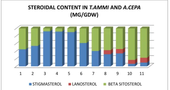
stigmasterol (0.240 mg/gdw) which was comparatively little less than that of the amount
of β- sitosterol (0.295 mg/gdw) in the seeds of
T.ammi (Table 6.2; Fig. 6.1) The maximum
amount of stigmasterol was present in four weeks old tissue of T.ammi (0.249 mg/gdw) whereas the
highest content of β- sitosterol was observed in six weeks old tissue of A. cepa (0.315 mg/gdw) However, lanosterol, was present only in the tissue of A. cepa which was maximum in six weeks old tissue (0.039 mg/gdw).
FIG. 2: Graph showing the steroidal content in A.cepa and T.ammi
Phytosterol such as β-sitosterol and stigmasterol have been reported in tissue cultures of Artemisea scoparia, Datura metel, Emblica officinalis and Trigonella foenum-graecum (Khanna, 1987) and
βsitosterol and stigmasterol in the tissue of C.
arietinum, S. indicum and Momordia charantia
(Khanna, 1984). Only β-sitosterol and
stigmasterol.It has been reported in tissue culture
of S. xanthocarpum (Heble et al., 1968)
Helianthus annus (Sharma, 1975), S. aviculare
(Gaur, 1978) and Trigonella corniculata (Jain,
1979) .Lanosterol, stigmasterol and β-sitosterol along with cholesterol have been observed in tissue of Sesamum indicum (Jain and Khanna, 1973) and M. Charantia (Khanna and Mohan, 1973) Smoczklewicz et. al., (1982) have reported
βsitosterol from bulbs of Allium cepa. Whereas
Cholesterol, campesterol. β-sitosterol and
stigmasterol have been identified from the bulbs of A. sativum (Stoyamo et.al., 1981) Claus et. al., (1980) and Catalano et. al., (1983) have shown
the presence of β-sitosterol and stigmasterol in umbelliferous vegetables.
In the present study, however, three phytosterols
as β-sitosterol , stigmasterol and lanosterol have been observed in the tissue of A. cepa whereas
only two phytosterols, β-sitosterol , stigmasterol have been identified from plant parts and tissue culture of T. ammi.
1 2 3 4 5 6 7 8 9 10 11
STEROIDAL CONTENT IN T.AMMIAND A.CEPA
(MG/GDW)
Vol. 1 No. 6 2013 www.phytojournal.com Page | 47 Fig 3:GLC curve of isolated and standard β-sitosterol , stigmasterol and lanosterol from Allium cepa L. tissue culture.
Fig 4:GLC curve of isolated and standard β-sitosterol , stigmasterol from Trachysoermum ammi Sprague.
Vol. 1 No. 6 2013 www.phytojournal.com Page | 48 Fig 6. Infra-red spectra of isolated and standard stigmasterol.
Fig.7. Infra-red spectra of isolated and standard β-sitosterol.
4. References
1. HEBLE M.R. , NARAYANA SWAMI, S. AND M.S.
CHADHA, 1968. Dios genin and B-sitosterol;
isolation from Solanum xanthocarpum tissue
culture. Science, 161 : 1145
2. JAIN, S.C. AND P. KHANNA, 1973. Production of
sterols from Sesamum Indicum L. tissue culture.
Indian J. Pharm. 35 : 163-164.
3. KAUL, 13. AND E.J. STABA, 1968. Dioscorea,
tissue culture, I. Biosynthesis and isolation of diosgenin from Dioscorea deltodea callus and suspension cells. Lloydia, 31 : 171-179
4. KHANNA P.1984. Useful metabolites from plant
tissue culture. Fifty plant species. In: Sixteen philip R. White Memorial Lecture, January 2, Ranchi, Bihar(India).
5. KHANNA P.1987. Antimicrobials and other
useful metabolites from plant tissue culture, Keynote address DAE Symposium on plant Microbe Interaction, Nov. 11-13. Isolation and identification of steroids from plant tissue culture. Proc. Intl. Symp. On Morphogenesis of plant cell Organ and Tissue Culture, held in New Delhi, 17-22. Nov.
6. KHANNA P., T.N. NAG, S.C. JAIN AND S. C. JAIN AND S. MOHAN,1975. Extraction of Momordica Chaarantia L. Indian J. Exp. Biol. 11: L 58-60.
7. SMOCZKIEWICZOWA, M.A., J. LUTOMSKI,
NITCHKE, 1981. Chemical and pharmacolgocial characterization of Allium cepa L. Herbapol. 27(2): 169-185.
8. STOYANOVA IVANOVA, B. , TSUTSULUVA, A.
AND R. CAPUTTO, 1981. On the hydrocarbon and sterol composition in the scales and the fleshy part of Allium sativum L. bulb. Riv. Ital. Eppos, 62(7): 373-6
9. TOMITA, Y., A. VOMORI AND H. MINATO,1970.
Steroidal sapogenin and sterols in tissue culture of Dioscorea tokoro. Phytochem. 9 : 111-114.
10. CLAUS, R., H.O. HOPPEN AND B. PHROMONO,
1979. Steroid identification in vegetables. Experientia 35 (12): 1674-1675.