Vol-7, Special Issue-Number4-June, 2016, pp523-529 http://www.bipublication.com
Research Article
Evaluation of Bond Strength and Quality of Fiber Posts Cemented With Two
Cements in Asymmetric Dental Root Canal
Atefeh Ramezani1, Ehsan Moudi2, Zahra Sadat Madani3, Forouzesh shirgaee talari1,
Hossein pourkhalili1 and Abdolhamid Alhavaz*4 1Prosthodontic resident, Department of Prosthodontics,
Babol University of Medical Sciences, Babol, Iran
2
Department of Oral and Maxillofacial Radiology, Dental School, Babol University of Medical Sciences, Babol, Iran
3
Department of Endodontics, Dental School, Babol University of Medical Sciences, Babol, Iran.
4
Dental Materials Research Center, Department of Prosthodontics, Babol University of Medical Sciences, Babol, Iran.
*Corresponding Author: Abdolhamid Alhavaz, Email:com.yahoo@alahvaz
ABSTRACT:
Background and Objective:Debonding is one of the most common causes of failures in post fibers used in the root canalat interface of dentin-fiberpost. The purpose of this study is to evaluate the interface of the fibers post in the root canal with appropriate and inappropriate compliance with CBCT and its push-out bond strength with two types of resin cement used in the mandibular premolars.
Materials and Methods:Forty (40)Mandibular Premolarteeth which were extracted were useddue to theorthodontic problems. After endodontic, the teeth were randomly classified into two groups including teeth with post space in compliance with the fiber post and a group of posts space wider than fiber post. Thereafter,each group wassub-divided into two groups according to the used cement: panaviaF2.0 (Kuraray Medical Inc., Osaka, Japan), Rebilda DC(Voco, and Germany) and finally, four groups were created [P.a:canal with appropriate adaptation + panavia F2.0, P.in:canal with inappropriate adaptation + panavia F2.0, R.a:canal with appropriate adaptation + Rebilda DC, R.in:canal with inappropriate adaptation + Rebilda DC]. Data analysis was carried out using ANOVA, Post hoc Tukey test, Chi-square test (p <0.05).
Results: The bond strength was significantly affected by the analyzed root area (p-value = 0.03) and there was a significant difference between two canals with appropriate and inappropriate compliance with the same type of cement (p-value = 0.05). In addition, the bond strength was not affected by cement type (p-value = 0.67) and the area of the voids was higher in P.in groups. Nevertheless, in R.a group, no void was observed.
Conclusion: The bond strength was affected by the post space but it was not affected by cementation techniques. As a result of this, applicator of Rebilda cement reduces the voids in the root canal with appropriate compliance.
Keywords: Rebilda DC, panavia-Fluoro 2.0, Dental cement, Fiber glass, Cone Beam Computed Tomography, Bond strength.
[I] INTRUDUCTION
Reconstruction of endodontically-treated teeth hasbeen studied extensively, but there are challenges on some issues[1]. Metal cast posts may increase the risk of root fracture due to corrosion or wedging effects. For this reason, fibre
the root canal[3]. The ideal fitness of post is a critical factor that is responsible for theappropriate thickness of cement.Poor fitness led to thick cement, especially in the coronal of the root canaldue to the void or bubble increase that lead to the debonding[4]. Debonding can be developed in the fiber posts fordifferent reasons like preparing the post surface, dentin bonding components, cement, and application of adhesives, and also polymerization methods. Different morphology in various regions of the root canal leads to different qualities of the bond in the coronal, middle and apical regions[3]. There is still no agreement on the ideal thickness of cement and also the effect of void on the bond strength of fiber posts [5]. One of the clinical problems for the dentists in the restoration of endodontically treated teethis unfitness of diameters of post space and that of the post.Although using the drills with appropriate size by the manufacturers create a perfect compliance of post with the wall of the root canal,but the teeth root canals have different shapes and therefore, the thickness of resin cement around posts can vary[6].Several studies about the defects at the dentin-cementjunctionare known to be related to how the method was utilized for application of cement[7]. According to the previous studies,the push-out test is preferable so as to evaluate the bond strength than other cases, such as shear-test, since in this test, fracture occurs in parallel at the dentin junction and adhesives cause real shear-test[8].In several studies,the SEM or analysis of digital images have been utilized for reviewing the junctionof dentin- glass fiber post[5]. The purpose of this study was to evaluate the of push-out bond strength of two different types of cements and also observe the CBCT for the three-dimensional assessment of dentin-junction glass fiber post in root canal with appropriate and inappropriatecompliance.
[II] MATERIALS AND METHODS
In the present research, 40 mandibular premolars teeth were selected (teeth were stored in saline) and then, two mesiodistal and buccolingual views
used within 15 s after mixing) andthen canal walls were exposed using a brush to blend smeary.The additions were removedby completely using the air pore andpaper points. Thereafter Panavia was prepared on the completely dried pad based on the instructions.After that, the post was dipped in the dough and then it was placed with a gentle vibration to prevent formation of air bubbles in the canal. Thereafter the canal was treated with Halogen light-cure for 3-2 s so as to remove the additional cement. At the end, it was cured for 20s from coronal side. In the relationship with the Rebilda DC cement, after inoculation of the root canal walls with primer, Futurabond DC (Voco,Germany)was placed using a good brush,
cement,andthe applicator in the
canal.Thereafter,the posts were placed in the canal according to the manufacturer's instructions. Then each group was cured for 24h in distilled water maintained at 37°C. The samples were sent for CBCT analysis and CBCT scans were obtained using the Newtom5G system(Verona, Italy), witha potential difference of110 kVp, tube follow of 3.46 mA,duration of 6 × 68.4 and 6×6 field of view. The thickness of each voxel sizewas 0.3 Mm,the thickness of each slice was0.1 mm, and the spaces of each slice was 0.5. Images were evaluated by NNT viewer software in the axial plane.
Using the cutting tool,somesamples with thickness of 1mm were prepared for conducting push-out test in threeareasnamelycoronal, middle, and apical.To provide push-out test, the universal testing machine (Zwick) was utilized. In the coronal section, the cuttings were marked using the markers. After fixing the sample on the device so that the apical side was toward the power importing tool, the force was entered with a speed of 1 mm/min on the sample.The sample surface was calculated usingthe following formula:(r1 + r2) √ (r1 + r2) ² + h²,π: 3.14, r1= radius of the coronal portion of the post, r2= radius of the apical part, h = height of the post during the cross-cutting; then the applied force was converted from N to MPa:
Bonding strength = debonding force/total bonding area
After applying the push-out test, the samples were evaluated using stereo microscope with a magnification of 40 × so as to determine the type of failure.The types of failure were categorized in 5 categories.Type1: adhesive failure between dentin & cement (ADC), Type 2: adhesive failure between post & cement (APC), Type 3: cohesive failure withinthe cement (CC), Type 4: cohesive failure within the post (CP), Type 5: mixed type (M).
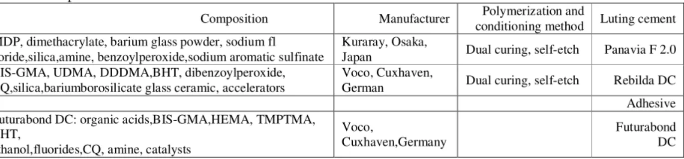
Table 1: Composition of the materials use
Luting cement
Polymerization and conditioning method
Manufacturer
Composition
Panavia F 2.0
Dual curing, self-etch Kuraray, Osaka,
Japan MDP, dimethacrylate, barium glass powder, sodium fl
uoride,silica,amine, benzoylperoxide,sodium aromatic sulfinate
Rebilda DC
Dual curing, self-etch Voco, Cuxhaven,
German BIS-GMA, UDMA, DDDMA,BHT, dibenzoylperoxide,
CQ,silica,bariumborosilicate glass ceramic, accelerators
Adhesive
Futurabond DC Voco,
Cuxhaven,Germany Futurabond DC: organic acids,BIS-GMA,HEMA, TMPTMA,
BHT,
ethanol,fluorides,CQ, amine, catalysts
[III] RESULTS
The bond strength was statistically significant in the coronal, middle, apical and separately between the groups (Coronal: value=0.000, C: p-value=0.003, apical: p-value =0.006) (Table 2) in [Figure.1]. While the bond strength wasn’t significantly related to the coronal, middle, and
ofP.a and P.in was not significant (p=0.36), but the relationship was significant in the R.a and R.in (p=0.007). Nevertheless,in the apical area of P.a and P.inwas significant (p=0.032), while in the Rebilda DC cement, itwas not significant (p=0.073).In the coronal and medial areas of R.a group, no voids were observed.The area of void
observed in the coronal area was higher in the coronal and middle areas of theP.a group.While at the apical area, void surface area was higher using theP.in group. Among all samples, the most types of the fractures were Type1 adhesive failure between dentin & cement (ADC) in [Figure- 2].
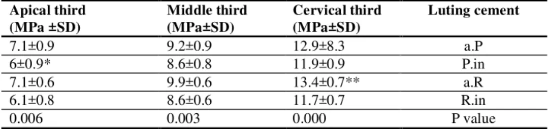
Table 2: Mean push-out bond strength values (MPa) with their standard deviations
Several cases where Tukey’s post-hoc analysis was used showed no significant difference (Alpha = 0.05), (lowest *, the most **).
Figure-1: The push-out bond strength in three areas (1) coronal, (2) middle, (3) apical
Figure-2: failure type
Luting cement Cervical third
(MPa±SD) Middle third
(MPa±SD) Apical third
(MPa ±SD)
P . a 12.9±8.3
9.2±0.9 7.1±0.9
in . P 11.9±0.9
8.6±0.8 6±0.9*
R . a 13.4±0.7**
9.9±0.6 7.1±0.6
in . R 11.7±0.7
8.6±0.6 6.1±0.8
P value 0.000
[IV] DISCUSSION
In this study, bond strength of glass fiber post was evaluated with two types of resin cementsand also in canals with appropriate and inappropriate compliance using the push-out test.In this study, it was found that bond strength in the coronal area was more in all groups and then in the middle area, while in the apical area, the strength was less in other parts. This test is based on stress shear at the dentin-cement junction similar to the post-cement junction.
The push-out test is a reliable technique for precise measurement of the bond strength of fiber posts to root dentin compared to modified routine tests of microtensile. For this reason,the push-out method is actually a real shear test[8]. In a study that was carried out by Pereira et al, it was found that the bond strength was different in various areas of the root[9]. Ebrahimi et al concluded that the bonding strength of fiber posts in the coronal area is more affected by their adhesive system polymerization methods[3]. Dua et al achieved similar results with this research while in the study; it wasshown that the bond strength is affected by the type of cement[10]. In the study of Pereira and Foxton, significant differences were not observed in bond strength in different parts of the roots[9]. Nevertheless, in a study carried out byRengo, it was concluded that difficulty in clearing the apical region of the posts can explain the low bond strength in the apical area[5]. According to a study carried out by Ebrahimi, dual-cure adhesives in the apical portion of the adhesive bond strength than light – cure.It was revealed that due to the reduction of the intensity of light in the apical area, lower degree of conversion is less in optical adhesives[3]. So,the difference had less rooted bond strength in different areaswhich wasbecause of less dependence of the light equitability of bond strength in different parts of the roots[3].
In this study, the cement dual –cure was used so as to remove the effect of light transmission intensity.So,the reduction of bond strength can be as a result of the reduced density in the apical area
of dentinal tubules in the apical area.In a study carried out by Musharraf et al, it was concluded that as a result of the increased density of dentinal tubules in the coronal,apical tubules were more than the bond strength in the coronal area[1]. The two different methods usedin cementation in this study showed that the bond strength is not affected by cementation techniques because the differences in the two types of cement bond strength were not significant and/or with the void area in the coronal was more and was ineffective in reducing the bond strength in the coronal area. Nevertheless, according to the study of watskeet al,conventional cementation method creates the voids[7]. Nevertheless,according to a study carried out by Bitter, void cannot be a weakness for the lower strength but can act as stress relaxation[11].In this research, incompliance of posts reduced bond strength in canals with poor compliance,similar to study of Uezonoglue et al,it was shown that the bond strength decreases with increasing thickness of cement,and they concluded that reduction of shrinkage in the thinner cement causes less stress on the joints. They also showed that by increasing the thickness of the cement, there is the probability of creating more voids that could weaken the cement and eventually debonding[6].While Peretz et al believe that by increasing the thickness of the cement, bond strength will not reduce[12].
cement (ADC) which is similar to Bitter study. Dua also carried out a study which revealed that most of the failure of the type of adhesive was between cement and dentin[10].
The purpose of this research is to evaluate the voids in the cement space of CBCT. The advantages of CBCT is that it is a non-destructive method for samples, and in fact, there is the possibility of sample analysis without losing any information while in the traditional methods,the parts of the dental tissue or material during cutting and preparation for testing arelost and only a limited number of samples can be usedfor observation[5].In this research,it was revealed that in the root canal with appropriate compliance, the void were less but there were bubbles,which can be due to the variety of root form, despite the fact that the use of appropriate drills in compliance with post is appropriate. In addition, the root canals with Rebilda DC cement had less voids.This could be as a result of the use of applicator for cementation of posts in the root canal which prevents trapping of air bubbles.According toWatzke,defects at the cement junction depend on the cementing methods and application of applicator aid which caused more homogencity of the cement in the root canal[7]. It is noteworthy that using the application in dual-cure cements in root canals is not due to its faster setting before correct arrival of the post[14].On the other hand,Ferrari et al reported that using microbrush in root canal creates a uniform bond in the canal[15]. Rengo et al concluded that the volume of voids in the coronal area regardless of the post position is greater and based on that, void size in the coronal was greater. The bond strength is greater in the coronal area which is due to the effect of the voids as stress absorber and it was also reported that it is because of the volume of large voids in the coronal area which is also due to high cement volume[5].Dua study also showed thatthe use of spiral Lentoral reduces the voids of cement[10]. In a study carried out by Bitter et al, it was also shown that though the voids was higher in Rebilda DCcement, but it was not lower
in bond strength compared to other cements, because void could compensate for the damaging effect of C-factor in stress relaxation[11].
[V] CONCLUSION
Bond strength is affected by the post space but will not be affected by cementation techniques. The applicator of Rebilda DC cement reduces voids in root canal with appropriate adaptation . The presence of void in the coronal area had no effect on reducing bond strength.
FINANCIAL DISCLOSURE:This study was sponsored by the Babol University of Medical Sciences.
ACKNOWLEDGEMENT
The authors would like to acknowledge Doctor Mrs.SorayyaKhafri who help in the statistical work. We would also like to acknowledge Babol Research Center, School of Dentistry, Dental Materials Research Center, Tehran University of Medical Sciences, as well Doctor Ehsan Moudi ofRadiology center that helped us in this study.
REFERENCES
1. Mosharraf, R. and A. Haerian, Push-out bond strength of a fiber post system with two resin cements. Dental research journal, 2011. 8(Suppl1): p. S88.
2. Schwartz, R.S. and J.W. Robbins, Post placement and restoration of endodontically treated teeth: a literature review. Journal of endodontics, 2004. 30(5): p. 289-301.
3. Ebrahimi, S.F., et al., Effect of polymerization mode of two adhesive systems on push-out bond strength of fiber post to different regions of root canal dentin. Dental research journal, 2014. 11(1): p. 32.
4. Grandini, S., et al., A one step procedure for luting glass fibre posts: an SEM evaluation. International endodontic journal, 2004. 37(10): p. 679-686.
with oval and circular posts. Clinical oral investigations, 2014. 18(2): p. 571-578.
6. Uzunoğlu, E., S.A. Türker, and Z. Yilmaz,
Influence of cement type and thickness on polyfiber post adhesion. Journal of conservative dentistry: JCD, 2014. 17(3): p. 255.
7. Watzke, R., R. Frankenberger, and M. Naumann, Probability of interface imperfections within SEM cross-sections of adhesively luted GFP. dental materials, 2009. 25(10): p. 1256-1263.
8. Perdigao, J., G. Gomes, and V. Augusto, The effect of dowel space on the bond strengths of fiber posts. Journal of Prosthodontics, 2007. 16(3): p. 154-164.
9. Pereira, J., et al., Evaluation of push‐out bond strength of four luting agents and SEM observation of the dentine/fibreglass bond interface. International endodontic journal, 2013. 46(10): p. 982-992.
10. Dua, A., D. Dua, and O. Wali, Effect of three resin cements on micro push-out bond strength of fiber-reinforced composite post to radicular dentin at different root levels. International Journal of Clinical Dental Science, 2015. 6.
11. Bitter, K., et al., Analysis of resin-dentin interface morphology and bond strength evaluation of core materials for one stage post-endodontic restorations. PloS one, 2014. 9: p. e86294.
12. Perez, B., et al., Does the thickness of the resin cement affect the bond strength of a fiber post to the root dentin? The International journal of prosthodontics, 2005. 19(6): p. 606-609. 13. Zhang, L., et al., Effect of post‐space treatment
on retention of fiber posts in different root regions using two self‐etching systems. European journal of oral sciences, 2008. 116(3): p. 280-286.
14. Fakiha, Z., A. Al‐Aujan, and S. Al‐Shamrani, Retention of cast posts cemented with zinc phosphate cement using different cementing techniques.Journal of Prosthodontics, 2001. 10(1): p. 37-41.