pH-Dependent Assembly and Segregation of
the Coiled-Coil Segments of Yeast Putative
Cargo Receptors Emp46p and Emp47p
Kentaro Ishii1☯, Hiroki Enda2☯, Masanori Noda3, Megumi Kajino2, Akemi Kim2,
Eiji Kurimoto2,4, Ken Sato5, Akihiko Nakano6,7, Yuji Kobayashi3, Hirokazu Yagi2, Susumu Uchiyama1,3, Koichi Kato1,2,8*
1Okazaki Institute for Integrative Bioscience, National Institutes of Natural Sciences, Okazaki, Aichi, Japan, 2Graduate School and Faculty of Pharmaceutical Sciences, Nagoya City University, Nagoya, Aichi, Japan, 3Department of Biotechnology, Graduate School of Engineering, Osaka University, Suita, Osaka, Japan, 4Faculty of Pharmacy, Meijo University, Nagoya, Aichi, Japan,5Department of Life Sciences, Graduate School of Arts and Sciences, The University of Tokyo, Meguro-ku, Tokyo, Japan,6Department of Biological Sciences, Graduate School of Science, The University of Tokyo, Bunkyo-ku, Tokyo, Japan,7Live Cell Super-Resolution Imaging Research Team, RIKEN Center for Advanced Photonics, Wako, Saitama, Japan, 8Institute for Molecular Science, National Institutes of Natural Sciences, Okazaki, Aichi, Japan
☯These authors contributed equally to this work. *[email protected]
Abstract
Emp46p and Emp47p are yeast putative cargo receptors that recycle between the endo-plasmic reticulum and the Golgi apparatus. These receptors can form complexes in a pH-dependent manner, but their molecular mechanisms remain unclear. Here, we successfully reproduced their interactionsin vitrosolely with their coiled-coil segments, which form stable
heterotetramers in the neutral condition but segregate at lower pH. Mutational data identi-fied a key glutamate residue of Emp46p that serves as the pH-sensing switch of their oligo-mer formation. Our findings elucidate the mechanisms of the dynamic cargo receptor interactions in the secretory pathway and the design framework of the environment-respon-sive molecular assembly and disassembly systems.
Introduction
Eukaryotic cells are equipped with a vesicular transport system for sorting and translocating cargo proteins in the secretory pathway. Protein transport in the early secretory pathway is mediated by COPII-coated vesicles, which bud from the endoplasmic reticulum (ER) and travel to the Golgi compartment [1]. Growing evidence indicates that several soluble secretory proteins require specific transmembrane cargo receptors as adaptors between these cargos and the COPII coats for selective packaging [2,3].
In mammalian cells, ERGIC-53 has been identified as a cargo receptor that recycles between the ER and the Golgi apparatus and contributes to the anterograde transport of several glyco-proteins, including the lysosomal glycoproteins cathepsin C and cathepsin Z and blood
a11111
OPEN ACCESS
Citation:Ishii K, Enda H, Noda M, Kajino M, Kim A, Kurimoto E, et al. (2015) pH-Dependent Assembly and Segregation of the Coiled-Coil Segments of Yeast Putative Cargo Receptors Emp46p and Emp47p. PLoS ONE 10(10): e0140287. doi:10.1371/ journal.pone.0140287
Editor:Maria Gasset, Consejo Superior de Investigaciones Cientificas, SPAIN
Received:July 26, 2015
Accepted:September 23, 2015
Published:October 8, 2015
Copyright:© 2015 Ishii et al. This is an open access article distributed under the terms of theCreative Commons Attribution License, which permits unrestricted use, distribution, and reproduction in any medium, provided the original author and source are credited.
Data Availability Statement:All relevant data are within the paper and its Supporting Information files.
as yet [8–11]. Like ERGIC-53, these yeast proteins share homologous CRDs and possess a
puta-tive coiled-coil segment followed by a transmembrane region with a C-terminal tail. Despite their structural homology with ERGIC-53, their CRDs are independent of Ca2+and their coiled-coil segments contain no cysteine residue. Immunoprecipitation of differently tagged recombinant proteins expressed in theemp46Δemp47Δcells indicated that, in the ER, Emp46p and Emp47p are monomeric and homo-oligomeric, respectively, and capable of interacting with each other [9]. These interactions are mediated through their coiled-coil segments. Emp47p is required for the ER exit of Emp46p; however, the converse is not true. Emp46–Emp47 interaction was not
detected in the Golgi fraction. On the basis of these findings, it has been suggested that Emp47p is a receptor for Emp46p, escorting it from the ER to the Golgi apparatus. However, molecular mechanisms of the assembly and disassembly of these proteins are largely unknown. Therefore, we herein characterize the putative interactions of the coiled-coil segments of Emp46p and Emp47p to provide a molecular basis of the dynamic processes involved in vesicular transport.
Results and Discussion
Homo-oligomeric states of Emp46p
CCand Emp47p
CCWe attempted to reproduce oligomer formation of Emp46p and Emp47p using their coiled-coil segments. The bacterially expressed coiled-coiled-coiled-coil segment of Emp46pCC(252–355) and
Emp47pCC(271–274) were subjected to circular dichroism (CD) spectroscopic analysis, which
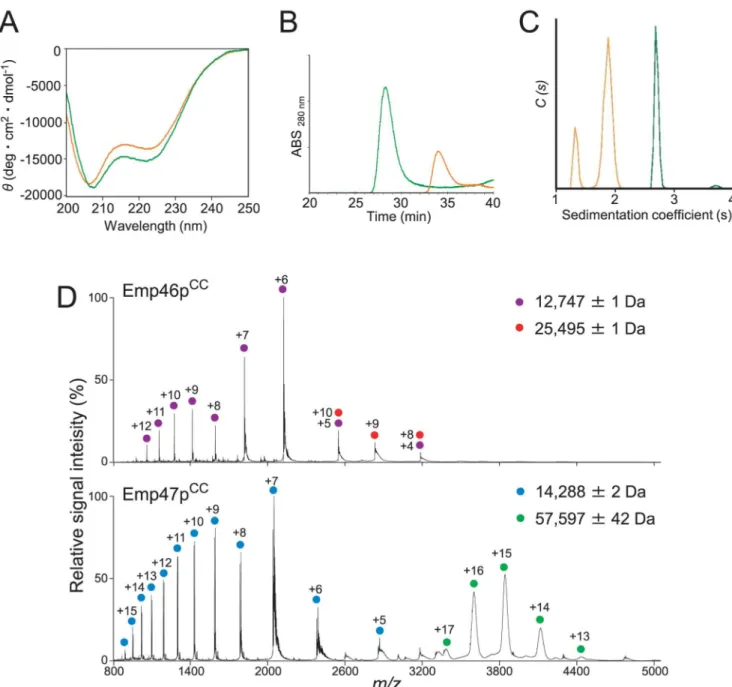
confirmed theα-helical structures of both recombinant proteins (Fig 1A). The oligomeric states of Emp46pCCand Emp47pCCwere subsequently characterized by biochemical and bio-physical methods. Gel filtration and chemical cross-linking experiments indicated that
Emp47pCC, but not Emp46pCC, formed a homotetramer (Fig 1BandS1 Fig). For more accurate determination of oligomeric states, Emp46pCCand Emp47pCCwere subjected to sedimentation velocity analytical ultracentrifugation (SV-AUC) and mass spectroscopy (MS) analyses. SV-AUC data showed that Emp46pCCexhibited two species, with peaks at 1.2 S and 1.9 S, from which their molecular weight (Mw) were estimated to be 12.7 and 21.2 kDa, respectively (Fig 1C). The populations of the two peaks for Emp46pCCwere concentration dependent (S2A Fig) and the s-value of the species with the higher s-value changed as the concentration changed. This is a typical result for an association—dissociation system with rapid dissociation
kinetics [12]. The MS analysis that consistently provided accurate molecular masses of the two species, both monomer and dimer, were detected for Emp46pCC(Fig 1D). Meanwhile, Emp47pCChad a single peak at approximately 2.7 S with apparently no concentration depen-dence, providing an Mwof 63.3 kDa (Fig 1CandS2B Fig). These data revealed that Emp46pCC underwent monomer—dimer equilibrium, whereas Emp47pCCformed a stable tetramer,
Hetero-oligomeric coiled-coil interactions between Emp46p
CCand
Emp47p
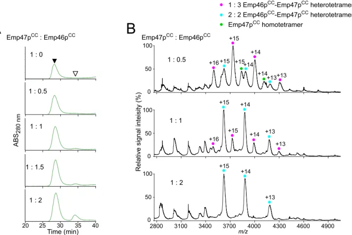
CCNext, we attempt to evaluate the potential interaction between Emp46pCCand Emp47pCC. In gel filtration of Emp47pCCtitrated with Emp46pCCunder a neutral condition, that is, pH 7.4, less than one molar equivalent of Emp46pCCco-eluted with Emp47pCC, indicating the forma-tion of hetero-oligomers with Emp47pCC(Fig 2A). Chemical cross-linking experiments Fig 1. Characterization of oligomeric states of Emp46pCCand Emp47pCC.(A) CD spectra of Emp46pCC
(orange) and Emp47pCC(green). (B) Gel filtration profiles of Emp46pCC(orange) and Emp47pCC(green) at pH 7.4. (C) Distribution of sedimentation coefficients derived from sedimentation velocity analytical ultracentrifugation (SV-AUC) experiments. The orange line indicates Emp46pCC(75μM), whereas the green line indicates Emp47pCC(75μM). (D) Mass spectra of Emp46pCC(top) and Emp47pCC(bottom) at pH 7.4. Peaks corresponding to the monomer and dimer of Emp46pCCare indicated by purple and red dots with charge states, respectively. Peaks corresponding to the monomer and tetramer of Emp47pCCare indicated by blue and green dots with charge states, respectively.
detected a covalently linked heterodimer on SDS-PAGE (S1 Fig). MS data demonstrated that Emp46pCCand Emp47pCCformed heterotetramers with a 1:3 or 2:2 stoichiometry; the later increased with increasing quantities of Emp46pCC(Fig 2B). All of these data indicate that the homotypic and heterotypic interactions involving Emp46p and Emp47p, which were previ-ously observed by immunoprecipitation, are well reproducedin vitrosolely with their coiled-coil segments and characterized more quantitatively.
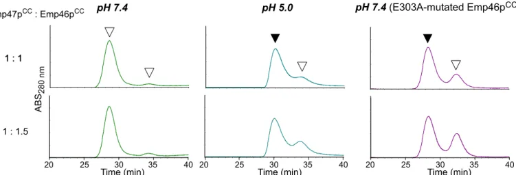
In mass spectra measured at pH 5.0, the homo-oligomeric and hetero-oligomeric forms of the Emp46pCCand Emp47pCCwere detected with the identical stoichiometric numbers with those observed at pH 7.0 (S3andS4Figs). However, the quantitative comparison of the MS data was difficult in terms of relative incidence of the complex species because relative ioniza-tion efficiencies of protein complexes are generally different under different pH condiioniza-tions. For quantitative estimation of the relative population of the complexes, we employed gel filtration. The results clearly demonstrated that the Emp46pCC–Emp47pCCheterotetramer formation
was impaired at pH 5.0 (Fig 3). Emp46p and Emp47p have been reported to form complexes in the ER but segregate in the Golgi compartment. H+pumping by v-ATPase causes progressive acidification from the ER through the Golgi to thetrans-Golgi network with typical pH values of ER,cis-Golgi, andtrans-Golgi network that are reported to be 7.2, 6.4, and 5.4, respectively. Fig 2. Characterization of complex formation between Emp46pCCand Emp47pCCat pH 7.4.(A) Gel-filtration profiles of the mixture of Emp46pCCand Emp47pCCat 1:0, 1:0.5, 1:1, 1:1.5, and 1:2 molar ratios. White and black arrows indicate the elution positions of Emp46pCCalone and oligomers involving Emp47pCC(and Emp46pCC), respectively. (B) Mass spectra of the mixtures of Emp46pCCand Emp47pCCat pH 7.4. The concentration of Emp47pCCwas fixed and that of Emp46pCCwas varied at the molar ratio indicated. Peaks corresponding to 1:3 and 2:2 heterotetramers of Emp46pCCand Emp47pCCare indicated by magenta and cyan dots with charge states, respectively. Peaks corresponding to the homotetramer of Emp47pCCare indicated by a green dot with charge states.
The present data demonstrate that the Emp46pCC–Emp47CCinteraction is weakened by
lower-ing the solution pH. Based on these findlower-ings, we suggest that assembly and segregation of these cargo receptors during their transit from the ER to the Golgi compartment are at least in part ascribed to their pH-dependent coiled-coil interaction.
Key glutamate of pH-dependent segregation of Emp46p
CCand
Emp47p
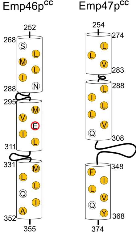
CCIn general, coiled-coil segments exhibit amphiphilicα-helices, which are characterized by the heptad repeat,HPPHPPP, of hydrophobic (H) and polar (H) residues, and are assembled to bury their hydrophobicH/Hsurface. A glutamate residue (Glu303) is located in the hydropho-bic surface of Emp46pCC(Fig 4). It is possible that this glutamate negatively contributes to the homo-oligomer formation of Emp46pCCbecause of the potential electrostatic repulsion caused by its negative charge under neutral conditions; however, this might be preferably accommo-dated in the tetrameric complex formed with Emp47pCC. On the basis of this idea, we exam-ined the possible effect of the alanine substitution of Glu303 in Emp46pCCon its oligomer formation. The E303A mutant of Emp46pCCmaintained the integrity of theα-helical structure (S5A Fig) and tended to form a homodimer in comparison with the wild type (S5B Fig). This mutation impaired the Emp46pCC–Emp47pCCinteraction even at pH 7.4 as shown by gel
fil-tration (Fig 3) and chemical cross-linking analysis (S5B Fig). Consistently, in native mass spec-tra, this mutant was detected only as the 1:3 Emp46pCC/Emp47pCCcomplex even under the condition where the wild-type Emp46pCCwas observed exclusively as the 2:2 complex with Emp47pCC(S6 Fig). These results indicate that Glu303 of Emp46pCCserves as the pH-sensing switch of assembly and segregation of Emp46p and Emp47p. The pH-dependent coiled-coil interaction is exemplified by macrophage scavenger receptor, in which the trimericα-helical coiled-coil domain exhibits conformational change depending on solution pH [13]. In this sys-tem, the C-terminal coil segment forms a random structure at neutral pH and a coiled-coil structure under acidic condition. Moreover, it has been reported that a glutamate residue located in the hydrophobic surface of this coiled-coil segment plays a critical role in this pH-dependent conformational transition [13]. Our results along with these data suggest possible mechanistic similarities shared among the pH-dependent coiled-coil interactions.
Fig 3. Gel filtration profiles of the mixture of Emp46pCCand Emp47pCCunder pH 7.4 (left) and 5.0 (center) and the mixture of Emp46pCCmutant
E303A and Emp47pCCunder pH 7.4 (right) at 1:1 and 1:1.5 molar ratios.White arrows indicate the elution positions of Emp46pCCor its E303A mutant. Black arrows indicate the elution positions of the hetero-oligomers involving Emp47pCC.
In summary, the present study successfully characterized the pH-dependent coiled-coil interactions of Emp46p and Emp47p, identifying the key residue that controls this interaction. These results contribute toward understanding the molecular mechanisms underlying the dynamic cargo receptor interactions in the yeast secretory pathway. Moreover, our findings Fig 4. Schematic of the coiled-coil segments of Emp46pCCand Emp47pCC.Residues consisting of a
hydrophobic core are represented. Hydrophobic residues are represented by yellow, whereas Glu303 is represented with a red circle.
will provide a framework for designing molecular assembly and disassembly systems mediated by coiled-coil interactions responsive to changes in environmental conditions.
Materials and Methods
Protein expression and purification
The coiled-coil segments of Emp46p and Emp47p were identified using COILS/PCOILS (http://toolkit.tuebingen.mpg.de/pcoils) [14]. Plasmid vectors that contained the genes of the coiled-coil segment of Emp46pCC(252–355) or Emp47pCC(254–374) produced by
amplifica-tion of polymerase chain reacamplifica-tion were constructed and cloned for their expression as a fusion protein with hexahistidine-tagged ubiquitin (His6-Ub) using a pET28a(+) vector (Novagene) or a His6-tagged protein using a pET28b(+) vector (Novagene). These vectors were trans-formed into theEscherichia colistrain BL21-CodonPlus. The cells were grown at 37°C in Luria—Bertani medium containing 15μg/mL of kanamycin. Protein expression was induced
by adding 0.5 mM isopropyl-βD-thiogalactopyranoside when absorbance reached 0.8 at 600 nm. After 3 h, cells were harvested, suspended in 50 mM Tris—HCl (pH 8.0), and
subse-quently, disrupted by sonication. After centrifugation, the His6-Ub-tagged or His6-tagged pro-teins were purified in a Chelating Sepharose Fast Flow column (GE Healthcare Life Sciences). The Emp46pCCand Emp47pCCwere enzymatically cleaved from the His6–Ub moiety using
recombinant glutathioneS-transferase-tagged yeast ubiquitin hydrolase-1, as described previ-ously [15], or cleaved from the His6-segment using thrombin protease (GE Healthcare), and then purified by chromatography using a gel filtration column (Superose 12 column 10/300 GL, GE Healthcare).
Circular dichroism measurements
The purified Emp46pCCand Emp47pCC(0.1 mg/mL) were dissolved in 50 mM Tris—HCl
buffer (pH 7.6) containing 0.15 M NaCl. Measurements of CD spectra were performed in a 1-mm quartz cuvette at room temperature using a spectropolarimeter (J-725, JASCO). After subtraction of the spectrum of the buffer alone, data were represented as mean residue ellipticities.
Gel filtration
The purified Emp47pCCprotein (70μM), either alone or in the presence of Emp46pCCor its
E303A mutants, was subjected to gel filtration using a Superose 12 column (GE Healthcare), equilibrated with 10 mM phosphate buffer (pH 7.4) containing 150 mM NaCl or 50 mM ace-tate buffer (pH 5.0) containing 150 mM NaCl.
Chemical cross-linking
The purified Emp46pCC(or its E303A mutant) and Emp47pCCproteins were dialyzed against 100 mM carbonate/bicarbonate buffer (pH 8.9). Next, dimethyl suberimidate dihydrochloride (DMS) was added to derive a final concentration of 0.1 mg/mL and incubated for 60 or 120 min at room temperature. The cross-linking reaction was quenched by adding 500 mM sodium phosphate buffer (pH 6) to adjust pH.
Analytical ultracentrifugation
SV-AUC experiments were performed in 10 mM mmol/L phosphate buffer (pH 7.4) contain-ing 150 mM NaCl uscontain-ing a Proteomelab XL-I Analytical Ultracentrifuge (Beckman—Coulter).
Mass spectrometry under non-denaturing conditions
The purified Emp46pCC(or its E303A mutant) and Emp47pCCproteins (100μM) were
buffer-exchanged into 200 mM ammonium acetate at pH 7.4 by passing through a Bio-Spin 6 column (Bio-Rad). The samples (43μM) were immediately analyzed by nanoflow electrospray using
in-house made gold-coated glass capillaries (approximately 2μL sample loaded per analysis).
The mixtures of Emp47pCC(33μM) and 17, 33, and 67μM Emp46pCCwere incubated at 25°C
for 30 min, and analyzed by electron spray ionization MS after the buffer exchange. Spectra were recorded on a SYNAPT G2-SiHDMS mass spectrometer (Waters, Manchester, UK) in positive ionization mode at 1.63 kV with 150 V sampling cone voltage and source offset volt-age, 0 V trap and transfer collision energy, and 5 mL/min trap gas flow. The spectra were cali-brated using 1 mg/mL cesium iodide and analyzed by Mass Lynx software (Waters).
Supporting Information
S1 Fig. Detection of oligomeric states of Emp46pCCandEmp47pCCby chemical cross
lin-ing analysis.Emp46pCC, Emp47pCC, and their mixture of Emp46pCCand Emp47pCCwere individually incubated with DMS for 60 min. The reaction mixtures were analyzed using SDS-PAGE.
(PDF)
S2 Fig. Sedimentation coefficient distributions of Emp46pCCand Emp47pCCat different concentrations.(A) Emp46pCC. (B) Emp47pCC. Solid line: 150μM, dashed line: 75μM, dotted
line: 15μM.
(PDF)
S3 Fig. Mass spectra of Emp46pCCand Emp47pCCat pH 5.0.Emp46pCC(top). Emp47pCC (bottom). Peaks corresponding to the monomer and dimer of Emp46pCCare indicated by pur-ple and red dots with charge states, respectively. Peaks corresponding to the monomer, trimer, and tetramer of Emp47pCCare indicated by blue, yellow, and green dots with charge states, respectively.
(PDF)
S4 Fig. Mass spectra of the mixtures of Emp46pCCand Emp47pCCat pH 5.0.The concentra-tion of Emp47pCCwas fixed and that of Emp46pCCwas varied at the molar ratios indicated. Peaks corresponding to 1:3 and 2:2 heterotetramers of Emp46pCCand Emp47pCCare indicated by magenta and cyan dots with charge states, respectively. Peaks corresponding to the homote-tramer of Emp47pCCare indicated by a green dot with charge states.
S5 Fig. Characterization of conformation and oligomeric states of Emp46pCCmutant E303A.(A) CD spectra of the wild-type (left) and E303A-mutated Emp46pCC(right). (B) DMS cross-linking of the wild-type and E303A-mutated Emp46pCCin the presence or absence of Emp47pCC. The proteins and their mixtures were individually incubated with DMS for 60 min. The reaction mixtures were analyzed using SDS-PAGE.
(PDF)
S6 Fig. Mass spectra of the mixtures of Emp47pCCand the E303-mutated Emp46pCCat pH 7.4.The concentration of Emp47pCCwas fixed and that of the mutated Emp46pCCwas varied at the molar ratios indicated. Peaks corresponding to 1:3 heterotetramers of Emp47pCCand the E303A-mutated Emp46pCCare indicated by magenta dots with charge states. Peaks corre-sponding to the homotetramer of Emp47pCCare indicated by a green dot with charge states. (PDF)
Acknowledgments
We thank to Mr. Takahiro Maruno (Osaka University) for his help in analytical ultracentrifu-gation during the early stage of this study.
Author Contributions
Conceived and designed the experiments: EK HY SU KK. Performed the experiments: KI HE MN MK AK HY. Analyzed the data: KI HE MN EK YK HY SU. Contributed reagents/materi-als/analysis tools: KS AN. Wrote the paper: KI MN HY EK AN SU KK.
References
1. Schekman R, Orci L. Coat proteins and vesicle budding. Science. 1996; 271(5255):1526–33. PMID: 8599108
2. Fiedler K, Simons K. A putative novel class of animal lectins in the secretory pathway homologous to leguminous lectins. Cell. 1994; 77(5):625–6. PMID:8205612
3. Itin C, Roche AC, Monsigny M, Hauri HP. ERGIC-53 is a functional mannose-selective and calcium-dependent human homologue of leguminous lectins. Mol Biol Cell. 1996; 7(3):483–93. PMID:8868475
4. Kamiya Y, Kamiya D, Yamamoto K, Nyfeler B, Hauri HP, Kato K. Molecular basis of sugar recognition by the human L-type lectins ERGIC-53, VIPL, and VIP36. J Biol Chem. 2008; 283(4):1857–61. PMID: 18025080
5. Appenzeller-Herzog C, Roche AC, Nufer O, Hauri HP. pH-induced conversion of the transport lectin ERGIC-53 triggers glycoprotein release. J Biol Chem. 2004; 279(13):12943–50. PMID:14718532
6. Kamiya Y, Yamaguchi Y, Takahashi N, Arata Y, Kasai K, Ihara Y, et al. Sugar-binding properties of VIP36, an intracellular animal lectin operating as a cargo receptor. J Biol Chem. 2005; 280(44):37178– 82. PMID:16129679
7. Kawasaki N, Ichikawa Y, Matsuo I, Totani K, Matsumoto N, Ito Y, et al. The sugar-binding ability of ERGIC-53 is enhanced by its interaction with MCFD2. Blood. 2008; 111(4):1972–9. PMID:18056485
8. Sato K, Nakano A. Emp47p and its close homolog Emp46p have a tyrosine-containing endoplasmic reticulum exit signal and function in glycoprotein secretion in Saccharomyces cerevisiae. Mol Biol Cell. 2002; 13(7):2518–32. PMID:12134087
9. Sato K, Nakano A. Oligomerization of a cargo receptor directs protein sorting into COPII-coated trans-port vesicles. Mol Biol Cell. 2003; 14(7):3055–63. PMID:12857885
10. Sato K, Nakano A. Reconstitution of coat protein complex II (COPII) vesicle formation from cargo-reconstituted proteoliposomes reveals the potential role of GTP hydrolysis by Sar1p in protein sorting. J Biol Chem. 2004; 279(2):1330–5. PMID:14627716