Injuries Associated with Femoral Shaft Fractures
with Special Emphasis on Occult Injuries
E. Carlos Rodriguez-Merchan, MD; Luis Moraleda, MD; Primitivo Gomez-Cardero, MD
Research performed at La Paz University Hospital, Madrid, Spain
Abstract
Background: Fractures of the femoral shaft are mostly the result of high-energy accidents that also cause multiple trauma injuries, in particular ipsilateral knee and hip injuries.
The purpose of this study was to investigate the incidence of injuries associated with femoral shaft fractures and how many of them were undetected.
Methods: We studied 148 patients (150 femoral shaft fractures) with an average age of 52 (range: 18-97). Femoral shaft
fractures were treated with antegrade intramedullary nailing in 118 cases (78.7%), and with open reduction and internal ixa -tion in 32 cases (21.3%). Unlocked reamed intramedullary nailing was performed in Winquist type I and type II fractures, while statically locked unreamed intramedullary nailing was carried out in Winquist type III and type IV fractures.
Results: There were 70 patients with associated injuries (46.4%). The associated injuries went undetected in 18 out of 70
patients (25.5%). Six femoral nonunions (4%) occurred in patients under 70 years of age (high-energy accidents) treated by open reduction and internal ixation.
Conclusion: Injuries associated with femoral shaft fractures were very frequent (46.4%) in our series, with 25.5%
unde-tected. Open reduction and internal ixation was a poor prognostic factor of nonunion in these fractures.
Keywords: Associated injuries, Diaphysis, Femur, Fracture, Treatment
Introduction
D
iaphyseal femur fractures are mostly the result of high-energy trauma, for which reason they endan-ger life itself, account for important handicaps, and are usually associated with multilevel injuries. Their most frequent sequelae are limb shortening, poor alignment and stiffness in the knee (1-4).The incidence of diaphyseal femur fractures ranges from 9.9 to 12 for every 100,000 persons/year: 60% occur in men and 40% in women. The average age is 25, with a maximum incidence peak among 15 and 24 years of age (1-4).The cause in the majority of cases is high-energy
trauma, mainly traffic accidents (80-90%). The fractures
caused by minor trauma occur in patients above 60 (1, 2). The considerable energy required to cause many of these fractures often also provoke injuries in other structures, above all in the ipsilateral hip and knee and they often go undiagnosed (1-4).
With respect to fracture type, numerous classifications
exist in the literature on the subject, based on fracture location and geometry, comminution, the seriousness of
injuries on soft tissue, and the absence of associated
inju-ries. However, in practice, none of these classifications are
broadly accepted.
The AO classification, which defines 27 diaphyseal femo -ral fracture patterns based on the location of the fracture (proximal, mid-shaft or distal), its anatomy (transverse or oblique) and the degree of comminution, does not have implications on therapy or prognosis.
However, the Winquist et al classification, based on de -gree of comminution, has therapeutic implications (5).
Four types are defined: type I, with non-existent or mini -mal comminution; type II, with comminution of less than 50% of the circumference of the femoral shaft; type III, with comminution affecting 50-100% of the circumfer-ence of both major fragments; and type IV, with circum-ferential shaft comminution and no contact between the cortical parts of the larger fragments after reduction. Types I and II resist shortening and malrotation with a non-locking intramedullary nail, whereas types III and IV require a distal and proximal locking nail.
The purpose of this study is to identify the incidence of
Corresponding Author: E Carlos Rodriguez-Merchan, Depart-ment of Orthopaedic Surgery, La Paz University Hospital, Paseo
de la Castellana 261, 28046-Madrid, Spain.
E-mail: ecrmerchan@gmx.es
Arch Bone Joint Surg. 2013; 1(2): 59-63. http://abjs.mums.ac.ir
the online version of this article abjs.mums.ac.ir
Table 4. Associated undetected injuries and the time of diagnosis
Associated injuries N=70
Undetected (n=18; 12%)
Emergency Post-Operative
Outpatient Clinic
Hip fracture 12
Femoral neck 0
Intertrochanteric 12 2 2
Supracondylar femoral
fracture 7
Ipsilateral tibial plateau
fracture 20 4 4
Patellar fracture 6 2 2
ACL tear 4 2
Nerve injury 10
Vascular injury 6
Ipsilateral acetabular
fracture 2
Phalanx fracture 4
Ipsilateral cuboid fracture 4
Table 2. Fracture types according to the classification of
Winquist et al (5)
Fracture pattern Winquinst & Hansen5
Transverse 37 (24.3%) Type I 89 (59.5%) Oblique 26 (17.6%) Type II 32 (21.6%) Spiral 44 (29.7%) Type III 20 (13.5%)
Butterfly 32 (21.6%) Type IV 9 (5.4%) Comminuted 5 (2.7%)
Segmental 6 (4.1%)
injuries associated with diaphyseal femur fractures and how many of them are not detected.
Materials and Methods
This is a case series study for which the data were ob-tained by revising the clinical cases of diaphyseal femur
fractures in patients above 18, who were admitted into
“La Paz” University hospital in a 10-year period
(2001-2010). Exclusion criteria were age under 18, peripros -thetic fracture, and patient not admitted to the hospital. Table 1 shows the collected data.
The fractures in this study were defined according to
fracture pattern (transverse, oblique, spiral, segmental or
comminuted) and according to the Winquist et al classifi -cation (5).
The descriptive statistics of the samples were done using the SPSS version 13.0 analysis software program (SPSS Inc. Chicago, IL).
Results
This study deals with 148 patients with diaphyseal fe -mur fractures (150 fracture cases). Average age was 51.95
(range: 19-97, SDE 25.5). There were 86 males (58.1%)
and 62 females (41.9%). There was high-energy
trauma-tism in 68 of the cases (45.9%). We divided the patients into two age groups, under and over 70 In the under 70
age group we found 100 patients with an average age of
36.3 (age range: 19-68, SD 13.4), 78 male (78%) and 22 female (22%). Additionally, there were 68 patients with high-energy traumatisms (68%). In the over 70 age group, we found 48 patients with an average age of 84.54 (age range: 72-97, SD 6.6), eight male (16.7%) and 40 female (83.3%). There was no case of high-energy trauma. Frac -ture type and treatment are shown in Tables 2 and 3.
Antegrade implantation of an intramedullary nail was
performed in 118 fractures (with the patient in the lat -eral position on a traction table), while 32 fractures were
stabilized with open reduction and internal fixation (plate
and screws).
With respect to the diagnosis of associated injuries, 70 patients suffered associated injuries (Table 4). In 18 pa
-tients (25.8 %) the injuries were not diagnosed initially.
These were: eight injuries not diagnosed in emergency but diagnosed in the course of the operation (four tibial plateau fractures, two ipsilateral hip fractures and two fractures of the lower pole of the ipsilateral patella); four injuries were diagnosed during the immediate postopera-tive period (two fractures of the lower pole of the ipsi-lateral patella and two ipsiipsi-lateral hip fractures); and six injuries were diagnosed during patient monitoring in the outward clinic (four ipsilateral tibial plateau fractures and
two ACL tears). Fourteen of the 18 patients with missed
injuries had suffered a high-energy trauma. Six femoral Table 1. Data collected in this series
Affiliation data, case number, age, sex and personal history
Fracture: date of fracture, type of fracture, type of treatment. Complications:
Delay of consolidation, nonunion, whether surgical treatment was necessary and what it consisted of.
Infection, type of infection (surgical wound, depth, pin infections) and whether surgery was required.
Defective consolidation: limb shortening, inward or outward angling, antecurvatum, recurvatum, rotation; and whether surgery was required.
Associated lesions:
Fracture on another level of the ipsilateral limb. Soft tissue injuries in ipsilateral knee.
Neurological or vascular injury in affected limb. Craneal trauma
Pulmonary injury Other injuries Missed / Not missed
Table 3. Type of treatment of femoral fractures in this series
Treatment type Total (N=150)
Antegrade intramedullary nail
(Unlocked in Winquist and Hansen´s type I and type II fractures; statically locked proximally and distally in Winquist and Hansen´s type III and type IV fractures)
118 (78.7%)
Open reduction and internal fixation (Plate and
Table 8. Mechanisms of nerve injuries in the context of diaphyseal
femoral fractures
Mechanism of Nerve Injuries
Sciatic nerve: distension (excessive or prolonged traction)
Common peroneal nerve: compression (leg in external rotation in a patient wearing a plaster splint)
Femoral nerve / Pudendal nerve: compression by the perineal support of the traction table
Table 6. Tibial plateau fractures associated to diaphyseal femoral fractures
Paul et al (12) 25 cases in five years (5 per year) Elmrini et al (13) 18 cases in eight years (2.2 per year)
Schiedts (14) 24 cases in five years (4.8 per year) Gregory et al (15) 47 cases in six years (7.8 per year) Current study 20 cases in 10 years (2 per year)
Table 7. Percentage of ACL tears associated to diaphyseal femoral fractures
Walling et al (16) 33% Moore et al (17) 5.3% Szalay et al (18) 11% Lakshman and Scotland (19) 52.3%
Walker and Kennedy (20) 48% Current study 2.7%
Table 5. Hip fractures associated to diaphyseal femoral fractures
Laporte et al (6) 5.6% All cases after high-energy traumatisms. One case was undetected (did not suffer avascular
necrosis or nonunion). Affirms that priority should be given to the diaphyseal fracture.
Watson and Moed (7) 6-9% (femoral neck)
Casey and Chapman (8) 73 patients All cases of non-union or avascular necrosis occurred in patients with delayed diagnoses and treatment of the hip fracture
Wiss et al (9) 2.5-5% (femoral neck) Describe 18% of cases with symptomatic nonunion of the hip fracture
Rockwood (10) One third of hip fractures are not detected initially. Priority should be given to the treatment of the hip fracture
Barquet et al (11) 13 patients
(intertrochanteric) Two cases were not detected
Current study 8% 83.3% in the context of high-energy traumatism. All cases in the intertrochanteric region
Figure 1. Patient with diaphyseal femoral fracture after a traffic accident. Six months later, after complaining from chronic knee pain, MRI of the ipsilateral knee was performed showing a fracture
of the lateral tibial plateau (A). The fracture was treated with open
reduction and internal fixation, with a satisfactory result (B, C: preoperative and postoperative x-rays).
nonunions (4%) occurred in patients under 70 years of
age (high-energy accidents) who were treated by open
reduction and internal fixation. We found 10 temporary
nerve injuries (four of the sciatic nerve, six of the pero-neal nerve) where in eight of them involved high-energy
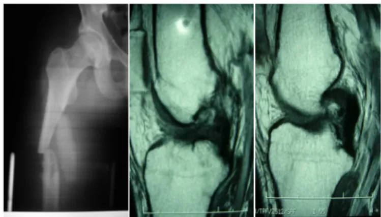
Figure 2. Patient with diaphyseal femoral fracture after a high-ener-gy accident (A). Eight months after the fracture, ipsilateral knee MRI was performed when the patient complained of knee instability. The absence of ACL (B) could be observed, suggesting chronic tearing (C). A bone-tendon-bone ACL patellar tendon autograft
reconstruc-tion was performed 1 month after diagnosis of the ACL tear.
trauma.
Diaphyseal femur fractures are accompanied by femoral neck fractures in 2.5% to 9% of the cases, with approxi-mately one-third of such cases remaining undiagnosed (Table 5). Moreover, ipsilateral fractures of the femur and tibia and tibial plateau fractures associated with diaphy-seal femoral fractures are uncommon (Table 6). However,
the frequency of knee laxity is at 53% (Table 7). Nerve
injuries due to medical error may occur in the context of
diaphyseal femur fractures (Table 8).
References implies a very high associated missed tibia fracture rate,
with the high probability that this rate is actually greater
(Figure 1). There were two cases of ACL tear (2.7%) in our
series, both after high-energy traumatisms (Figure 2).
Discussion
The treatment of choice for diaphyseal femur fractures is intramedullary nail stabilization, it achieves correct align-ment and high rates of bone healing (95-99%), has a low incidence of complications and permits early limb mobil-ity (21-23). Nonetheless, there are still some controver-sies with respect to diaphyseal femur fracture treatments. Also, since the mechanism causing the injuries in many cases is a high-energy accident, injuries associated with diaphyseal fractures are frequent.
The literature states that a diaphyseal femur fracture is accompanied by a femoral neck fracture in 2.5% to 9% of cases, with approximately one third of such cases remain-ing undiagnosed (6-12). In this series, all of the cases were fractures of the trochanteric region, with no femoral neck fracture having been diagnosed. In our series it is possible that either no femoral neck fracture associated with the diaphyseal fracture existed, or that the percentage of frac-ture undetected extended to 100% of the cases.
None of the ten cases of ipsilateral fracture of the femur and tibia in this series was associated with an ipsilateral knee injury, which is not consistent with the literature. Moreover, Schiedts states that knee instability is the main reason for the unsatisfactory results of his series (14).
Thus, this under-diagnosis may imply significant preva -lence.
Since the majority of diaphyseal femur fractures are caused by high-energy traumatisms, it is logical to think that knee ligaments may be injured due to their being sub-jected to great stress. In effect, the frequency of ipsilateral ACL knee lesions in a femur fracture varies between 5.3%
and 48% in the literature and the frequency of knee laxity reaches up to 53%. There are two cases of ACL tear (2.7%)
in our series, both after high-energy traumatisms. These
two cases represent 2.7% of the patients, a percentage significantly lower than those in the literature.
The examination of the knee in the presence of a mobile
fracture is difficult. De Campos et al examined the knees of 40 patients and conducted arthroscopy with closed dia-physeal femur fractures under anesthesia (24). More than
half of the patients with significant arthroscopy findings
showed effusion or laxity greater than degree I. This au-thor recommends a high index of suspicion for injuries in the ipsilateral knee of femur fracture patients. Dickson et al give details of injuries found in the magnetic resonance
of the ipsilateral knee during the first ten days after the
fracture and related it to the discoveries of examination
under anaesthesia, describing the sensitivity and specific -ity of the examination under anaesthesia for ACL, PCL, LCL and MCL lesions, respectively (25). They concluded that given a high index of suspicion, it is necessary to
under-take an examination of the knee under anaesthesia, with magnetic resonance being indicated if the said examina-tion is suggestive. In the histories of our patients, there is no record of any examination done under anaesthesia in
the surgical protocol of the final treatment of the femur
fracture, no exploratory arthroscopy and no MRI in the acute phase of the fracture. This, together with the low incidence of knee injuries in our series, leads us to think that perhaps there is under-diagnosis in our hospital with respect to these injuries.
Furthermore, injury of the superficial femoral artery in
approximately 2% of diaphyseal femur fractures has been described (26). Isaacson et al described five vascular in -juries associated with closed diaphyseal femur fractures
(27). Barr et al stated that the delay in the diagnosis of these injuries led to unsatisfactory results in a group of young patients (26). They found 15% of the patients
with significant hemodynamic abnormalities in the in -jured limb when their ankle/arm indices were subjected to Doppler measurements an average of 13 months after the fracture. There was one vascular injury in this series, which ended up in amputation.
Femoral and sciatic nerves are not usually injured in diaphyseal femur fractures and the majority of these in-juries occur with penetrating traumatisms. Nonetheless, nerve injuries may occur during fracture treatments. The majority of these nerve injuries are temporary. Rockwood
affirms that these injuries may be prevented by taking a
series of measures: cushioning the plaster splint around
the head of the fibula, revising the scrotum and groin of
the patient after positioning the perineal support to see that this is not exerting excessive traction, using traction only when necessary, applying traction to a distal femur
through a Steinmann pin in a way that permits knee flex -ion, or relaxing intraoperative traction once the pin has been put in place (10). We found 10 temporary nerve in-juries (four of the sciatic nerve, six of the peroneal nerve). Eight of them involved high-energy traumatism.
In conclusion, injuries associated with diaphyseal femur fractures were very frequent (46.4%). In 25.5% of our cases that had associated injuries, these were not detect-ed. It is necessary to pay special attention to the diagno-sis of possible hip and tibial fractures and to possible ACL injuries in the ipsilateral knee. The six nonunions of this series (4%) occurred in high-energy femoral shaft
frac-tures in patients younger than 70 years treated with open reduction and internal fixation.
E. Carlos Rodriguez-Merchan MD Luis Moraleda MD
Primitivo Gomez-Cardero MD
Department of Orthopaedic Surgery
La Paz University Hospital, Paseo de la Castellana 261
28046-Madrid, Spain
1. Salminen ST, Pihlajamaki HK, Avikainen VJ, Bostman ON. Population based epidemiologic and morphologic study of femoral shaft fractures. Clin Orthop Relat Res.
2000; 372: 241-9.
2. Regel G, Lobenhoffer P, Grotz M, Pape HC, Lehmann U, Tscherne H. Treatment results of patients with multi-ple trauma: an analysis of 3406 cases treated between
Trauma. 1995; 38: 70-8.
3. Bengner U, Ekbon T, Johnell O, Nilsson DE. Incidence of femur and tibial shaft fractures, epidemiology
1950-1983 in Malmo Sweden. Acta Orthop Scand. 1994; 61:
251-4.
4. Arneson TJ, Malton III LJ, Lewallen DG, O’Fallon WN. Epidemilogy of diaphyseal and distal femoral
frac-tures in Rochester Minnesota, 1965-1984. Clin Orthop Relat Res. 1988; 234:188-94.
5. Winquist RA, Hansen ST, Clawson DK. Closed in-tramedullary nailing of femoral fractures: a report of
five hundred and twenty cases. J Bone Joint Surg Am. 1984; 66-A: 529-39.
6. Laporte C, Benazet JP, Scemama P, Castelain C, Saillant G. Ipsilateral hip and femoral shaft fractures: compo-nents of therapeutic choice. Rev Chir Orthop
Repara-trice Appar Mot. 1999; 85: 24-32.
7. Watson JT, Moed BR. Ipsilateral femoral neck and shaft fractures: complications and their treatment. Clin
Or-thop Relat Res. 2002; 399: 78-86.
8. Casey MJ, Chapman MW. Ipsilateral concomitant frac-tures of the hip and femoral shaft. J Bone Joint Surg
Am. 1979; 61-A: 503-9.
9. Wiss DA, Sima W, Brien WW. Ipsilateral fractures of the femoral neck and shaft. J Orthop Trauma. 1992; 6: 159-66.
10. Nork SE. Femoral shaft fractures. In: Bucholz RW, Heckman JD, Court-Brawn CM, Tornetta P. Rockwood and Green’s Fractures in Adults. 5th ed. Philadelphia.
Lippincott Williams & Wilkins. 2009:1713-5.
11. Barquet A, Fernandez A, Leon H. Simultaneous ipsilat-eral trochanteric and femoral shaft fracture. Acta
Or-thop Scand. 1985; 56: 36-9.
12. Paul GR, Sawka MW, Whitelaw GP. Fractures of the ip-silateral femur and tibia: Emphasis on intra-articular and soft tissue injury. J Orthop Trauma. 1990; 4: 309-14.
13. Elmrini A, Elibrahimi A, Agoumi O, Boutayeb F, Mah-foud M, Elbardouni A, et al. Ipsilateral fractures of the
tibia and femur or floating knee. Int Orthop. 2006; 30:325-8.
14. Schiedts D, Mukisi M, Bouger D, Bastaraud H. Ipsilat-eral fractures of the femoral and tibial diaphyses. Rev
Chir Orthop Reparatrice Appar Mot. 1996; 82: 535-40.
15. Gregory P, DiCicco J, Karpin K, DiPasquale T, Hersco-vici D, Sanders R. Ipsilateral fractures of the femur and tibia: treatment with retrograde femoral nailing and undreamed tibial nailing. J Orthop Trauma. 1996; 10: 309-16.
16. Walling AK, Seradge H, Spiegel PG. Injuries to the knee ligaments with fractures of the femur. J Bone Joint Surg
Am. 1982; 64-A: 1324-7.
17. Moore TH, Patzakis MJ, Harvey P. Ipsilateral diaphy-seal femur fractures and knee ligament injuries. Clin
Orthop Relat Res. 1988; 232: 182-9.
18. Szalay MJ, Hosking OR, Annear P. Injury of the knee ligament associated with ipsilateral femoral shaft tures and with ipsilateral femoral and tibial shaft
frac-tures. Injury.1990; 21: 398-400.
19. Lakshman K, Scotland TR. The incidence of knee liga-ment injuries in 105 patients with lower limb
frac-tures. J Bone Joint Surg. 1985; 67 B: 151.
20. Walker DM, Kennedy JC. Occult knee ligament injuries associated with femoral shaft fractures. Am J Sports
Med. 1980; 8: 172-4.
21. Brumback RJ, Virkus WW. Intramedullary nailing of the femur: reamed versus nonreamed. J Am Acad
Or-thop Surg. 2000; 8: 83-90.
22. Bucholz RW, Jones A. Fractures of the shaft of the
fe-mur. J Bone Joint Surg Am. 1991; 73-A: 1561-6.
23. Wolinsky P, Tejwani N, Richmond JH, Koval Kj, Egol K, Stephen DJG. Controversies in intramedullary nailing of femoral shaft fractures. AAOS Instructional Course Lectures. 2002; 51: 291-303.
24. De Campos J, Vangsness CT, Merritt PO, Sher J. Ipsilat-eral knee injury with femoral fracture. Examination under anesthesia and arthroscopic evaluation. Clin
Orthop Relat Res. 1994; 300: 178-82.
25. Dickson KF, Galland MW, Barrack RL, Neitzschman HR, Harris MB, Myers L, et al. Magnetic resonance imaging of the knee after ipsilateral femur fracture. J Orthop
Trauma. 2002; 16: 567-71.
26. Barr H, Santes G, Stephenson I. Occult femoral artery injury in relation to fracture of the femoral shaft. J
Car-diovasc Surg. 1987; 28: 193-5.
27. Isaacson J, Louis DS, Costenbader JM. Arterial injury associated with closed femoral-shaft fracture. Report