Interleukin-19 Acts as a Negative Autocrine
Regulator of Activated Microglia
Hiroshi Horiuchi1, Bijay Parajuli1, Yue Wang1, Yasu-Taka Azuma2, Tetsuya Mizuno1, Hideyuki Takeuchi1*, Akio Suzumura1
1Department of Neuroimmunology, Research Institute of Environmental Medicine, Nagoya University, Furo-cho, Chikusa-ku, Nagoya, 464–8601, Japan,2Laboratory of Veterinary Pharmacology, Division of
Veterinary Science, Osaka Prefecture University Graduate School of Life and Environmental Science, Izumisano, Osaka, 598–8531, Japan
*htake@riem.nagoya-u.ac.jp
Abstract
Activated microglia can exert either neurotoxic or neuroprotective effects, and they play piv-otal roles in the pathogenesis and progression of various neurological diseases. In this study, we used cDNA microarrays to show that interleukin-19 (IL-19), an IL-10 family cyto-kine, is markedly upregulated in activated microglia. Furthermore, we found that microglia are the only cells in the nervous system that express the IL-19 receptor, a heterodimer of the IL-20Rαand IL-20Rβsubunits. IL-19 deficiency increased the production of such pro-in-flammatory cytokines as IL-6 and tumor necrosis factor-αin activated microglia, and IL-19 treatment suppressed this effect. Moreover, in a mouse model of Alzheimer’s disease, we observed upregulation of IL-19 in affected areas in association with disease progression. Our findings demonstrate that IL-19 is an anti-inflammatory cytokine, produced by activated microglia, that acts negatively on microglia in an autocrine manner. Thus, microglia may self-limit their inflammatory response by producing the negative regulator IL-19.
Introduction
Microglia are the macrophage-like resident immune cells that contribute to the maintenance of homeostasis in the central nervous system (CNS). Their functions include antigen presentation to initiate the immunological reaction, direct attack against non-self antigens, debris clearance, and support of neuronal circuit development [1–5]. Abnormal activation of microglia often damages the CNS, and microglial activation is a characteristic pathological hallmark of various neurological disorders, including neuroinflammatory and neurodegenerative diseases [6–12]. Activated microglia can exert either neurotoxic or neuroprotective effects, depending on their environmental conditions and the type and extent of stimulation [13–16], and can release both pro-inflammatory cytokines (e.g. interleukin-1β[IL-1β], IL-6, tumor necrosis factor-α
[TNF-α]) and anti-inflammatory cytokines (e.g. IL-10, transforming growth factor-β). Pro-inflammatory cytokines exacerbate neuroinflammation by fueling microglial activation, a11111
OPEN ACCESS
Citation:Horiuchi H, Parajuli B, Wang Y, Azuma Y-T, Mizuno T, Takeuchi H, et al. (2015) Interleukin-19 Acts as a Negative Autocrine Regulator of Activated Microglia. PLoS ONE 10(3): e0118640. doi:10.1371/ journal.pone.0118640
Academic Editor:Michelle L. Block, Indiana School of Medicine, UNITED STATES
Received:September 17, 2014
Accepted:January 12, 2015
Published:March 20, 2015
Copyright:© 2015 Horiuchi et al. This is an open access article distributed under the terms of the Creative Commons Attribution License, which permits unrestricted use, distribution, and reproduction in any medium, provided the original author and source are credited.
Data Availability Statement:All relevant data are within the paper.
whereas anti-inflammatory cytokines oppose those effects by suppressing microglial produc-tion of pro-inflammatory cytokines [17], suggesting that microglial activation is self-limiting. 19 is a member of the 10 family cytokines including 20, 22, 24, 26, IL-28A, IL-28B, and IL-29 [18]. At the primary sequence level, 19 is highly homologous to IL-20 and IL-24, and shares a receptor formed by heterodimerization of the IL-IL-20Rαand IL-20Rβ
subunits [19–21]. Previous studies showed that IL-19 contributes to the pathogenesis of auto-immune diseases such as psoriasis, asthma, and rheumatoid arthritis by upregulation of pro-inflammatory cytokines, including IL-6 and TNF-α[22–24]. However, the source and biological functions of IL-19 in the CNS have yet to be elucidated.
In this study, we found that microglia are the predominant producers of IL-19 in the CNS, as well as the only CNS cells to express functional IL-19 receptors. Furthermore, IL-19 sup-presses microglial production of pro-inflammatory cytokines. Our findings suggest that micro-glia self-limit their participation in neuroinflammation and neurodegeneration in various neurological diseases by producing a negative regulator, IL-19.
Methods
Reagents
Lipopolysaccharide (LPS) was purchased from Sigma-Aldrich (St. Louis, MO, USA). Recombi-nant mouse IL-19 protein was purchased from R&D Systems (Minneapolis, MN, USA).
Animals
All animal experiments were conducted under protocols approved by the Animal Experiment Committee of Nagoya University (approved numbers: 14017, 14018, and 14020). C57BL/6 (B6) mice were purchased from Japan SLC (Hamamatsu, Japan). IL-19-/-mice (B6 back-ground) [25,26] were obtained from Regeneron Pharmaceuticals, Inc. (Tarrytown, NY, USA). Transgenic mice expressing variants of human amyloid precursor protein (APP) with the K595N and M596L mutations and presenilin 1 (PS1) with the A264E mutation were purchased from the Jackson Laboratory (B6C3-Tg(APP695)3Dbo Tg(PSEN1)5Dbo/J; #003378) [27], and then backcrossed to C57BL/6J mice for more than 10 generations after purchase (the resultant strain is designated here as APP/PS1 Tg) [28].
Cells
Primary cultures were prepared from B6 mice or IL-19-/-mice. Primary microglia cultures were isolated from primary mixed glial-cell cultures prepared from newborn mice using the“shaking off”method (at 14 daysin vitro), as described previously [29]. The purity of the cultures was> 99%, as determined by anti-CD11b immunostaining. Cultures were maintained in Dulbecco’s modified Eagle’s minimum essential medium (DMEM) (Sigma-Aldrich) supplemented with 10% fetal bovine serum (FBS) (Equitech-Bio, Inc., Kerrville, TX, USA), 5μg/ml bovine insulin (Sigma-Aldrich), and 0.2% glucose. Primary astrocyte cultures were purified from primary mixed glial cultures by three to four repetitions of trypsinization and replating, as described pre-viously [30]. The purity of astrocytes was>95%, as determined by GFAP-specific immunostain-ing. Primary neuron cultures were prepared from the cortices of mouse embryos at embryonic day 17 (E17), as described previously [31]. Briefly, cortical fragments were dissociated into single cells in dissociation solution (Sumitomo Bakelite, Akita, Japan) and resuspended in nerve culture medium (Sumitomo Bakelite). Neurons were plated on polyethylenimine-coated glass coverslips (Asahi Techno Glass, Chiba, Japan) at a density of 5 × 10⁴cells/well (500μl/well) in 24-well
Competing Interests:Akio Suzumura is a PLOS
multidishes. The purity of the cultures was>95%, as determined by NeuN-specific immunos-taining. Neurons were used at 14 daysin vitrofor the following assessments.
RNA extraction, cDNA microarrays, and reverse
transcription-polymerase chain reaction (RT-PCR)
Microglia and astrocytes were seeded at a density of 1 × 10⁵cells/well (500μl/well) in 48-well plates. Neurons were plated on polyethylenimine-coated coverslip at a density of 5 × 10⁴cells/ well (500μl/well) in 24-well plates. Microglia were treated with 100 ng/ml LPS for 0–24 h or 1–1000 ng/ml for 6 h. Astrocytes were treated with 100 ng/ml LPS for 0–24 h or 1–1000 ng/ml for 12 h. Total RNA was extracted from microglia, astrocytes, neurons, hippocampi of APP/PS1 Tg mice and B6 mice (10- and 15-month-old) using the miRNeasy Mini Kit (Qiagen, Valencia, CA, USA). RNA was run on an Agilent 2100 Bioanalyzer to assess quality; only samples with excellent RNA quality (RNA Integrity Number [RIN]>7) were used. For microarray experi-ments, RNA samples were evaluated on the Agilent Mouse SurePrint G3 8 × 60K Microarray. The resultant signals were quantitated and analyzed using the Agilent GeneSpring 12.1 soft-ware. For RT-PCR experiments, RNA was reverse transcribed into cDNA using SuperScript III (Life Technologies, Carlsbad, CA, USA). Expression levels of genes encoding IL-19, IL-6,
TNF-α, hypoxanthine phosphoribosyltransferase 1 (HPRT1), and glyceraldehyde 3-phosphate dehy-drogenase (GAPDH) were measured using quantitative PCR (qPCR), which was performed on cDNA on a Rotor-Gene Q using the Rotor-Gene SYBR Green PCR Kit (Qiagen). Relative ex-pression levels were determined using theΔΔCTmethod; the genes of interest were normalized
to the geometric mean of HPRT1 and GAPDH. The following specific primer sets were used: Mouse IL-1 sense: 5’-TACAGAGACAGGGTGTTCCAGGAC-3’;
Mouse IL-19 antisense: 5’-GCATTGGTGGCTTCCTGACTGCAGT-3’; Mouse IL-6 sense: 5’-ACAAGTCGGAGGCTTAATTACACAT-3’; Mouse IL-6 antisense: AATCAGAATTGCCATTGCACAA-3’; Mouse TNF-αsense: GACCCTCACACTCAGATCATCTTCT-3’; Mouse TNF-αantisense: 5’-CCACTTGGTGGTTTGCTACGA-3’; Mouse HPRT1 sense: 5’-CCTAAGATGAGCGCAAGTTGAA-3’; Mouse HPRT1 antisense: 5’-CCACAGGACTAGAACACCTGCTAA-3’
Mouse GAPDH sense: 50-TGTGTCCGTCGTGGATCTGA-3’ Mouse GAPDH antisense: 50- CCTGCTTCACCACCTTCTTGA-3’ Assays were carried out in five independent trials.
Enzyme-linked immunosorbent assay (ELISA)
Microglia and astrocytes were seeded at a density of 1 × 10⁵cells/well in 48-well plates, and then treated with 100 ng/ml LPS for 0–24 h. Hippocampi of APP/PS1 Tg and B6 mice (15-month-old) were collected after transcardial perfusion with phosphate-buffered saline. Concentrations of IL-19, IL-6, and TNF-αin culture supernatant were determined using the appropriate ELISA kits (IL-19, eBioscience; IL-6 and TNF-α, R&D Systems). Assays were carried out in three independent trials.
Western blotting
7.5], 150 mM NaCl, 1% Nonident P-40, 2 mM EDTA, 0.1% SDS) containing phosphatase in-hibitor cocktail (Sigma-Aldrich) and protease inin-hibitor mixture (Roche, Mannheim, Ger-many). Fifty micrograms of cell lysate protein dissolved in Laemmli sample buffer were separated on 4–20% SDS-polyacrylamide gels (Mini-Protean TGX, Bio-Rad, Hercules, CA, USA), and transferred to Hybond-P polyvinylidene difluoride membranes (GE Healthcare, Buckingham, UK). The membranes were blocked with 5% skim milk in Tris-buffered saline containing 0.05% Tween-20 for 1 h at room temperature, and then incubated overnight at 4°C with rabbit anti-IL-20Rαpolyclonal antibodies (Merck Millipore, Billerica, MA, USA), rat anti-IL-20Rβmonoclonal antibody (eBioscience, San Diego, CA, USA), mouse anti-STAT3 monoclonal antibody (BD Pharmingen, Franklin Lakes, NJ, USA), rabbit anti-pSTAT3 poly-clonal antibodies (Cell Signaling Technology, Danvers, MA, USA), or mouse anti-β-actin monoclonal antibody (Sigma-Aldrich), followed by incubation with horseradish peroxidase—
conjugated secondary antibodies (GE Healthcare, Buckingham, UK) for 1 h at room tempera-ture. The signals were visualized using SuperSignal West Pico chemiluminescent substrate (Thermo Fisher Scientific), and quantitated using a CS Analyzer 3.0 system (Atto, Tokyo, Japan). Assays were carried out in five independent trials.
Cell proliferation assay
Microglia were treated with IL-19 (0–100 ng/ml) for 24 h. Cell proliferation was assessed using the 3-(4,5-dimethyl-thiazol-2yl)-5-(3-carboxymethoxyphenyl)-2-(4-sulfophenyl)-2H-tetrazo-lium (MTS) assay with the CellTiter 96 AQueous One Solution assay (Promega, Madison, WI, USA) as described previously [33]. Assays were carried out in five independent trials.
Statistical analysis
Statistical significance was analyzed using Student’st-test or one-way analysis of variance
(ANOVA) followed by post-hoc Tukey’s test, using GraphPad Prism version 6.0 (GraphPad Software, La Jolla, CA, USA).
Results
Activated microglia are the predominant producers of IL-19
First, we evaluated the mRNA expression profile of activated microglia using cDNA microar-rays. We found that 1,449 genes were significantly upregulated following treatment with LPS for 24 h. We identified IL-19 as the most upregulated gene (Table 1).
Next, we confirmed IL-19 expression in CNS cells using qPCR and ELISA. LPS stimulation significantly induced IL-19 mRNA and protein expression in microglia and astrocytes (Fig. 1), but not in neurons (data not shown). IL-19 mRNA expression levels in microglia and astro-cytes reached their peaks at 6 h and 12 h after stimulation with 100 ng/ml LPS, respectively (Fig. 1, A and D). Next, we evaluated IL-19 mRNA expression levels in microglia and astrocytes stimulated with LPS at graded doses (0–1000 ng/ml) for 6 h and 12 h, respectively. IL-19 mRNA reached a plateau at 100 ng/ml LPS (Fig. 1, B and E). Production of IL-19 in microglia and astrocytes increased in a time-dependent manner, and microglia produced approximately twice as much IL-19 as astrocytes (Fig. 1, C and F). These results indicate that IL-19 is predom-inantly produced by activated microglia in the CNS.
Functional IL-19 receptor is exclusively expressed in microglia
A recent study reported that IL-19 receptor is expressed in astrocytes, but not in microglia [34]. However, our data indicated that the IL-20Rαsubunit was exclusively expressed in microglia, whereas the IL-20Rβsubunit was ubiquitously expressed in neurons, astrocytes, and microglia (Fig. 2A). Moreover, LPS stimulation did not alter the expression levels of the IL-19 receptor subunits in astrocytes and microglia (Fig. 2B). Downstream signals of IL-19 reportedly mediate STAT3 phosphorylation [20,21,35]. Indeed, Western-blotting analysis confirmed that treatment with IL-19 induced STAT3 phosphorylation in microglia (Fig. 2C). Although a previous study also showed that IL-19 suppresses the production of IL-6 and TNF-αin LPS-stimulated astro-cytes [34], in our hands IL-19 did not affect the levels of production of these cytokines by LPS-stimulated astrocytes (Fig. 3, A and B). These data show that microglia exclusively express the functional receptor of IL-19, and are thus the primary potential targets of IL-19 in the CNS.
IL-19 does not activate microglia in resting state
Next, we investigated whether IL-19 affects the proliferation of microglia. The MTS assay re-vealed that stimulation with IL-19 itself did not affect microglial proliferation (Fig. 4). Similar-ly, treatment with IL-19 did not induce microglial production of pro-inflammatory cytokines such as IL-6 and TNF-α(Fig. 5, A and B).
IL-19 suppresses the production of pro-inflammatory factors in activated
microglia
Next, we investigated whether IL-19 affects the functions of activated microglia. Additional treatment of IL-19 did not significantly suppress the production of IL-6 and TNF-αin stimulated microglia, but ablation of IL-19 increased the production of these cytokines in LPS-stimulated microglia (Fig. 5, C and D), and these effects were reversed by addition of IL-19 (Fig. 5, C and D). These results indicate that IL-19 may serve to limit microglial activation by suppressing pro-inflammatory cytokine production.
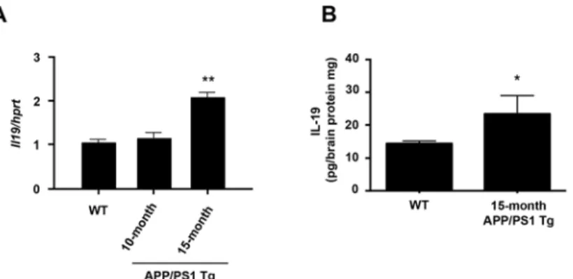
In APP/PS1 Tg mice, IL-19 is upregulated in affected areas in
association with disease progression
Finally, to assess the involvement of IL-19 in neurodegenerative diseases, we investigated the expression pattern of IL-19 in a mouse model of AD using APP/PS1 Tg mice. mRNA
Table 1. Upregulated genes in activated microglia (Top 10).
Gene name Fold Change (vs. untreated)
interleukin-19 7287.7
schlafen 4 5064.0
cDNA sequence U90926 4117.9
urate (5-hydroxyiso-)hydrolase 2273.6
interleukin-6 1852.3
solute carrier organic anion transporter family, member 2b1 1502.6
nitric oxide synthase 2, inducible 1022.8
interleukin-1 family, member 6 980.2
prostaglandin E synthase 915.8
immunoresponsive gene 1 908.5
Gene expression levels are expressed as fold change relative to the untreated control (n = 3).
expression levels of IL-19 gradually increased in the hippocampi of APP/PS1 Tg mice
(Fig. 6A). Protein levels of IL-19 were also significantly elevated in the diseased mice (Fig. 6B).
Discussion
In this study, we showed that activated microglia secrete IL-19 and are the only CNS cells to ex-press functional IL-19 receptor. Although a recent paper [34] reported that astrocytes express IL-19 and its receptor, our findings revealed that microglia are both the main source and target of IL-19 in the CNS. Previous studies reported that IL-19 is derived primarily from macrophages, and to a lesser extent from B cells and epithelial-lineage cells including epithelial cells, endothelial cells, and keratinocytes [36–38]. Our data also demonstrated that microglia, the macrophage-like cell in the CNS, produce more IL-19 than astrocytes, which belong to the epithelial lineage.
Previous reports showed that monocyte-lineage cells release IL-19 upon pro-inflammatory stimulation by factors such as LPS, IL-6, IL-17, and TNF-α[39,40]. IL-19 induces monocytes to produce IL-6 and TNF-α[22,39] and triggers T cells to release T-helper type 2 cytokines
Fig 1. Activated microglia are the predominant source of IL-19 in the CNS.(A) qPCR data for IL-19 in microglia treated with LPS (100 ng/ml) for 0–24 h. (B) qPCR data for IL-19 in microglia treated with LPS (0– 1000 ng/ml) for 6 h. (C) ELISA data for IL-19 in culture supernatant of microglia treated with LPS (100 ng/ml) for 24 h. (D) qPCR data for IL-19 in astrocytes treated with LPS (100 ng/ml) for 0–24 h. (E) qPCR data for IL-19 in astrocytes treated with LPS (0–1000 ng/ml) for 12 h. (F) ELISA data for IL-19 in culture supernatant of astrocytes treated with LPS (100 ng/ml) for 24 h. Microglia released approximately twice as much IL-19 as astrocytes.*,p<0.05.**,p<0.01.***,p<0.001. Values are means±SD (n = 5).
Fig 2. Microglia are the only cells in the CNS that express functional IL-19 receptor.(A) Western blotting data for receptors of IL-19. Only microglia express both the IL-20Rαand IL-20Rβsubunits. Neu, neurons; Ast, astrocytes; Mi, microglia. (B) Western blotting data for receptors of IL-19. LPS stimulation did not change the expression levels of IL-19 receptor subunits. Skin was used as the positive control. NT, untreated; LPS, LPS-treated. (C) Western-blotting analysis of STAT3 phosphorylation in microglia treated with 100 ng/ml IL-19. IL-19 induced STAT3 phosphorylation, the main downstream signal of the IL-19 receptor. All quantitative data are expressed as means±SD (n = 3).*,p<0.01vs. 0 min.**,p<0.001vs. 0 min.
doi:10.1371/journal.pone.0118640.g002
Fig 3. IL-19 does not suppress production of pro-inflammatory cytokines in LPS-stimulated astrocytes.Astrocytes were treated with LPS (100 ng/ml) and recombinant IL-19 (100 ng/ml) for 24 h. (A) Production of IL-6 and (B) TNF-α. Treatment with IL-19 did not microglial production of IL-6 and TNF-α. Values are means±SD (n = 3). N.S., not significant.
Fig 4. IL-19 does not affect microglial proliferation.Microglial proliferation was evaluated by MTS assay. Treatment with 1–100 ng/ml IL-19 for 24 h did not affect microglial proliferation. Values are means±SD (n =
5).
doi:10.1371/journal.pone.0118640.g004
Fig 5. IL-19 suppresses production of pro-inflammatory cytokines in activated microglia.(A and B) Microglia were treated with recombinant IL-19 (100 ng/ml) for 24 h. Production of (A) IL-6 and (B) TNF-α. Stimulation with IL-19 itself did not induce microglial production of IL-6 and TNF-α. (C and D) Microglia from B6 mice and IL-19-/-mice were incubated in the presence of 100 ng/ml LPS, with or without 100 ng/ml IL-19, for 24 h. Production of (C) IL-6 and (D) TNF-α. IL-19 deficiency increased the production of pro-inflammatory factors in activated microglia, and this effect was reversed by addition of IL-19.*,p<0.001. Data indicate
means±SD (n = 3). N.D., not detected.
including IL-4, IL-5, IL-10, and IL-13 [23]. Therefore, IL-19 is recognized as an important player in the pathogenesis of systemic inflammatory diseases such as psoriasis, asthma, rheumatoid ar-thritis, and sepsis [22–24,41]. On the other hand, IL-19 also exerts anti-inflammatory activities in inflammatory bowel disease and vascular inflammatory diseases [25,42]. In contrast to its pro-inflammatory properties in monocytes, IL-19 attenuates the production of IL-6 and TNF-α
in macrophages [25]. Our data also demonstrated that IL-19 has an anti-inflammatory activity in microglia. Activation of STAT3 is a key downstream step in IL-19 receptor signaling [20,21,35], and our data also showed that stimulation with IL-19 activates STAT3 in microglia, as shown previously in macrophages [25]. Moreover, IL-19 reportedly suppresses inflammatory responses in vascular smooth muscle cells via downregulation of human antigen R, which pro-motes transcription of pro-inflammatory factors [42]. Thus, the pleiotropic properties of IL-19 in monocyte-lineage cells may depend on signaling downstream of the IL-19 receptor.
Activated microglia play a pivotal role in the pathogenesis and progression of various neu-rological disorders such as trauma, stroke, inflammation, epilepsy, and neurodegenerative dis-eases by releasing neurotoxic factors including glutamate, nitric oxide, and reactive oxygen species [6–12,43]. Microglia also exacerbate neuroinflammation associated with leukocyte infil-tration by secreting pro-inflammatory cytokines and chemokines including TNF-α,
interferon-γ(IFN-γ), IL-1β, IL-6, IL-12, IL-17, and IL-27 [5,43–47]. These microglial inflammatory and neurotoxic factors fuel the spiral of microglial activation and sustain chronic neuroinflamma-tion and neurodegeneraneuroinflamma-tion [1,5,43,48]. On the other hand, activated microglia also utilize self-limiting systems. IFN-γ, a well-known microglia stimulatory factor, is released from acti-vated microglia [44,47] but it also alleviates microglia-mediated neuroinflammation in manner that depends on activation-induced cell death [49], providing a possible mechanism for relapse and remission in multiple sclerosis. Our data suggest that IL-19 may participate in a novel mechanism by which microglia self-limit their inflammatory response. Unlike IFN-γ, IL-19 did not affect microglial survival. Furthermore, supplemental IL-19 did not exert additional sup-pressive effects on pro-inflammatory cytokine production by activated microglia, probably be-cause these cells had already released a saturating amount of IL-19. Therefore, IL-19 may be a weaker negative regulator of microglia than IL-10. Mouse model of AD exhibited gradual upre-gulation of IL-19 in the affected areas as disease progressed. Further studies are needed to
Fig 6. In a mouse model of AD, IL-19 is upregulated in affected regions in association with disease progression.(A) qPCR data for 19 in the hippocampi of APP/PS1 Tg and B6 mice. (B) ELISA data for IL-19 in the hippocampi of APP/PS1 Tg and B6 mice. IL-IL-19 expression level gradually increased as disease progressed. WT, wild-type B6 mice.*,p<0.01vs. WT.**,p<0.001vs. WT. Values are means±SD (n = 3).
clarify whether additional administration of IL-19 could effectively slow and halt the progres-sion of neurodegenerative diseases.
In summary, we identified IL-19 as a novel anti-inflammatory cytokine released from acti-vated microglia. In the CNS, microglia are the only cells that express functional IL-19 receptor; therefore, microglia are both the main source and target of IL-19. Ablation of IL-19 increased microglial production of pro-inflammatory factors, and this effect was reversed by addition of IL-19. Our findings suggest that microglia self-limit their participation in neuroinflammation and neurodegeneration by producing a negative regulator, IL-19.
Author Contributions
Conceived and designed the experiments: HH HT AS. Performed the experiments: HH BP HT. Analyzed the data: HH HT AS. Contributed reagents/materials/analysis tools: YW YA TM. Wrote the paper: HH HT AS.
References
1. Block ML, Zecca L, Hong JS. Microglia-mediated neurotoxicity: uncovering the molecular mechanisms. Nat Rev Neurosci. 2007; 8(1): 57–69. PMID:17180163
2. Boche D, Perry VH, Nicoll JA. Review: activation patterns of microglia and their identification in the human brain. Neuropathol Appl Neurobiol. 2013; 39(1): 3–18. doi:10.1111/nan.12011PMID:
23252647
3. Kempermann G, Neumann H. Neuroscience. Microglia: the enemy within? Science. 2003; 302(5651): 1689–1690. PMID:14657479
4. Kreutzberg GW. Microglia: a sensor for pathological events in the CNS. Trends Neurosci. 1996; 19(8): 312–318. PMID:8843599
5. Takeuchi H. Neurotoxicity by microglia: Mechanisms and potential therapeutic strategy. Clin Exp Neu-roimmunol. 2010; 1(1): 12–21.
6. Bruijn LI, Miller TM, Cleveland DW. Unraveling the mechanisms involved in motor neuron degeneration in ALS. Annu Rev Neurosci. 2004; 27: 723–749. PMID:15217349
7. Cagnin A, Brooks DJ, Kennedy AM, Gunn RN, Myers R, Turkheimer FE, et al. In-vivo measurement of activated microglia in dementia. Lancet. 2001; 358(9280): 461–467. PMID:11513911
8. Eikelenboom P, Bate C, Van Gool WA, Hoozemans JJ, Rozemuller JM, Veerhuis R, et al. Neuroinflam-mation in Alzheimer’s disease and prion disease. Glia. 2002; 40(2): 232–239. PMID:12379910 9. McGeer PL, McGeer EG. Inflammatory processes in amyotrophic lateral sclerosis. Muscle Nerve.
2002; 26(4): 459–470. PMID:12362410
10. Nelson PT, Soma LA, Lavi E. Microglia in diseases of the central nervous system. Ann Med. 2002; 34 (7–8): 491–500. PMID:12553496
11. Orr CF, Rowe DB, Halliday GM. An inflammatory review of Parkinson’s disease. Prog Neurobiol. 2002; 68(5): 325–340. PMID:12531233
12. Pavese N, Gerhard A, Tai YF, Ho AK, Turkheimer F, Barker RA, et al. Microglial activation correlates with severity in Huntington disease: a clinical and PET study. Neurology. 2006; 66(11): 1638–1643. PMID:16769933
13. Jimenez S, Baglietto-Vargas D, Caballero C, Moreno-Gonzalez I, Torres M, Sanchez-Varo R, et al. In-flammatory response in the hippocampus of PS1M146L/APP751SL mouse model of Alzheimer’s dis-ease: age-dependent switch in the microglial phenotype from alternative to classic. J Neurosci. 2008; 28(45): 11650–11661. doi:10.1523/JNEUROSCI.3024-08.2008PMID:18987201
14. Nakanishi H, Wu Z. Microglia-aging: roles of microglial lysosome- and mitochondria-derived reactive oxygen species in brain aging. Behav Brain Res. 2009; 201(1): 1–7. doi:10.1016/j.bbr.2009.02.001
PMID:19428609
15. Sawada M. Neuroprotective and toxic changes in microglia in neurodegenerative disease. Parkinson-ism Relat Disord. 2009; 15 Suppl 1: S39–41. doi:10.1016/S1353-8020(09)70011-2PMID:19131042 16. Wu Z, Tokuda Y, Zhang XW, Nakanishi H. Age-dependent responses of glial cells and leptomeninges during systemic inflammation. Neurobiol Dis. 2008; 32(3): 543–551. doi:10.1016/j.nbd.2008.09.002
17. Sawada M, Suzumura A, Hosoya H, Marunouchi T, Nagatsu T. Interleukin-10 inhibits both production of cytokines and expression of cytokine receptors in microglia. J Neurochem. 1999; 72(4): 1466–1471. PMID:10098850
18. Sabat R, Wallace E, Endesfelder S, Wolk K. IL-19 and IL-20: two novel cytokines with importance in in-flammatory diseases. Expert Opin Ther Targets. 2007; 11(5): 601–612. PMID:17465720
19. Fickenscher H, Hor S, Kupers H, Knappe A, Wittmann S, Sticht H. The interleukin-10 family of cyto-kines. Trends Immunol. 2002; 23(2): 89–96. PMID:11929132
20. Dumoutier L, Leemans C, Lejeune D, Kotenko SV, Renauld JC. Cutting edge: STAT activation by IL-19, IL-20 and mda-7 through IL-20 receptor complexes of two types. J Immunol. 2001; 167(7): 3545–3549. PMID:11564763
21. Parrish-Novak J, Xu W, Brender T, Yao L, Jones C, West J, et al. Interleukins 19, 20, and 24 signal through two distinct receptor complexes. Differences in receptor-ligand interactions mediate unique bi-ological functions. J Biol Chem. 2002; 277(49): 47517–47523. PMID:12351624
22. Liao YC, Liang WG, Chen FW, Hsu JH, Yang JJ, Chang MS. IL-19 induces production of IL-6 and TNF-alpha and results in cell apoptosis through TNF-TNF-alpha. J Immunol. 2002; 169(8): 4288–4297. PMID:
12370360
23. Liao SC, Cheng YC, Wang YC, Wang CW, Yang SM, Yu CK, et al. IL-19 induced Th2 cytokines and was up-regulated in asthma patients. J Immunol. 2004; 173(11): 6712–6718. PMID:15557163 24. Sakurai N, Kuroiwa T, Ikeuchi H, Hiramatsu N, Maeshima A, Kaneko Y, et al. Expression of IL-19 and
its receptors in RA: potential role for synovial hyperplasia formation. Rheumatology (Oxford). 2008; 47 (6): 815–820. doi:10.1093/rheumatology/ken061PMID:18397956
25. Azuma YT, Matsuo Y, Kuwamura M, Yancopoulos GD, Valenzuela DM, Murphy AJ, et al. Interleukin-19 protects mice from innate-mediated colonic inflammation. Inflamm Bowel Dis. 2010; 16(6): 1017–1028. doi:10.1002/ibd.21151PMID:19834971
26. Ellison S, Gabunia K, Richards JM, Kelemen SE, England RN, Rudic D, et al. IL-19 Reduces Ligation-Mediated Neointimal Hyperplasia by Reducing Vascular Smooth Muscle Cell Activation. Am J Pathol. 2014; 184(7): 2134–2143. doi:10.1016/j.ajpath.2014.04.001PMID:24814101
27. Borchelt DR, Ratovitski T, van Lare J, Lee MK, Gonzales V, Jenkins NA, et al. Accelerated amyloid de-position in the brains of transgenic mice coexpressing mutant presenilin 1 and amyloid precursor pro-teins. Neuron. 1997; 19(4): 939–945. PMID:9354339
28. Takeuchi H, Mizoguchi H, Doi Y, Jin S, Noda M, Liang J, et al. Blockade of gap junction hemichannel suppresses disease progression in mouse models of amyotrophic lateral sclerosis and Alzheimer’s dis-ease. PLoS One. 2011; 6(6): e21108. doi:10.1371/journal.pone.0021108PMID:21712989
29. Suzumura A, Mezitis SG, Gonatas NK, Silberberg DH. MHC antigen expression on bulk isolated mac-rophage-microglia from newborn mouse brain: induction of Ia antigen expression by gamma-interferon. J Neuroimmunol. 1987; 15(3): 263–278. PMID:3110208
30. Liang J, Takeuchi H, Doi Y, Kawanokuchi J, Sonobe Y, Jin S, et al. Excitatory amino acid transporter expression by astrocytes is neuroprotective against microglial excitotoxicity. Brain Res. 2008; 1210: 11–19. doi:10.1016/j.brainres.2008.03.012PMID:18410911
31. Takeuchi H, Mizuno T, Zhang G, Wang J, Kawanokuchi J, Kuno R, et al. Neuritic beading induced by activated microglia is an early feature of neuronal dysfunction toward neuronal death by inhibition of mi-tochondrial respiration and axonal transport. J Biol Chem. 2005; 280(11): 10444–10454. PMID:
15640150
32. Doi Y, Takeuchi H, Horiuchi H, Hanyu T, Kawanokuchi J, Jin S, et al. Fingolimod phosphate attenuates oligomeric amyloid beta-induced neurotoxicity via increased brain-derived neurotrophic factor expres-sion in neurons. PLoS One. 2013; 8(4): e61988. doi:10.1371/journal.pone.0061988PMID:23593505 33. Takeuchi H, Jin S, Wang J, Zhang G, Kawanokuchi J, Kuno R, et al. Tumor necrosis factor-alpha
in-duces neurotoxicity via glutamate release from hemichannels of activated microglia in an autocrine manner. J Biol Chem. 2006; 281(30): 21362–21368. PMID:16720574
34. Cooley ID, Chauhan VS, Donneyz MA, Marriott I. Astrocytes produce IL-19 in response to bacterial challenge and are sensitive to the immunosuppressive effects of this IL-10 family member. Glia. 2014; 62(5): 818–828. PMID:24677051
35. Preimel D, Sticht H. Molecular modeling of the interleukin-19 receptor complex. Novel aspects of recep-tor recognition in the interleukin-10 cytokine family. J Mol Model. 2004; 10(4): 290–296. PMID:
15243778
37. Hsing CH, Li HH, Hsu YH, Ho CL, Chuang SS, Lan KM, et al. The distribution of interleukin-19 in healthy and neoplastic tissue. Cytokine. 2008; 44(2): 221–228. doi:10.1016/j.cyto.2008.06.007PMID:
18809337
38. Wolk K, Kunz S, Asadullah K, Sabat R. Cutting edge: immune cells as sources and targets of the IL-10 family members? J Immunol. 2002; 168(11): 5397–5402. PMID:12023331
39. Gallagher G, Dickensheets H, Eskdale J, Izotova LS, Mirochnitchenko OV, Peat JD, et al. Cloning, ex-pression and initial characterization of 19 (IL-19), a novel homologue of human interleukin-10 (IL-interleukin-10). Genes Immun. 2000; 1(7): 442–450. PMID:11196675
40. Hsing CH, Hsieh MY, Chen WY, Cheung So E, Cheng BC, Chang MS. Induction of interleukin-19 and interleukin-22 after cardiac surgery with cardiopulmonary bypass. Ann Thorac Surg. 2006; 81(6): 2196–2201. PMID:16731153
41. Hsing CH, Chiu CJ, Chang LY, Hsu CC, Chang MS. IL-19 is involved in the pathogenesis of endotoxic shock. Shock. 2008; 29(1): 7–15. PMID:18246602
42. Cuneo AA, Herrick D, Autieri MV. Il-19 reduces VSMC activation by regulation of mRNA regulatory fac-tor HuR and reduction of mRNA stability. J Mol Cell Cardiol. 2010; 49(4): 647–654. doi:10.1016/j. yjmcc.2010.04.016PMID:20451530
43. Takeuchi H, Suzumura A. Gap junctions and hemichannels composed of connexins: potential thera-peutic targets for neurodegenerative diseases. Front Cell Neurosci. 2014; 8: 189. doi:10.3389/fncel. 2014.00189PMID:25228858
44. Kawanokuchi J, Mizuno T, Takeuchi H, Kato H, Wang J, Mitsuma N, et al. Production of interferon-gamma by microglia. Mult Scler. 2006; 12(5): 558–564. PMID:17086900
45. Kawanokuchi J, Shimizu K, Nitta A, Yamada K, Mizuno T, Takeuchi H, et al. Production and functions of IL-17 in microglia. J Neuroimmunol. 2008; 194(1–2): 54–61. doi:10.1016/j.jneuroim.2007.11.018
PMID:18295350
46. Sonobe Y, Yawata I, Kawanokuchi J, Takeuchi H, Mizuno T, Suzumura A. Production of IL-27 and other IL-12 family cytokines by microglia and their subpopulations. Brain Res. 2005; 1040(1–2): 202–207. PMID:15804439
47. Wang X, Suzuki Y. Microglia produce IFN-gamma independently from T cells during acute toxoplasmo-sis in the brain. J Interferon Cytokine Res. 2007; 27(7): 599–605. PMID:17651021
48. Orellana JA, Saez PJ, Shoji KF, Schalper KA, Palacios-Prado N, Velarde V, et al. Modulation of brain hemichannels and gap junction channels by pro-inflammatory agents and their possible role in neuro-degeneration. Antioxid Redox Signal. 2009; 11(2): 369–399. doi:10.1089/ars.2008.2130PMID:
18816186