Cytokine Inhibitors and Pain Control
Inibidores de Citocinas e Controle da Dor
Waldiceu A. Verri Jr.(1), Thiago M. Cunha(1), Stephen Poole(2), Sérgio H. Ferreira(1), Fernando Q. Cunha(1)
Recebido em 17/03/07. Aprovado, após revisão, em 25/09/07. Conflits of interest: nothing to disclose. 1. Department of Pharmacology, School of Medicine of Ribeirao Preto University of Sao Paulo, Brazil.
2. Division of Immunology and Endocrinology, National Institute for Biological Standards and Control, England, UK.
Corresponding author: Fernando de Queiroz Cunha, Faculdade de Medicina de Ribeirão Preto (USP), Av. Bandeirantes, 3.900, Ribeirão Preto, SP, Brazil, CEP 14049-900, phone: + 55 16 3602-3205, fax: + 55 16 3633-0021, e-mail: [email protected]
RESUMO
Os autores fazem uma revisão sobre evidências que demonstram o papel de citocinas em modelos experimentais de dor, discutindo possíveis terapias com alvo em citocinas para controle da dor.
Palavras-chave: hiperalgesia, dor inflamatória, citocinas,
noci-cepção, analgesia.
ABSTRACT
The authors describe the evidences supporting the role of cytokines in experimental pain, discussing possible approaches for pain control using cytokine-targeting therapies.
Keywords: hyperalgesia, inflammatory pain, cytokines,
nocicep-tion, analgesia.
PAIN
The present review describes the evidences supporting the role of cytokines in experimental pain, discussing pos-sible approaches for pain control using cytokine-targeting therapies such as soluble receptors, receptor antagonists, neutralizing antibodies, and drugs that inhibit cytokine production.
Pain is a nociceptive sensation which perception causes unpleasant emotions. It is accepted that sensitization of the primary sensory neurons is essential to inflammatory pain. Nonetheless, this sensitization of the nociceptor was, for a long time, regarded as a result of the excitatory action of various inflammatory mediators (a “soup of inflammatory mediators”) released in the site of the inflamed or damaged tissues(1). However, this hypothesis was challenged by the
discovery of the non-steroidal anti-inflammatory drugs me-chanism of action by Vane’s group(2) and the demonstration
that, in man(3) and in animals(3), eicosanoids do not cause
overt pain but sensitize the pain receptors.
Likewise, other inflammatory mediators like endo-thelin(4,5), sympathetic amines(6,7), substance P(8-10), NGF(11,12)
as well as bradykinin(13) also possess the same nociceptor
sensitizing property. These mediators act directly on neu-ronal receptors, triggering molecular mechanisms that ultimately facilitate the electrical activity of the neuronal
membrane. Although our views of the molecular mecha-nisms of nociceptor sensitization are not complete, there is general agreement that the G-protein coupled receptor stimulation by the inflammatory mediators activates the enzyme adenylate cyclase with production of cAMP. This substance, in turn, triggers the activation of a group of protein kinases (PKA and PKC), which in turn lead to phosphorylation of ion channels in the membrane. As a result, there is a facilitation of the inward sodium current by TTX resistant Na+ channels, facilitation of inward Ca++
currents and inhibition of the outward K+ currents. This is
probably the basic peripheral mechanism of hyperalgesia, when a previously slight or ineffective thermal, mechanical or chemical stimulation becomes painful.
exudates or inflamed tissues give a distorted picture of the evolution of the pathological process and even suggest a disorganized soup of cells and mediators. In fact, sequen-tial release of inflammatory mediators or cellular events observed after challenge by inflammatory stimuli will be observed only with sequential temporal determination. Most of the time, it is this temporal and physio-pathological hierarchy that allows the researcher to discover the site of action of old drugs or to propose targets for new drug development. The cascade of hypernociceptive cytokines described in this review is one of these physio-pathological hierarchic processes that our lab helped to discover in the last two decades(14). Cytokines in general act as
interme-diate mediators, releasing other cytokines as well as final mediators. In some instances however, cytokines may act as a final hyperalgesic mediator and have been described as being involved in the development of neuronal damage, contributing to the symptom described in man as allody-nia. In allodynia (“allo” = different) the perceived pain is different than that expected by the type of stimulus applied (touch, for example, may produce an intense pricking pain). Contrasting, in hyperalgesia, the perceived “quality” of the pain induced by the triggering stimulus (e.g. mechanical or thermal) can be anticipated by the patient. In the present review we use the term “hypernociception” to describe any increase in sensitivity of the primary nociceptor neuron in animal models, considering that the terms allodynia and hyperalgesia have been developed for use in clinical practice rather than for experimental studies.
PRO-INFLAMMATORY CYTOKINES AND
PERIPHERAL INFLAMMATORY PAIN
Cytokines are defined as proteins produced and released in a coordinated sequence by cells in response to a variety of inflammatory stimuli, such as parasites, viruses, bacteria and their products, or in response to other cytokines(15,16).
In general, cytokines constitute a link between cellular injuries or immunological recognition of non-self and the local or systemic signs of inflammation(17-20).
Although chemokines are considered cytokines (che-motactic cytokines), they belong to a particular group of cytokines with specific chemical and functional properties. They are usually smaller than the other cytokines (8–10 kDa) and direct the recruitment of leukocytes from the blood stream to the extravascular tissues by acting on receptors that are differentially expressed on leukocyte subsets. Today, besides their role in leukocyte recruitment
during inflammation, other functions have been attributed to the chemokines such as angiogenesis, modulation of the immune response and fever(21).
In inflammation, the resident cells such as dendritic cells, macrophages, mast cells and lymphocytes are tissue cells that release cytokines after recognizing the inflammatory stimuli. These cytokines play an essential role in the development of inflammatory pain as well as other inflammatory events, such as leukocyte migration. The principal cytokines des-cribed as participating in the development of inflammatory pain were Interleukin (IL)-1β, Tumor Necrosis Factor (TNF)-α, IL-6, and the chemokines IL-8, Chemokine-Induced Neutrophil Chemoattractant (CINC)-1 and Keratinocyte-derived Chemokine (KC)(14,22-30). Recently, it
has been demonstrated that IL-15, IL-18 and IL-12 also induce inflammatory hypernociception(19,31-33). The role of
these cytokines and chemokines as well as their interactions is discussed below.
CASCADE OF CYTOKINES MEDIATES
INFLAMMATORY HYPERNOCICEPTION
Following the discovery of cytokines, many efforts were applied to characterizing their function. In the late 1980s, the participation of cytokines in the induction of inflam-matory signals such as the recruitment of white blood cells, acute phase protein release, fever, and the increase of per-meability of blood vessels(34,35) had already been described.
With the community’s new understanding of these cytokine properties, IL-1β entered the inflammatory pain scene.
Different cell types including macrophages, monocytes and glial cells produce IL-1β. In turn, this cytokine induces the production of other inflammatory mediators(36). The
demonstration that IL-1β stimulates the production of prostaglandins(37,38) and that prostaglandins ultimately
sen-sitize the nociceptor(39,40), prompted the investigation of the
IL-1β hypernociceptive role and mechanism. In a seminal finding, it was demonstrated the IL-1β importance to the genesis of inflammatory hypernociception in experimental animals(22). The intraplantar injection of IL-1β produces
a severe mechanical hypernociception, which depends on prostanoid production since the local pre-treatment with indomethacin (COX inhibitor) blocked its effects. Moreover, it was observed that IL-1β via prostaglandins production mediates inflammatory hypernociception indu-ced by local (rat paw) administration of carrageenin (Cg) or LPS(22). In fact, the Cg- or LPS-induced mechanical
Figure 1 – Inflammatory stimulus-induced cytokine cascades mediate mechanical hypernociception in rats. The scheme represents the coordinated release of mediators triggered by the different inflammatory stimuli. Ag (antigen), Cg (carrageenin), CINC-1 (cytokine-induced neutrophil chemoattractant-1), ET-1 (endothelin-1), IL (Interleukin), LPS (lipopolysaccharide), PGE2 (prostaglandin E2), SA (sympathetic amines)(4,22-24,29,31,32,45-50).
local treatment with antibodies against IL-1β(24). This
partial inhibition suggests that another hypernociceptive pathway besides the IL-1β/prostanoids is involved in the mechanical inflammatory hypernociception induced by Cg. As mentioned earlier, it seems that sympathetic amines account for the other component of inflammatory pain, at least in this experimental model.
Studying the role of chemokines in inflammatory hyper-nociception, it was found that whereas IL-1β induces the release of prostanoids, the neutrophil chemoattractant che-mokine IL-8/CXCL8 (human cheche-mokines) or CINC-1/ CXCL1 (rat chemokines) mediates the participation of sympathetic components of inflammatory hypernocicep-tion. In fact, the IL-8/CXCL8 or CINC-1/CXCL-induced mechanical hypernociception is inhibited by β−adrenergic receptor antagonists, but not by COX inhibitors. Moreo-ver, antiserum anti-IL-8/CXCL8 also partially inhibited (50%) the Cg-induced hypernociception in rats(23). The
CXC chemokines hypernociceptive mechanism seems to depend on the tissue evaluated. For instance, in contrast with the hypernociception induced by the administration of IL-8 in cutaneous hindpaw tissue, in rat knee joints IL-8 induces mechanical hypernociception that is prevented by IL-1ra(41).
Following these observations, another pro-inflamma-tory cytokine was in evidence. TNF-α was considered as a key molecule in the initiation of the inflammatory process. Generally, it is the first cytokine released in response to inflammatory stimuli such as a bacterial infection(42-44).
Mo-reover, the possible involvement of TNF-α in the initiation of inflammatory hypernociception was raised since TNF-α stimulates the release of IL-1β and CXC chemokines.
Once again, our group was the first to demonstrate the hypernociceptive effect and mechanisms of TNF-α in
the hind paw of rats. The TNF-α hypernociception was
partially inhibited by indomethacin (COX inhibitor) and atenolol (β-adrenoreceptor antagonist), and abolished by the co-treatment with these drugs, suggesting that prosta-noids and sympathetic amines mediate the TNF-α-induced hypernociception. Furthermore, treatment with anti-IL-1β or anti-IL-8/CINC-1 antiserum partially inhibited
TNF-α-induced hypernociception, and the combination of both antisera completely abolished TNF-α effects, suggesting that IL-1β and IL-8/CINC-1 also mediate TNF-α-induced hypernociception. In addition, the mechanical hypernoci-ceptive effects of IL-1β and IL-8/CINC-1 are inhibited by indomethacin and atenolol, respectively. These data suggest that TNF-α induces hypernociception in rats via
two independent and parallel pathways: 1. TNF-α – IL-1β Prostanoids; 2. TNF-α– CINC-1 sympathetic amines (Fi-gure 1). Moreover, Cg- or LPS-induced hypernociception is abolished by the pretreatment with anti-TNF-α antise-rum. Thus, TNF-α plays a pivotal role in the Cg and LPS induction of hypernociception in rats acting via the two parallel pathways described above(24,45).
In the same manner as TNF-α, IL-1β and IL-8/CINC, IL-6 also produces mechanical hypernociception in rats, which was inhibited by indomethacin, anti-IL-1β antise-rum or IL-1ra. Regarding the cytokine cascade underlying the IL-6-induced inflammatory hypernociception, it was demonstrated that antiserum against IL-6 inhibits
TNF-α-induced mechanical hypernociception in rats(24). Thus, it
seems that TNF-α, IL-6 and IL-1β sequentially precede the release of prostanoids to induce hypernociception in rats. These experiments strongly supported our above-mentioned hypothesis that in rats there is a cascade of cytokines constituting a link between injuries and the release of primary hypernociceptive mediators. This concept allows us to understand why the inhibition of one (IL-1β or TNF-α) or several (glucocorticoids) cytokines inhibits hypernociception induction. The clinical success of anti-TNF-α in rheumatoid arthritis also exemplifies this concept(51), which opposes the
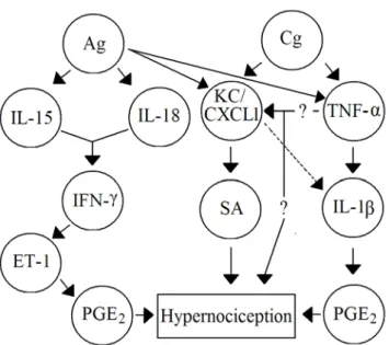
Figure 2 –Cascades of cytokines mediate mechanical hypernociception induced by non-immune and immune inflammation in mice. Ag (antigen), Cg (carrageenin), ET-1 (endothelin-1), IFN-γ (Interferon-γ), IL (Interleukin), KC/CXCL1 (Keratinocyte-derived chemokine), PGE2 (prostaglandin E2), SA (sympathetic amines), TNF-α
(Tumour necrosis factor-α)(19,30,33).
Other groups also find a sequential role of cytokines in different hypernociceptive models in rats. For instance, in a model of peripheral sensitization, the intraperitoneal administration of lithium chloride or LPS sensitizes the rat to the tail flick test. It has been hypothesized that this sen-sitization is mediated by sequential release of TNF-α and IL-1β that activates the subdiaphragmatic vagal afferents(25).
In a different model, TNF-α trigger IL-1β mediated thermal and mechanical hypernociception induced by intraplantar administration of complete Freund’s adjuvant (CFA)(29). In
this model, IL-1βinduces the release of nerve growth factor (NGF), rather than stimulating prostanoid production. In fact, antibodies against NGF inhibited the hypernociception induced by CFA and IL-1β. In addition, IL-1ra inhibited
CFA- and IL-1β-, but not NGF-induced mechanical and
thermal hypernociception. Therefore, suggesting the follo-wing sequential release of these mediators: CFA – TNF-α – IL-1β – NGF nociceptor sensitization(26,29).
The relevance of a biological experimental concept depends on its confirmation in more than one model and animal species. In this regard, our hypothesis was further sup-was further sup-ported by recent data showing that mechanical inflammatory hypernociception in mice is also mediated by a peripheral cascade of cytokines(30). In mice, the Cg inflammatory
sti-mulus induces the release of TNF-α and KC/CXCL1 that
trigger subsequent release of IL-1β followed by prostanoid production. KC/CXCL1 also stimulates the sympathetic component of inflammatory hypernociception (Figure 2).
In addition to experimental models, the contribution of TNF-α to inflammatory pain has also been clinically suggested. For instance, TNF-α was associated with musculoskeletal pain syndromes and pain states associated with nucleus pulposus herniation(52,53). Furthermore, the
pain associated with mandibular movement and tenderness (allodynia) on posterior palpation of the temporomandi-bular joint has been related to the level of TNF-α in the synovial fluid(54).
Thus, accumulating evidences in the literature suggests a role of cytokines as intermediary hypernociceptive media-tors, mostly in the peripheral tissues, supporting that these molecules are likely a therapeutic target for controlling inflammatory pain states. In this regarding, our group de-veloped several peptides based on the amino acid sequence structure of IL-1β. Among them, the tripeptide KD(P)T consistently inhibited IL-1β and inflammatory mechanical hypernociception. Recently, a selective IL-1 receptor anta-gonist that differs from naturally occurring IL-1ra by the presence of a methionine group named Anakinra (Amgen
Inc.) was developed. Anakinra was tested in models of in-flammatory diseases and is under clinical evaluation(55,56).
Besides IL-1β, TNF has also become a target for the treatment of inflammatory diseases including arthritis. Drugs such as infliximab (chimeric anti-TNF-α antibody),
etanercept (p75 TNF-α receptor / immunoglobulin G
fu-sion protein), and recently, adalimumab (fully humanized monoclonal anti-TNF-α antibody) are available. These anti-TNF-α therapies were shown to be effective in diffe-rent diseases that are generally associated with pain such as uveitis(57), skin and joint manifestations of psoriasis(58),
and mainly in rheumatoid arthritis(59-62).
Despite the great amount of data concerning the no-ciceptive role of TNF-α and the beneficial effect of anti-TNF drugs on inflammatory diseases as mentioned above, there are few studies suggesting that these drugs inhibit hypernociception induction. Etanercept as well as anti-TNF antibodies reduced experimental inflammation and neuropathy-induced hypernociception(63-65) and infliximab
therapy significantly reduced pain scores in rheumatoid arthritis(51,66). It is noteworthy that chronic anti-TNF
therapies may present side effects, including in some cases heart failure and also immune suppression, which may lead to serious infections such as tuberculosis(67,68).
the-rapies must be considered. Taking this into account, some already-known drugs emerge as an alternative for inhibition of cytokines. In this sense, we investigated the potential of pentoxifylline and thalidomide for reducing inflammatory hypernociception. In fact, thalidomide and pentoxifylline reduced hypernociception associated with inflammation by a mechanism dependent on the inhibition of cytokine production, mainly at the peripheral site. Therefore, the cost-benefit relationship of pentoxifylline and thalidomide prompt the use of these drugs or drugs with similar mecha-nisms of action to control inflammatory pain.
ROLE OF IL-15 AND IL-18 IN TH-1
IMMUNE INFLAMMATION-INDUCED
HYPERNOCICEPTION
Th1 immune inflammatory responses are involved in the development of different diseases including rheuma-toid arthritis, myocarditis, encephalomyelitis, diabetes, and lupus(69-72). Indeed, these inflammatory diseases are
frequently accompanied by hyperalgesia. Thus, the deve-accompanied by hyperalgesia. Thus, the deve-lopment of inflammatory nociception models that mimic these diseases is of great importance, because they would allow rational studies of the pathophysiology of those conditions and, therefore, better and more specific thera-pies. In this regard, models such as the antigen-induced inflammatory hypernociception in previously immunized animals resemble the immune inflammation triggered by Th1 responses(19).
It has been demonstrated that cytokines (TNF-α, IL-1β, IL-8/CINC-1 and IL-6) also play an important role in hypernociception induced by an immune challenge. In fact, specific anti-rat TNF-α, anti-rat IL-1β and anti-human IL-8 antiserum or IL-1ra decreased the antigen-induced hypernociception, and the levels of these cytokines were elevated in the paw after the administration of the antigen in immunized rats and mice(73)(Figures 1 and 2). Besides the
role of these well-known hyperalgesic cytokines described above, other cytokines (IL-15, IL-18) seem important during hypernociception resulting from Th1 immune inflammatory responses.
IL-18 and IL-15 share a role in the development of RA, in the driving of Th1 responses, and in the induction of interferon-gamma (IFN-γ) production(69-71,74). Supporting
these evidences, IL-15 and IL-18-targeting therapies ameliorate collagen-induced arthritis, which is a classical model of RA(75-77). Based on this experimental finding, it
was performed a clinical trial (phase I-II) using a
monoclo-nal anti-IL-15 antibody for RA treatment. This approach presented significant beneficial effect on RA severity in RA patients(78).
Taking this evidence into account, we have recently reported that IL-15 and IL-18 mediate Th1-like-induced inflammatory hypernociception via the sequential release of IFN-γ, endothelin-1 (ET-1) and PGE2. The hypernoci-ception induced by IL-15 and IL-18 is independent of each other, suggesting that they are acting synergistically(19,33).
Therefore, IL-15 and IL-18 targeting therapies might also be of importance for down-regulating adaptive inflamma-tion-induced hypernociception, probably with lower inci-dence of side effects such as propensity to infections(67,68).
Besides IL-15 and IL-18, IL-12 is also an important cytokine for arthritis development(70,79). Its pronociceptive
effect was demonstrated in rats. IL-12 injection induces mechanical hypernociception, which is mediated by ET-1 action on ETB receptors, and is not dependent of prosta-noids, sympathetic amines or leukotrienes in rats(32). This
effect is important since human volunteers reported pain in the site of IL-12 injection during clinical trials for cancer treatment or vaccine adjuvant(32).
CYTOKINES AND DIRECT NOCICEPTORS
SENSITIZATION
Despite the evidences that the hypernociceptive effects of cytokines are indirect as described above, it has been reported that sensory neurons express cytokine receptors. Therefore, these cytokines might also directly sensitize the nociceptor during inflammation. In effect, some research groups demonstrated that cytokines such as TNF-α evoke action potentials in nociceptive neurons when applied topi-cally to peripheral axons in vivo, and increases the sensitivity to mechanical and chemical stimuli(27,80,81). Investigating the
TNF mechanism to provoke this direct effect in neurons, Jin & Gereau(82) investigated the effect of this cytokine
on TTX-R Na+ activity. It was demonstrated that TNF-α
acting on TNFR1 enhances TTX-R Na+ currents in primary afferent nociceptive neurons by activating the neuronal MAP kinase (p38) pathway.
The direct effect of IL-1β, IL-6 and chemokines has also been demonstrated. For instance, IL-1β and IL-6 together with their soluble receptor (sIL-6R) are able to sensitize the sensory neurons to heat(83,84). The IL-6 effect
was due to the activation of Janus tyrosine kinase and the PKC intracellular signaling pathway(83,84). Moreover,
anandamide by Gi-protein-, phospholipase C-, and PKC- dependent mechanism in vitro(85).
Nevertheless, it is important to be careful in the inter-pretation of these results and other possibilities should be considered: 1) although the cytokines are acting directly on sensory neurons, they could produce secondary mediators, such as prostanoids, which finally sensitized the nocicep-tors. Actually, Nicol et al. (27), demonstrated: 1) that TNF-α
enhances the sensitivity of cultured neurons to capsaicin in a COX products-dependent manner; 2) that cytokines act on cells other than sensory neurons, such as dorsal root ganglion satellite cells, that in turn produce direct sensitizing mediators; and 3) the relevance of the direct effect of cytokines to the genesis of inflammatory hypernociception. In this context, Parada et al. (86) showed that the effective attenuation of
TNFR1 expression in peripheral sensory neurons by intra-thecal treatment with antisense oligodeoxynucleotides to this receptor did not alter either TNF-α- or Cg-induced acute mechanical hypernociception, when injected in rat paws. Therefore, it seems that TNF-acting on sensory neuron mem-brane TNFR1 is not necessary to the onset of peripheral acute inflammatory hypernociception. Thus, similar experiments have to be performed with IL-6 and IL-1β to determine the importance of their direct effect on the sensitive neuron for the genesis of inflammatory hypernociception.
CHRONIC INFLAMMATORY PAIN MEDIATED
BY CYTOKINES
The contribution of cytokines to the genesis of acute inflammatory pain has been extensively demonstrated, as mentioned above. However, its role in chronic inflamma-tory pain states is still unclear.One problem is that there are few experimental models of chronic inflammatory pain. Although some acute inflammatory models could become chronic such as the CFA-induced inflammation, it is still an unsolved acute inflammation. The clinically important chronic pain cases are when the lesion is already resolved but the pain persists.
In an attempt to study these cases we developed an experimental model to investigate the mechanism of the phenomenon mentioned above. Actually, a persistent hypernociceptive state can be induced by successive daily injections of PGE2 or dopamine. After 14 daily injections of the stimulus (PGE2 or dopamine), the sensitivity of the nociceptor does not return to its basal level but, instead, reaches a plateau, which persists for more than thirty days(87). Other studies have reported that a persistent
hypernociception can also be triggered by the persistent presence of stimuli(88).
In line with the PGE2- or dopamine-induced persis-tent hypernociception, it was demonstrated that repeated intraplantar injections of IL-1β, IL-8 or TNF-α also cause persistent mechanical hypernociception that is prevented by daily treatment with indomethacin and atenolol. In fact, the daily treatment with indomethacin or atenolol inhibits 50% of the persistent hypernociception induced by TNF-α, and the combination of indomethacin and atenolol blo-cked the onset of the process. These results suggest that, as in the case of acute hypernociception, the persistent hypernociception induced by IL-1β and IL-8 is due to the endogenous release of eicosanoids and sympathetic amines, respectively. Moreover, both mediators play a role in the development of the persistent hypernociception induced by TNF-α(89).
In addition to the data generated using the model of persistent hypernociception described above, the possible role of TNF-α in “chronic pain” was further supported by the results obtained using hypernociceptive priming. In this animal model, the injection of Cg induces an inflammatory hypernociception lasting hours to days, which produces a “primed” state lasting several weeks. During this time, injection of PGE2 induces hypernociception that is
marke-dly enhanced and prolonged compared to PGE2-induced
hypernociception in normal “unprimed” rats(90).
Studying the genesis of the priming state, Parada et al.(86)
showed that intrathecal administration of ODN antisense to TNFR1, which reduced the TNFR1 mRNA in sensory neurons, but not in the peripheral tissue, attenuates either Cg- or TNF-α-induced priming without affecting the
acute hypernociception. The mechanism by which TNF-α
induces chronic hypernociceptive priming is not completely clear, but it has been suggested that this cytokine induces an increase of PKCεlevels in the nociceptor(91). Moreover,
the modulation of TTX-R Na+ currents via p38 activation could also be responsible for amplifying the latter effect of PGE2 on this channel activity during chronic priming.
PHARMACOLOGICAL INHIBITION OF
INFLAMMATORY HYPERNOCICEPTION
BY DRUGS THAT BLOCK THE RELEASE
OF CYTOKINES
(1.1.-1.6.), there are additional approaches to inhibit cytokines effects. The inhibition of cytokines production is an efficient therapy for preventing hypernociception. Therefore, the following sections (2.1.- 2.3.) will focus in drugs that act by this mechanism.
GLUCOCORTICOIDS
The main mechanism described for glucocorticoids is related to the modulation of nuclear factor κB (NFκB). This transcription factor was identified in the nuclei of mature B lymphocytes as a transcription factor that binds to a 10 bp DNA element in the kappa immunoglobulin light-chain enhancer(92). The NF-κB is a key player in
controlling both innate and adaptive immunity. NF-κB is present in the cytoplasm in association with inhibitory proteins that are known as inhibitors of NF-κB (IκBs). After activation by a large number of inducers, the IκB proteins become phosphorylated, ubiquitylated and, subsequently, degraded by the proteasome. The degradation of IκB allows the translocation of NF-κB proteins to the nucleus and bind their cognate DNA binding sites to regulate the transcription of a large number of genes, including pro-inflammatory cytokines, chemokines, adhesion molecules, matrix metalloproteinases (MMPs), COX-2 and inducible nitric oxide synthase (iNOS)(93).
Furthermore, there are accumulating evidence that NF-κB activity is also regulated by the direct modification of NF-κB proteins through phosphorylation and, perhaps, acetylation. Several investigators have shown that IL-1
and TNF induce NF-κB phosphorylation and activation
by pathways that are distinct from those leading to IκB degradation and NF-κB nuclear translocation(93).
A main mechanism by which transcription factors regu-late gene expression is to bring histone acetyltransferases (HATs) and histone deacetylases (HDACs) to target sites to modify histone acetylation. Histoneacetylation status influences the folding and functional state of the chromatin fibre and modulates the accessibility of DNA to the trans-criptional machinery for gene expression. Previous studies have shown that NF-κB interacts with HATs, to positively regulate gene expression, and with HDACs to negatively regulate transcription(93).
It is well recognized that steroid hormones are im-portant inhibitor of NF-κB. The mechanism of action of glucocorticoids (GC) involves the binding of GC with the GC receptor (GR) in the cell cytoplasm. The complex GC-GR can directly bind to the NF-κB, inhibiting its action.
In addition, the complex GC-GR can migrate to the cell nucleus and bind to GC-responsive genes (GREs), inhibi-ting the production of pro-nociceptive cytokines (TNF-α and IL-1β) and affecting their transport. In addition the complex GC-GR increases the IκB synthesis, which in turn binds to NF-κB, inhibiting its translocation to the nucleus(94).
Taking into account the evidence in the literature, we investigated whether glucocorticoids have antinociceptive effects. It was found that the hypernociceptive responses to intraplantar injections of carrageenin, bradykinin, TNF-α, IL-1β, and IL-6 were inhibited by pretreatment with de-xamethasone. However, the hypernociceptive responses to IL-8, PGE2 and dopamine were not affected, which is in agreement with the cascade of cytokines described in rats. Dexamethasone also inhibited TNF-α release by a murine macrophage cell-line stimulated with LPS. Furthermore, there is evidence that the antinociceptive effects of dexame-thasone are, at least in part, mediated by annexin-1 (pre-viously designated lipocortin-1). The lipocortin-1-mediated glucocorticoids antinociceptive effect was demonstrated by Ferreira et al.(95), using the rat paw constant pressure test.
The inhibition of hypernociceptive responses to injections of bradykinin and IL-1β by dexamethasone was reversed by antiserum anti-lipocortin-1, injected subcutaneously, 24h and 1h before hypernociceptive substances. This was supported by the finding that the hypernociceptive res-ponses to injections of bradykinin, TNF-α and IL-1β, but not responses to PGE2, were inhibited by pretreatment with lipocortin-1(2-26). Further supporting the lipocortin-1-mediated dexamethasone effects, lipocortin-1(2-26) partially inhibited TNF-α release by a murine macrophage cell-line stimulated with LPS, and antiserum anti-lipocortin-1 par-tially reversed the inhibition by dexamethasone. Therefore, these data are consistent with a partial role for endogenous lipocortin-1 in mediating the antinociceptive effect of dexamethasone. It is probable that GC-GREs-induced inhibition of NF-κB activity mediates the other component of the dexamethasone antinociceptive activity.
In agreement with the proposed mechanisms of action of glucocorticosteroids, dexamethasone inhibits not only the expression of inflammatory mediators, but also the expression of receptors such as the B1 (bradykinin recep-tor), which are upregulated during the hypernociceptive response to formalin(96) and Cg. Additionally, both thermal
Furthermore, there is evidence that corticosteroid mo-dulation (pituitary-adrenal axis) may have a role in regu-lating the stress-induced analgesia, and this may implicate the interactions of the corticosteroids with pain-inhibiting systems(98). Both exogenous (pharmacological) and
endoge-nous (physiological) glucocorticoids suppress the hyperno-ciception and the up-regulation of spinal preprodynorphin mRNA induced by sustained inflammation provoked by CFA. The results also suggest that spinal preprodynorphin mRNA suppression may partially underlie the inhibition of the CFA-induced hypernociception(99).
Besides the glucocorticosteroids, there are also other inhibitors of NF-κB action, especially IκB kinase inhibitors (IKK). The IKK inhibitors prevent the ubiquination of IκB, the first step in the activation of NF-κB. Thus, it would be expected that inhibition of IKK activity may prevent injury-, infection-, or stress-induced upregulation of various proinflammatory genes and may thereby reduce hypernoci-ception and inflammation. In cell culture experiments, the IKK inhibitor S1627 inhibited IL-1β-stimulated nuclear translocation and DNA-binding of NF-κB. The effects of S1627 on hypernociception were also evaluated(100).
S1627 reversed thermal and mechanical hypernociception, and the inflammatory paw edema in the zymosan-induced paw inflammation model. S1627 also significantly reduced tactile and thermal hypernociception in the CCI model of neuropathic pain. However, the drug had no effect on acute inflammatory nociception induced by formalin and did not affect responses to heat and tactile stimuli in naive animals. In agreement with this S1627 prevented the zymosan-induced nuclear translocation of NF-κB in the spinal cord and the upregulation of NF-κB-responsive genes including COX-2, TNF-α and IL-1β.
THALIDOMIDE
Thalidomide (α-N-phthalylglutamic-acid-amine) was developed as an anti-emetic drug to be used mainly during pregnancy. However, the use of thalidomide is restricted due to its teratogenic effects. It is the drug of choice for the treatment of erythema nodosum leprosum, an acute inflammatory complication occurring in 30% of leproma-tous leprosy patients, usually in association with initiation of multidrug therapy(101). The research on thalidomide
me-chanisms of action demonstrated that it selectively inhibits TNF-α production by human alveolar macrophages(102) and
human monocytes stimulated by E. coli or Mycobacterium leprae products(103,104). In fact, thalidomide inhibits TNF-α
production by enhancing its mRNA degradation(102).
Fur-thermore, thalidomide does not affect the production of other cytokines by LPS-stimulated monocytes(103).
The specific and effective inhibitory action of thalidomi-de on TNF-α production ensures the demonstration of its effectiveness for the treatment of different diseases such as graft-versus-host disease(105), oral aphthous ulcers in patients
with human immunodeficiency virus infection(106),
refrac-tory rheumatoid arthritis(107), and neutrophil migration to
the synovial cavity during immune inflammation(108).
Considering the evidence on the potential therapeutic use of thalidomide in diseases associated with TNF-α, we investigated whether it would have analgesic effects. It was found that thalidomide prevents the Cg-induced me-chanical hypernociception in rats and acetic acid induced-abdominal contortions in mice. Nevertheless, it did not affect the mechanical hypernociceptive effect of TNF-α or PGE2. These results reinforce the idea that the inhi-bition of inflammatory hypernociception by thalidomide may be associated with the selective inhibition of TNF-α mRNA expression(48,104). Moreover, the analgesic effect
of thalidomide is peripheral and indirect, neither affec-ting edema nor inducing antihypernociceptive cytokines such as IL-4 and IL-10. The potential therapeutic use of thalidomide to control pain has been also demonstrated on the hypernociception induced by CCI in rats(109) and
carrageenin-induced chronic hypernociceptive priming, in which phenomena TNF-α has a pivotal role(86). Thus,
there is consistent evidence for the potential usefulness of thalidomide in the management of inflammatory pain associated with TNF-α.
PENTOXIFYLLINE
Pentoxifylline was originally developed for the treatment of vascular diseases, and its pharmacokinetics and pharma-codynamics ensure its safety. Similar to thalidomide, the main mechanism of action of pentoxifylline is the reduction
of TNF-α production by inhibiting mRNA expression by
more than 50%(110-112). Such an effect has been detected in
vivo in patients with systemic inflammatory manifestation of erythema leprosum nodosum(112) and in vitro in peripheral
blood mononuclear cells(110,112). Furthermore,
pentoxifylli-ne concentration-dependently inhibited IL-2 and IFN-γ concomitantly with the inhibition of TNF-α in peripheral blood mononuclear cells(113). Nevertheless, the inhibition
of IL-1β(104,114,115) and IL-6(112,116) remains controversial.
cytokines such as IL-10(112), and in vitro, pentoxifylline may
augment IL-10 production in unstimulated whole blood cells or inhibit its production(117). Thus, it is possible that
the modulation/increase of IL-10 production by pentoxi-fylline may also account for its anti-hypernociceptive effect depending on the experimental model.
Because TNF-α and other cytokines have a crucial role in the development of different inflammatory diseases, pentoxifylline is a potential drug for the treatment of glomerulonephritis(118), leprae(112), streptozotocin-induced
diabetes mellitus(119), and sepsis(116,120).
Concerning inflammatory pain, it has been demonstra-ted in humans that the pre-treatment with pentoxifylline diminished the opioid requirement in the early post-cho-lecystectomy operative period, as well as TNF-α and IL-1β serum levels. In contrast, a recent study using the same procedure described above demonstrated that the post-treatment with pentoxifylline did not modify the serum levels of TNF-α and IL-6 or the opioid requirement(121).
These data suggest the importance of the pharmacokinetics of pentoxifylline for its action.
Furthermore, besides the demonstrations of the effective-ness of pentoxifylline in humans, there is consistent evidence of its antinociceptive activity in experimental models of nociception. In rats, pentoxifylline inhibits mechanical hyper-nociception administered pre or post-injury(122). Pentoxifylline
is also efficacious in different models of inflammatory noci-ception such as the writhing response induced by acetic acid and zymosan, zymosan-induced articular hypernociception in rats, and carrageenin-, bradykinin- and TNF-α-induced mechanical hypernociception. However, pentoxifylline did not inhibit iloprost (PGI2 analogue), IL-1β or PGE2 effects, suggesting that the mechanism of action underlying this drug’s effect is upstream of IL-1β release. No central effects of pentoxifylline were detected. Thus, pentoxifylline could be useful for the treatment of inflammatory pain(50).
Besides the inhibition of TNF-α and other cytokines, it has been reported that pentoxifylline inhibits in vivo
the endotoxin-induced NF-κB activation in intestine of rats during sepsis. Pentoxifylline may downregulate NF-κB expression via PKC inhibition(123). Although there
is no direct evidence that such a mechanism accounts for pentoxifylline’s antinociceptive actions, it is probably an additional beneficial effect since the NF-κB activation ulti-mately leads to pro-inflammatory cytokine production(124).
Moreover, as pentoxifylline unspecifically inhibits PKC and PKA(123) this may also contribute to the control of pain since
there is clearly evidence for the participation of PKCε in hypernociceptive priming(86,91) and PKA in
hypernocicep-tion induchypernocicep-tion(125,126).
It is important to mention that although specific cytoki-ne inhibitors (anankira, etacytoki-nercept, infliximab) are available, the high cost of these therapies must be considered. Taking this into account, the cost-benefit relationship of pentoxi-fylline and thalidomide prompt the use of these drugs.
CONCLUSIONS
This review reinforces the crucial role of cytokines and chemokines mediating inflammatory pain in the majority of the described experimental nociceptive models. Its central goal was to show the concept that cytokine/chemokine cascades link the inflammatory stimuli and the release of the final mediators (such as prostaglandins, sympathetic amines) ultimately responsible for the nociceptor sensitization. In addition, cytokines/chemokines may also directly sensitize the nociceptors. Thus, substances that block the synthesis or action of the nociceptive cytokines/chemokines could be useful for inflammatory pain treatment.
ACKNOWLEDGMENTS
The authors wish to express their appreciation to Ieda Regina dos Santos Schivo, Sérgio Roberto Rosa and Giuliana Bertozi Francisco for excellent technical assist-ance. This work was supported by grants from FAPESP and CNPq.
References
1. Lynn B: Textbook of pain. 2. ed. Edinburgh: Churchill Livingstone, 1984.
2. Vane JR: Inhibition of prostaglandin synthesis as a mechanism of action for aspirin-like drugs. Nat New Biol 231: 232-5, 1971. 3. Ferreira SH: Prostaglandins, aspirin-like drugs and analgesia.
Nat New Biol 240: 200-3, 1972.
4. Ferreira SH, Romitelli M, de Nucci G. Endothelin-1 participation in overt and inflammatory pain. J Cardiovasc Pharmacol 13: S220-2, 1989.
5. Zhou QL, Strichartz G, Davar G: Endothelin-1 activates ET(A) receptors to increase intracellular calcium in model sensory neurons. Neuroreport 12: 3853-7, 2001.
7. Hannington-Kiff FG: Textbook of pain 3. ed. Edinburg: Churchill Livingstone, 1989.
8. Henry JL: Effects of substance P on functionally identified units in cat spinal cord. Brain Res 114: 439-51, 1976.
9. Nakamura-Craig M, Gill BK: Effect of neurokinin A, substance P and calcitonin gene related peptide in peripheral hyperalgesia in the rat paw. Neurosci Lett 124: 49-51, 1991.
10. Xu XJ, Dalsgaard CJ, Wiesenfeld-Hallin Z: Spinal substance P and N-methyl-D-aspartate receptors are coactivated in the induction of central sensitization of the nociceptive flexor reflex. Neuroscience 51: 641-8, 1992.
11. Lewin GR, Mendell LM: Nerve growth factor and nociception. Trends Neurosci 16: 353-9, 1993.
12. Lewin GR, Ritter AM, Mendell LM: Nerve growth factor-induced hyperalgesia in the neonatal and adult rat. J Neurosci 13: 2136-48, 1993.
13. Staszewska-Barczak J, Dusting GJ: Sympathetic cardiovascular reflex initiated by bradykinin-induced stimulation of cardiac pain receptors in the dog. Clin Exp Pharmacol Physiol 4: 443-52, 1977. 14. Verri WA Jr., Cunha TM, Parada CA, et al.: Hypernociceptive
role of cytokines and chemokines: Targets for analgesic drug development? Pharmacol Ther 112: 116-38, 2006a.
15. Aggarwal BB, Puri, RK: Human cytokines their role in disease and therapy. 1st ed. Ann Arbour: Blackwell Science, 1995. 16. Vilcek J: The cytokines handbook. 1st ed. London: Academic
Press, 2003.
17. Dinarello CA: Proinflammatory cytokines. Chest 118: 503-8, 2000.
18. Hopkins SJ: The pathophysiological role of cytokines. Leg Med 5: S45-57, 2003.
19. Verri WA Jr., Cunha TM, Parada CA, et al.: IL-15 mediates immune inflammatory hypernociception by triggering a sequential release of IFN-gamma, endothelin, and prostaglandin. Proc Natl Acad Sci U S A 103: 9721-9725, 2006b.
20. Conti B, Tabarean I, Andrei C, Bartfai T: Cytokines and fever. Front Biosci 9: 1433-49, 2004.
21. Rossi D, Zlotnik A: The biology of chemokines and their receptors. Annu Rev Immunol 18: 217-42, 2000.
22. Ferreira SH, Lorenzetti BB, Bristow AF, Poole S: Interleukin-1 beta as a potent hyperalgesic agent antagonized by a tripeptide analogue. Nature 334: 698-700, 1988.
23. Cunha FQ, Lorenzetti BB, Poole S, Ferreira SH: Interleukin-8 as a mediator of sympathetic pain. Br J Pharmacol 104: 765-7, 1991.
24. Cunha FQ, Poole S, Lorenzetti BB, Ferreira SH: The pivotal role of tumour necrosis factor alpha in the development of inflammatory hyperalgesia. Br J Pharmacol 107: 660-4, 1992. 25. Watkins LR, Wiertelak EP, Goehler LE, et al.: Characterization
of cytokine-induced hyperalgesia. Brain Res 654:15-26, 1994. 26. Safieh-Garabedian B, Poole S, Allchorne A, Winter J, Woolf
CJ: Contribution of interleukin-1 beta to the inflammation-induced increase in nerve growth factor levels and inflammatory hyperalgesia. Br J Pharmacol 115: 1265-75, 1995.
27. Nicol GD, Lopshire JC, Pafford CM: Tumor necrosis factor enhances the capsaicin sensitivity of rat sensory neurons. J Neurosci 17: 975-82, 1997.
28. Lorenzetti BB, Veiga FH, Canetti CA, et al.: Cytokine-induced neutrophil chemoattractant 1 (CINC-1) mediates the sympathetic component of inflammatory mechanical hypersensitivitiy in rats. Eur Cytokine Netw 13: 456-61, 2002.
29. Woolf CJ, Allchorne A, Safieh-Garabedian B, Poole S: Cytokines, nerve growth factor and inflammatory hyperalgesia: the contribution of tumour necrosis factor alpha. Br J Pharmacol 121: 417-24, 1997.
30. Cunha TM, Verri WA Jr., Silva JS, et al.: A cascade of cytokines mediates mechanical inflammatory hypernociception in mice. Proc Natl Acad Sci U S A 102: 1755-60, 2005.
31. Verri WA Jr., Schivo IR, Cunha TM, et al.: Interleukin-18 induces mechanical hypernociception in rats via endothelin acting on ETB receptors in a morphine-sensitive manner. J Pharmacol Exp Ther 310: 710-7, 2004.
32. Verri WA Jr., Molina RO, Schivo IR, et al.: Nociceptive effect of subcutaneously injected interleukin-12 is mediated by endothelin (ET) acting on ETB receptors in rats. J Pharmacol Exp Ther 315: 609-15, 2005.
33. Verri WA Jr., Cunha TM, Parada CA, et al.: Antigen-induced inflammatory mechanical hypernociception in mice is mediated by IL-18. Brain Behavior and Immunity 21:535-43, 2007. 34. Dinarello CA: Interleukin 1 as mediator of the acute-phase
response. Surv Immunol Res 3: 29-33, 1984.
35. Granstein RD, Margolis R, Mizel SB, Sauder DN: In vivo inflammatory activity of epidermal cell-derived thymocyte activating factor and recombinant interleukin 1 in the mouse. J Clin Invest 77: 1020-7, 1986.
36. Dinarello CA: Interleukin-1, interleukin-1 receptors and interleukin-1 receptor antagonist. Int Rev Immunol 16: 457-99, 1998. 37. Bernheim HA, Gilbert TM, Stitt JT: Prostaglandin E levels in
third ventricular cerebrospinal fluid of rabbits during fever and changes in body temperature. J Physiol 301: 69-78, 1980. 38. Zucali JR, Dinarello CA, Oblon DJ, et al.: Interleukin 1
stimulates fibroblasts to produce granulocyte-macrophage colony-stimulating activity and prostaglandin E2. J Clin Invest 77: 1857-63, 1986.
39. Ferreira SH, Nakamura M: I - Prostaglandin hyperalgesia, a cAMP/ Ca2+ dependent process. Prostaglandins 18: 179-90, 1979. 40. Handwerker HO: Advances in pain research and therapy. 1st ed.
New York: Raven Press, 1976.
41. Davis AJ, Perkins MN: The involvement of bradykinin B1 and B2 receptor mechanisms in cytokine-induced mechanical hyperalgesia in the rat. Br J Pharmacol 113: 63-8, 1994. 42. Beutler B, Cerami A: Tumor necrosis, cachexia, shock, and
inflammation: a common mediator. Annu Rev Biochem 57: 505-18, 1988.
43. Tartaglia LA, Goeddel DV: Two TNF receptors. Immunol Today 13: 151-3, 1992.
44. Ware CF: Network communications: lymphotoxins, LIGHT, and TNF. Annu Rev Immunol 23: 787-819, 2005.
45. Poole S, Cunha FQ, Selkirk S, Lorenzetti BB, Ferreira SH: Cytokine-mediated inflammatory hyperalgesia limited by interleukin-10. Br J Pharmacol 115: 684-8, 1995.
47. Cunha JM, Cunha FQ, Poole S, Ferreira SH: Cytokine-mediated inflammatory hyperalgesia limited by interleukin-1 receptor antagonist. Br J Pharmacol 130: 1418-24, 2000.
48. Ribeiro RA, Vale ML, Ferreira SH, Cunha FQ: Analgesic effect of thalidomide on inflammatory pain. Eur J Pharmacol 391: 97-103, 2000.
49. Cunha JM, Rae GA, Ferrreira, SH, Cunha, FQ: Endothelins induce ETB receptor-mediated mechanical hypernociception in rat hindpaw: roles of cAMP and protein kinase C. Eur J Pharmacol 501: 87-94, 2004.
50. Vale ML, Benevides VM, Sachs D, et al.: Antihyperalgesic effect of pentoxifylline on experimental inflammatory pain. Br J Pharmacol 143: 833-44, 2004.
51. Rankin EC, Choy EH, Kassimos D, et al.: The therapeutic effects of an engineered human anti-tumour necrosis factor alpha antibody (CDP571) in rheumatoid arthritis. Br J Rheumatol 34: 334-42, 1995.
52. Schafers M, Sorkin LS, Sommer C: Intramuscular injection of tumor necrosis factor-alpha induces muscle hyperalgesia in rats. Pain 104: 579-88, 2003.
53. Onda A, Yabuki S, Kikuchi S: Effects of neutralizing antibodies to tumor necrosis factor-alpha on nucleus pulposus-induced abnormal nociresponses in rat dorsal horn neurons. Spine 28: 967-72, 2003.
54. Nordahl S, Alstergren P, Kopp S: Tumor necrosis factor-alpha in synovial fluid and plasma from patients with chronic connective tissue disease and its relation to temporomandibular joint pain. J Oral Maxillofac Surg 58: 525-30, 2000.
55. den Broeder AA, de Jong E, Franssen MJ, et al.: Observational study on efficacy, safety, and drug survival of anakinra in rheumatoid arthritis patients in clinical practice. Ann Rheum Dis 65: 760-2, 2006.
56. Vila AT, Puig L, Fernandez-Figueras MT, et al.: Adverse cutaneous reactions to anakinra in patients with rheumatoid arthritis: clinicopathological study of five patients. Br J Dermatol 153: 417-23, 2005.
57. Smith JA, Thompson DJ, Whitcup SM, et al.: A randomized, placebo-controlled, double-masked clinical trial of etanercept for the treatment of uveitis associated with juvenile idiopathic arthritis. Arthritis Rheum 53: 18-23, 2005.
58. Tobin AM, Kirby B: TNF alpha inhibitors in the treatment of psoriasis and psoriatic arthritis. BioDrugs 19: 47-57, 2005. 59. Murray KM, Dahl SL: Recombinant human tumor necrosis
factor receptor (p75) Fc fusion protein (TNFR:Fc) in rheumatoid arthritis. Ann Pharmacother 31: 1335-8, 1997.
60. Weinblatt ME, Kremer JM, Bankhurst AD, et al.: A trial of etanercept, a recombinant tumor necrosis factor receptor:Fc fusion protein, in patients with rheumatoid arthritis receiving methotrexate. N Engl J Med 340: 253-9, 1999.
61. Moreland LW: The role of cytokines in rheumatoid arthritis: inhibition of cytokines in therapeutic trials. Drugs Today 35: 309-19, 1999.
62. Haraoui B: The anti-tumor necrosis factor agents are a major advance in the treatment of rheumatoid arthritis. J Rheumatol 72: 46-47, 2005.
63. Sommer C, Lindenlaub T, Teuteberg P, et al.: Anti-TNF-neutralizing antibodies reduce pain-related behavior in two
different mouse models of painful mononeuropathy. Brain Res 913: 86-9, 2001a.
64. Sommer C, Schafers M, Marziniak M, Toyka KV: Etanercept reduces hyperalgesia in experimental painful neuropathy. J Peripher Nerv Syst 6: 67-72, 2001b.
65. Inglis JJ, Nissim A, Lees DM, et al.: The differential contribution of tumour necrosis factor to thermal and mechanical hyperalgesia during chronic inflammation. Arthritis Res Ther 7: R807-16, 2005.
66. Maini RN, Breedveld FC, Kalden JR, et al.: Therapeutic efficacy of multiple intravenous infusions of anti-tumor necrosis factor alpha monoclonal antibody combined with low-dose weekly methotrexate in rheumatoid arthritis. Arthritis Rheum 41: 1552-63, 1998. 67. Bickston SJ, Lichtenstein GR, Arseneau KO, Cohen RB,
Cominelli F: The relationship between infliximab treatment and lymphoma in Crohn’s disease. Gastroenterology 117: 1433-7, 1999.
68. Cush JJ: Unusual toxicities with TNF inhibition: heart failure and drug-induced lupus. Clin Exp Rheumatol 22: S141-7, 2004. 69. Fehniger TA, Caligiuri MA: Interleukin 15: biology and relevance
to human disease. Blood 97: 14-32, 2001.
70. Brombacher F, Kastelein RA, Alber G: Novel IL-12 family members shed light on the orchestration of Th1 responses. Trends Immunol 24: 207-12, 2003.
71. Strengell M, Matikainen S, Siren J, et al.: IL-21 in synergy with IL-15 or IL-18 enhances IFN-gamma production in human NK and T cells. J Immunol 170: 5464-9, 2003.
72. Cunha JM, Sachs D, Canetti CA, et al.: The critical role of leukotriene B4 in antigen-induced mechanical hyperalgesia in immunised rats. Br J Pharmacol 139:1135-45, 2003.
73. Watford WT, Hissong BD, Bream JH, et al.: Signaling by IL-12 and IL-23 and the immunoregulatory roles of STAT4. Immunol Rev 202: 139-56, 2004.
74. Nakanishi K, Yoshimoto T, Tsutsui H, Okamura H: Interleukin-18 is a unique cytokine that stimulates both Th1 and Th2 responses depending on its cytokine milieu. Cytokine Growth Factor Rev 12: 53-72, 2001.
75. Ruchatz H, Leung BP, Wei XQ, McInnes IB, Liew FY: Soluble IL-15 receptor alpha-chain administration prevents murine collagen-induced arthritis: a role for IL-15 in development of antigen-induced immunopathology. J Immunol 160: 5654-60, 1998.
76. Gracie JA, Forsey RJ, Chan WL, et al.: A proinflammatory role for IL-18 in rheumatoid arthritis. J Clin Invest 104: 1393-401, 1999.
77. Wei XQ, Leung BP, Arthur HM, McInnes IB, Liew FY: Reduced incidence and severity of collagen-induced arthritis in mice lacking IL-18. J Immunol 166: 517-21, 2001.
78. Baslund B, Tvede N, Danneskiold-Samsoe B, et al.: Targeting interleukin-15 in patients with rheumatoid arthritis: a proof-of-concept study. Arthritis Rheum 52: 2686-92, 2005.
79. Leung BP, McInnes IB, Esfandiari E, Wei XQ, Liew FY: Combined effects of IL-12 and IL-18 on the induction of collagen-induced arthritis. J Immunol 164: 6495-502, 2000. 80. Sorkin LS, Xiao WH, Wagner R, Myers RR: Tumour necrosis
81. Junger H, Sorkin LS: Nociceptive and inflammatory effects of subcutaneous TNFalpha. Pain 85: 145-51, 2000.
82. Jin X, Gereau RW: Acute p38-mediated modulation of tetrodotoxin-resistant sodium channels in mouse sensory neurons by tumor necrosis factor-alpha. J Neurosci 26: 246-55, 2006. 83. Obreja O, Rathee PK, Lips KS, Distler C, Kress M: IL-1 beta
potentiates heat-activated currents in rat sensory neurons: involvement of IL-1RI, tyrosine kinase, and protein kinase C. Faseb J 16: 1497-03, 2002.
84. Obreja O, Biasio W, Andratsch M, et al.: Fast modulation of heat-activated ionic current by proinflammatory interleukin 6 in rat sensory neurons. Brain 128: 1634-41, 2005.
85. Zhang N, Inan S, Cowan A, et al.: A proinflammatory chemokine, CCL3, sensitizes the heat- and capsaicin-gated ion channel TRPV1. Proc Natl Acad Sci U S A 102: 4536-41, 2005. 86. Parada CA, Yeh JJ, Joseph EK, Levine JD: Tumor necrosis factor
receptor type-1 in sensory neurons contributes to induction of chronic enhancement of inflammatory hyperalgesia in rat. Eur J Neurosci 17: 1847-52, 2003.
87. Ferreira SH, Lorenzetti BB, De Campos DI: Induction, blockade and restoration of a persistent hypersensitive state. Pain 42: 365-71, 1990.
88. Covey WC, Ignatowski TA, Knight PR, Spengler RN: Brain-derived TNFalpha: involvement in neuroplastic changes implicated in the conscious perception of persistent pain. Brain Res 859:113-22, 2000.
89. Sachs D, Cunha FQ, Poole S, Ferreira SH: Tumour necrosis factor-alpha, interleukin-1beta and interleukin-8 induce persistent mechanical nociceptor hypersensitivity. Pain 96: 89-97, 2002. 90. Aley KO, Messing RO, Mochly-Rosen D, Levine JD: Chronic
hypersensitivity for inflammatory nociceptor sensitization mediated by the epsilon isozyme of protein kinase C. J Neurosci 20: 4680-5, 2000.
91. Parada CA, Reichling DB, Levine JD: Chronic hyperalgesic priming in the rat involves a novel interaction between cAMP and PKCepsilon second messenger pathways. Pain 113: 185-90, 2005.
92. Sen R, Baltimore D: Inducibility of kappa immunoglobulin enhancer-binding protein Nf-kappa B by a posttranslational mechanism. Cell 47: 921-8, 1986.
93. Li Q, Verma IM: NF-kappaB regulation in the immune system. Nat Rev Immunol 2: 725-34, 2002.
94. Goulding NJ: Corticosteroids – a case of mistaken identity? British Journal of Rheumatology 37: 477-80, 1998.
95. Ferreira SH, Cunha FQ, Lorenzetti BB, et al.: Role of lipocortin-1 in the anti-hyperalgesic actions of dexamethasone. Br J Pharmacol 121: 883-8, 1997.
96. Campos MM, Mata LV, Calixto JB: Expression of B1 kinin receptors mediating paw edema and formalin-induced nociception. Modulation by glucocorticoids. Canadian Journal of Physiology and Pharmacology 73: 812-81, 1995.
97. Xie W, Liu X, Xuan H, Luo S, Zhao X, Zhou Z, Xu J: Effect of betamethasone on neuropathic pain and cerebral expression of NF-kappaB and cytokines. Neurosci Lett 393: 255-9, 1996. 98. Mousa S, Miller CH Jr, Couri D: Corticosteroid modulation
and stress-induced analgesia in rats. Neuroendocrinology 33: 317-9, 1981.
99. Zhang RX, Lao L, Qiao JT, Malsnee K, Ruda MA: Endogenous and exogenous glucocorticoid suppresses up-regulation of preprodynorphin mRNA and hyperalgesia in rats with peripheral inflammation. Neuroscience Letters 359: 85-8, 2004.
100. Tegeder I, Niederberger E, Schmidt R, et al.: Specific Inhibition of IkappaB kinase reduces hyperalgesia in inflammatory and neuropathic pain models in rats. J Neurosci 24: 1637-45, 2004. 101. Sarno EN, Grau GE, Vieira LM, Nery JA: Serum levels of tumour
necrosis factor-alpha and interleukin-1 beta during leprosy reactional states. Clin Exp Immunol 84: 103-8, 1991.
102. Tavares JL, Wangoo A, Dilworth P, Marshall B, Kotecha S, Shaw RJ: Thalidomide reduces tumour necrosis factor-alpha production by human alveolar macrophages. Respir Med 91: 31-9, 1997.
103. Sampaio EP, Sarno EN, Galilly R, Cohn ZA, Kaplan G: Thalidomide selectively inhibits tumor necrosis factor alpha production by stimulated human monocytes. The Journal of Experimental Medicine 173: 699-703, 1991.
104. Moreira AL, Sampaio EP, Zmuidzinas A, Frindt P, Smith KA, Kaplan G: Thalidomide exerts its inhibitory action on tumor necrosis factor alpha by enhancing mRNA degradation. The Journal of Experimental Medicine 177: 1675-80, 1993. 105. Vogelsang GB, Farmer ER, Hess AD, et al.: Thalidomide for
the treatment of chronic graft-versus-host disease. N Engl J Med 326: 1055-8, 1992.
106. Jacobson JM, Greenspan JS, Spritzler J, et al.: Thalidomide for the treatment of oral aphthous ulcers in patients with human immunodeficiency virus infection. National Institute of Allergy and Infectious Diseases AIDS Clinical Trials Group. N Engl J Med 336: 1487-93, 1997.
107. Gutierrez-Rodriguez O, Starusta-Bacal P, Gutierrez-Montes O: Treatment of refractory rheumatoid arthritis the thalidomide experience. J Rheumatol 16: 158-63, 1989.
108. Bombini G, Canetti C, Rocha FA, Cunha FQ: Tumour necrosis factor-alpha mediates neutrophil migration to the knee synovial cavity during immune inflammation. Eur J Pharmacol 496: 197-204, 2004.
109. Sommer C, Schmidt C, George A: Hyperalgesia in experimental neuropathy is dependent on the TNF receptor 1. Exp Neurol 151: 138-42, 1998.
110. Strieter RM, Remick DG, Ward PA, et al.: Cellular and molecular regulation of tumor necrosis factor-alpha production by pentoxifylline. Biochem Biophys Res Commun 155: 1230-6, 1988.
111. Doherty GM, Jensen JC, Alexander HR, Buresh CM, Norton JA: Pentoxifylline suppression of tumor necrosis factor gene transcription. Surgery 110: 192-8, 1991.
112. Sampaio EP, Moraes MO, Nery JA, Santos AR, Matos HC, Sarno EM: Pentoxifylline decreases in vivo and in vitro tumour necrosis factor-alpha (TNF-alpha) production in lepromatous leprosy patients with erythema nodosum leprosum (ENL). Clin Exp Immunol 111: 300-8, 1998.
113. Funk JO, Ernst M, Schonharting MM, Zabel P: Pentoxifylline exerts synergistic immunomodulatory effects in combination with dexamethasone or cyclosporin A. Int J Immunopharmacol 17: 1007-16, 1995.
cells by pentoxifylline and dexamethasone: dissociation of acivicin-induced TNF-alpha and IL-1 beta mRNA expression from acivicin-induced monocytoid differentiation. Blood 79: 3337-43, 1992.
115. Tilg H, Eibl B, Pichl M, et al.: Immune response modulation by pentoxifylline in vitro. Transplantation 56: 196-201, 1993. 116. Koo DJ, Yoo P, Cioffi WG, Bland KI, Chaudry IH, Wang P:
Mechanism of the beneficial effects of pentoxifylline during sepsis: maintenance of adrenomedullin responsiveness and downregulation of proinflammatory cytokines. J Surg Res 91: 70-6, 2000. 117. Marcinkiewicz J, Grabowska A, Lauterbach R, Bobek M:
Differential effects of pentoxifylline, a non-specific phospho-diesterase inhibitor, on the production of IL-10, IL-12 p40 and p35 subunits by murine peritoneal macrophages. Immunophar-macology 49: 335-43, 2000.
118. Chen YM, Ng YY, Lin SL, Chiang WC, Lan HY, Tsai TJ: Pentoxifylline suppresses renal tumour necrosis factor-alpha and ameliorates experimental crescentic glomerulonephritis in rats. Nephrol Dial Transplant 19: 1106-15, 2004.
119. Gunduz Z, Canoz O, Per H, et al.: The effects of pentoxifylline on diabetic renal changes in streptozotocin-induced diabetes mellitus. Ren Fail 26: 597-605, 2004.
120. Schade UF: Pentoxifylline increases survival in murine endotoxin shock and decreases formation of tumor necrosis factor. Circ Shock 31: 171-81, 1990.
121. Szczepanik AM, Wordliczek J, Serednicki W, Siedlar M, Czupryna A: Pentoxifylline does not affect nociception if administered postoperatively. Pol J Pharmacol 56: 611-6, 2004.
122. Wordliczek J, Szczepanik AM, Banach M, et al.: The effect of pentoxifiline on post-injury hyperalgesia in rats and postoperative pain in patients. Life Sci 66: 1155-64, 2000.
123. Biswas DK, Ahlers CM, Dezube BJ, Pardee AB: Pentoxifylline and other protein kinase C inhibitors down-regulate HIV-LTR NF-kappa B induced gene expression. Mol Med 1: 31-43, 1994.
124. Ji Q, Zhang L, Jia H, Xu J: Pentoxifylline inhibits endotoxin-induced NF-kappa B activation and associated production of proinflammatory cytokines. Ann Clin Lab Sci 34: 427-36, 2004. 125. L ynn, B, O’Shea, NR: Inhibition of forskolin-induced
sensitisation of frog skin nociceptors by the cyclic AMP-dependent protein kinase A antagonist H-89. Brain Research 780: 360-2, 1998.