Comparison of the direct fluorescence assay and
real-time polymerase chain reaction for the detection of
influenza virus A and B in immunocompromised
patients
Ana Helena Perosa, Aripuana˜ S. A. Watanabe, Sandra B. Guatura, Ellen R. M. Silva, Celso Granato, Nancy Bellei
Federal University of Sa˜o Paulo, Medicine Department, Infections Disease Unit, Clinical Virology Laboratory, Sa˜o Paulo/SP, Brazil.
OBJECTIVE: This study evaluated the diagnostic performance of two methods for the detection of influenza virus in immunocompromised transplant patients.
METHODS:A total of 475 respiratory samples, 236 from patients in a hematopoietic stem cell transplantation program and 239 from kidney transplant patients, were analyzed by a direct fluorescence assay and the Centers for Disease Control real-time polymerase chain reaction protocol for influenza A and B detection.
RESULTS:Influenza detection using either method was 7.6% in the hematopoietic stem cell transplant group and 30.5% in the kidney transplant patient group. Influenza detection by real-time polymerase chain reaction yielded a higher positive rate compared with fluorescence than that reported by other studies, and this difference was more pronounced for influenza A. The fluorescence assay sensitivity, specificity, positive and negative predictive values, and kappa coefficient were 17.6%, 100%, 1, 0.83, and 0.256, respectively, and lower detection rates occurred in the kidney transplant patients.
CONCLUSIONS:The real-time polymerase chain reaction performance and the associated turnaround time for a large number of samples support the choice of this method for use in different routine diagnostic settings and influenza surveillance in high-risk patients.
KEYWORDS: Influenza Virus; Diagnostic Methods; Hematopoietic Stem Cell Transplant; Kidney Transplant Recipients.
Perosa AH, Watanabe AS, Guatura SB, Silva ER, Granato C, Bellei N. Comparison of the direct fluorescence assay and real-time polymerase chain reaction for the detection of influenza virus A and B in immunocompromised patients. Clinics. 2013;68(9):1206-1209.
Received for publication onJanuary 4, 2013;First review completed onFebruary 3, 2013;Accepted for publication onApril 18, 2013
E-mail: nbellei@uol.com.br
Tel.: 55 11 5081-5394
& INTRODUCTION
Laboratory detection of influenza in immunocompromised patients is crucial for the diagnosis of respiratory illness because the clinical presentation in these patients is often nonspecific. Influenza A and B are associated with high morbidity and mortality in transplant recipients, especially hematopoietic stem cell transplant (HSCT) patients (1,2). Kidney transplant (KT) recipients present a high rate of acute allograft dysfunction when infected with influenza A(H1N1)pdm09 (3). Although influenza vaccination is recommended for immunocompromised patients,
immuno-suppressive drugs alter multiple immunological mechanisms, and those patients may have a poor vaccine response (4).
In recent years, progress has been made in vaccination strategies and antiviral treatment, but the benefits of this progress are related to improved diagnostic methods. Several laboratory techniques are sed in the diagnosis of influenza virus that differ with respect to sensitivity, cost, and turn-around time. The direct fluorescence assay (DFA) is com-monly performed in some medical centers (5) because it can provide rapid results and has the advantage of detecting seven respiratory viruses, allowing immediate treatment decisions to be made. Real-time polymerase chain reaction (qPCR) has been widely used since the 2009 pandemic and is the most appropriate method for influenza virus detection due to its high sensitivity and specificity, rapid turnaround time, and ability to differentiate seasonal and pandemic strains.
The aim of this study was to evaluate the diagnostic performance of DFA and qPCR in the detection of influenza virus infection among immunocompromised transplant patients.
Copyrightß2013CLINICS– This is an Open Access article distributed under the terms of the Creative Commons Attribution Non-Commercial License (http:// creativecommons.org/licenses/by-nc/3.0/) which permits unrestricted non-commercial use, distribution, and reproduction in any medium, provided the original work is properly cited.
No potential conflict of interest was reported.
DOI:10.6061/clinics/2013(09)05
CLINICAL SCIENCE
& PATIENTS AND METHODS
Study population and specimen collection
This was a retrospective study. The samples included in this analysis were collected from two groups of patients treated at Sa˜o Paulo Hospital:
– Kidney transplant group: kidney transplant outpati-ents with a clinical diagnosis of acute respiratory illness (ARI) of probable viral etiology were consid-ered eligible after undergoing physician evaluation at the renal transplant clinic from July 2002 to December 2004;
– HSCT group: patients with HSCT or hematological malignancies in the transplant program treated at the hematology outpatient clinic or hematology ward from March 2008 to December 2011 were included. Patients with ARI were included according to pre-vious evaluation by an infectious disease specialist, and all samples were submitted for routine diagnosis by DFA at our laboratory.
ARI was considered if acute symptoms of coryza, sore throat, or cough, with or without fever, or general symptoms, including myalgia, chills, and headache, were reported. Nasal washes were collected using 10 mL of lactated Ringer’s solution (5 mL/nostril) and transported to the Clinical Virology Laboratory. DFA was performed immediately, and an aliquot was frozen at -80
˚
C for further molecular analysis.The use of data and all procedures performed in this study were approved by the Ethics Committee of Sa˜o Paulo Hospital and the Federal University of Sa˜o Paulo. Written informed consent was obtained before enrollment. A questionnaire, which included questions about the presence of respiratory and general symptoms, time between onset of symptoms and sample collection, pre-existing conditions, and comorbidities, was utilized to obtain epidemiological and clinical data for the study.
Influenza detection
DFA was performed as previously described (6) using the Simulfluor Respiratory Screen and Panel kit (Chemicon Int., USA). Nucleic acid was extracted from 140mL of aliquots that had been stored at -80
˚
C, using the QIAmp Viral RNA kit (Qiagen, Germany), according to the manufacturer’s recommendations. We utilized the CDC qPCR protocol for influenza A(H1N1)pdm09 (7), with minor modifications to detect influenza B, using the AgPath-ID TM One StepRT-PCR kit (Ambion, USA) with a model ABI 7500 system (Applied Biosystems, USA). The primers used for detection targeted conserved regions of the matrix gene segment of influenza A and the non-structural (NS) gene segment of influenza B (8). RnaseP (a constitutive gene) was used as an internal control.
Statistical analysis
Statistical analysis was performed using a two-by-two table in OpenEpi (www.openepi.com), with the diagnostic qPCR as the gold standard. A chi-square test was conducted to assess the possible associations between the categorical variables, with a two-tailed significance level ofp,0.05.
& RESULTS
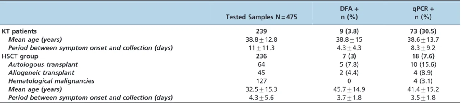
The present study evaluated 475 samples collected from 239 KT patients and 236 samples from the HSCT group. In the HSCT group, 46% (109/236) were HSCT patients and 54% (127/236) were hematological patients in a HSCT program. The vaccination rate was 8% (19/239) in the KT patients and 0.4% (1/236) in the HSCT group. The mean period between symptom onset and sample collection from KT patients was longer than that in the HSCT group (p,0.05). Table 1 provides the epidemiological and clinical characteristics of the patient groups.
In the KT group, cough was reported by 76.6% of patients, coryza by 77.8% of patients, and sore throat by 34.3% of patients. Fever and general symptoms were reported by only 18.8 and 57.3% of patients, respectively. In the HSCT group, the frequencies of cough, coryza, and sore throat were 58.9, 71.2, and 18.2%. Fever was reported by 42.4% of patients, and general symptoms were reported by 27.5% of patients.
Overall, 19.1% (91/475) of the samples from the 2 patient groups were influenza-positive by qPCR, and 3.4% (16/475) were positive by DFA. A positivity rate of 30.5% (73/239) was obtained in the KT group, whereas influenza was detected by any method in 7.6% (18/236) of the patients in the HSCT group. Influenza was detected in almost all months, but detection was more frequent during flu season months. In the HSCT group, influenza occurrence in already transplanted patients was 12.8% (14/109), and in patients in the transplant program, the percentage was 3.1% (4/127). Table 1 shows the comparative results obtained for each assay according to the patient group.
Seventy-five samples were qPCR-positive but DFA-negative, and no sample was found to be positive exclusively by the DFA. DFA sensitivity, specificity, positive
Table 1 -Epidemiological and clinical characteristics and influenza detection by DFA and qPCR according to patient group.
Tested Samples N = 475
DFA+
n (%)
qPCR+
n (%)
KT patients 239 9 (3.8) 73 (30.5)
Mean age (years) 38.8¡12.8 38.8¡15 38.6¡13.7
Period between symptom onset and collection (days) 11¡11.3 4.3¡4.3 8.3¡9.2
HSCT group 236 7 (3) 18 (7.6)
Autologous transplant 64 5 (7.8) 10 (15.6)
Allogeneic transplant 45 2 (4.4) 4 (8.9)
Hematological malignancies 127 0 4 (3.1)
Mean age (years) 32.5¡15.3 45.7¡14.9 41.4¡15.2
Period between symptom onset and collection (days) 4.3¡5.6 3.7¡1.8 3.5¡1.8
CLINICS 2013;68(9):1206-1209 Influenza and transplant patients
Perosa AH et al.
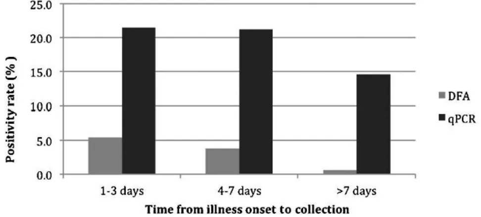
predictive value (PPV), negative predictive value (NPV) and kappa coefficient were 17.6%, 100%, 100%, 83.7% and 0.256, when we used qPCR as gold standard. The low detection of DFA was observed even in samples collected within a few days of symptom onset (Figure 1). The duration between symptom onset and sample collection for samples that tested positive only by qPCR was, on average, 4 days longer than that for the DFA-positive samples (8vs. 4 days), and the mean cycle threshold (CT) values of the negative and positive
samples according to DFA were 34.4 (range, 20 to 38¡3.6)
and 26.5 (range, 19 to 36¡5.2), respectively (p,0.05). Among the KT patients, fever was associated with influenza positivity by DFA (table 2) and qPCR (Table 3). In the HSCT group, this association was not observed.
In the KT patients, influenza A was detected by qPCR in 26.3% (63/239) of samples and by DFA in 2.1% (5/239) of samples. Influenza B was detected in 4.2% (10/239) and 1.7% (4/239) of samples by qPCR and DFA, respectively. In this group, the influenza A and B positivity rates (by any method), respectively, according to the years of study, were 60% and 7.5% in 2002, 59% and 0% in 2003, and 13.4% and 4% in 2004. In the HSCT group, influenza A was detected in 4.7% (11/ 236) of samples by qPCR and in 2.5% (6/236) of samples by DFA. The influenza B positivity rates were 3% (7/236) and 0.4% (1/236), respectively. In this group, the influenza A and B positivity rates (by any method), respectively, were 4.3% and 6.4% in 2008, 7.8% and 0% in 2009, 0% and 5.9% in 2010, and 2% and 0% in 2011. Influenza A(H1N1)pdm09 was not detected during the study period (2009-2011).
& DISCUSSION
This study compared the efficiency of DFA and qPCR in the detection of influenza in immunocompromised patients. Nosocomial outbreaks of influenza are common, and
immunocompromised patients are at significant risk. For the diagnosis of influenza in these patients, it is necessary to use a rapid and sensitive test that enables early identification of influenza cases and facilitates appropriate management deci-sions, such as patient isolation and antiviral treatment initiation. The comparison of the methods was conducted using 475 samples collected from KT recipients and patients in a HSCT program with respiratory infection. We detected influenza in 19.1% of the samples from the two groups of patients, which is in agreement with other Brazilian studies that have reported influenza A and B viruses as important causes of respiratory viral disease in immunocompromised and pediatric patients (9,10). The incidence of influenza infection among HSCT recipients is variable and is related to influenza epidemics in addition to the prevention measures adopted by each center. Our influenza detection rate in the HSCT group (7.6%) was lower but similar to that found in other studies (11,12).
Real-time PCR greatly enhanced the accuracy of influenza detection compared with DFA alone, detecting an addi-tional 75 positive samples. This difference was higher than that reported by Kuypers et al. (13), who obtained a four-fold greater number of positive samples by PCR than by DFA. Previous studies have demonstrated that qPCR is particularly efficient for the detection of influenza virus compared with traditional techniques in samples obtained more than four days after experimental infection (14).
In our study, a low rate of DFA detection occurred even in samples collected within a few days of symptom onset. However, the duration between symptom onset and sample collection for discordant samples (qPCR+/DFA-) was four
days longer than that for positive DFA samples. As high CT
values are representative of a low viral load, the fact that the CTvalues of these discordant samples are higher than those
in DFA-positive samples may be related to a lower viral load. It has been demonstrated recently that most of the
Figure 1 -Influenza positivity rate according to the time from illness onset to specimen collection.
Table 2 -Univariate logistic regression analysis of the association between the presence of fever and positivity by DFA in KT patients.
Variable
DFA+
n (%) p-value OR (95% CI)
No fever (n = 194)
3 (1.5) 0.0002* 9.79 (2.35-40.85)
Fever (n = 45) 6 (13.3)
OR = odds ratio; CI = confidence interval; * Significant atp,0.05.
Table 3 -Univariate logistic regression analysis of the association between the presence of fever and positivity by qPCR in KT patients.
Variable
qPCR+
n (%) p-value OR (95% CI)
No fever (n = 194) 49 (25.2) 0.0002* 3.38 (1.73-6.6)
Fever (n = 45) 24 (53.3)
OR = odds ratio; CI = confidence interval; * Significant atp,0.05.
Influenza and transplant patients
Perosa AH et al. CLINICS 2013;68(9):1206-1209
respiratory viruses not detected by DFA have low copy numbers of viral nucleic acid (13). It is important to consider that viral shedding in immunocompromised patients can be longer, and viral detection can occur over prolonged periods.
Differences in the duration between symptom onset and sample collection, studied periods, and physician suspicion between the two groups of patients could be responsible for the lower detection by DFA in KT patients. HSCT patients are considered to be a group at high risk for complications arising from respiratory infections, resulting in prompt clinical examination and sample collection for laboratory investigation.
Fever was reported by 30.5% of the studied patients and by only 37% of patients with influenza infection, which confirms that clinical influenza presentation in immunocompromised patients may be atypical due to medications that modulate the inflammatory response (15). Nevertheless, the presence of fever was strongly associated with influenza detection by any method.
We had the highest positivity rate in HSCT group (7.8%) in 2009. These cases were detected before the influenza pandemic; thus, none of the cases were typed as A(H1N1)pdm09. After the announcement of the pandemic in Brazil, our center adopted rigorous control measures to prevent nosocomial infection and its consequent impact in high-risk patients. Previous studies have demonstrated the effectiveness of isolation of suspected cases, use of masks and gloves in the HSCT unit, and respiratory virus testing of symptomatic patients (2,16).
In conclusion, the labor-intensive nature of the DFA, the need for highly trained technical personnel to perform the assay, and the low sensitivity of the assay have reduced its utility. Alternatively, the performance and turnaround time of qPCR when analyzing a higher number of samples support the choice of this method for different routine diagnostic settings and influenza surveillance in high-risk patients.
& ACKNOWLEDGMENTS
The authors acknowledge the financial support of the Conselho Nacional de Desenvolvimento Cientı´fico e Te´cnolo´gico (CNPq) and FAPESP.
& AUTHOR CONTRIBUTIONS
Perosa AH, Granato C, and Bellei N participated in the data analysis, writing, and review of the manuscript. Perosa AH, Watanabe AS, Guatura SB, and Silva ER participated in the laboratory analysis.
& REFERENCES
1. Ison MG, Hayden FG. Viral infections in immunocompromised patients: what’s new with respiratory viruses? Curr Opin Infect Dis. 2002; 15(4):355-67, http://dx.doi.org/10.1097/00001432-200208000-00002. 2. Nichols WG, Guthrie KA, Corey L, Boeckh M. Influenza infections after
hematopoietic stem cell transplantation: risk factors, mortality and the effect of antiviral therapy. Clin Infect Dis. 2004;39(9):1300-6, http://dx. doi.org/10.1086/425004.
3. Freitas TV, Ono G, Correˆa L, Gomes PS, Galante NZ, Tedesco-Silva H, et al. Clinical manifestations and evolution of infection by influenza A (H1N1) in kidney transplant recipients. J Bras Nefrol. 2011;33(2):136-41, http://dx.doi.org/10.1590/S0101-28002011000200004.
4. Crespo M, Collado S, Mir M, Cao H, Barbosa F, Serra C, et al. Efficacy of influenza A H1N1/2009 vaccine in hemodialysis and kidney transplant patients. Clin J Am Soc Nephrol. 2011;6(9):2208-14, http://dx.doi.org/10. 2215/CJN.02160311.
5. Shah JN, Chemaly RF. Management of RSV infections in adult recipients of hematopoietic stem cell transplantation. Blood. 2011;117(10):2755-63, http://dx.doi.org/10.1182/blood-2010-08-263400.
6. Carraro E, Neto DF, Benfica D, Perosa AH, Granato CF, Bellei NC. Applications of a duplex reverse transcription polymerase chain reaction and direct immunofluorescence assay in comparison with virus isolation for detection of influenza A and B. Diagn Microbiol Infect Dis. 2007;57(1):53-7. 7. Centers for Disease Control and Prevention [on line] 2009. CDC protocol of realtime RT-PCR for influenza A (H1N1). (http://www.who.int/csr/ resources/publications/swineflu/CDCRealtimeRTPCR_SwineH1Assay-2009_20090430.pdf)
8. Selvaraju SB, Selvarangan R. Evaluation of three influenza A and B real-time reverse transcription-PCR assays and a new 2009 H1N1 assay for detection of influenza viruses. J Clin Microbiol. 2010;48(11):3870-5, http://dx.doi.org/10.1128/JCM.02464-09.
9. Vidal LR, Siqueira MM, Nogueira MB, Raboni SM, Pereira LA, Takahashi GR, et al. The epidemiology and antigenic characterization of influenza viruses isolated in Curitiba, South Brazil. Mem Inst Oswaldo Cruz. 2008;103(2):180-5.
10. Raboni SM, Nogueira MB, Tsuchiya LR, Takahashi GA, Pereira LA, Pasquini R, et al. Respiratory tract viral infections in bone marrow transplant patients. Transplantation. 2003;76(1):142-6, http://dx.doi. org/10.1097/01.TP.0000072012.26176.58.
11. Renaud C, Campbell AP. Changing epidemiology of respiratory viral infections in hematopoietic cell transplant recipients and solid organ transplant recipients. Curr Opin Infect Dis. 2011;24(4):333-43, http://dx. doi.org/10.1097/QCO.0b013e3283480440.
12. Machado CM, Boas LS, Mendes AV, da Rocha IF, Sturaro D, Dulley FL, et al. Use of Oseltamivir to control influenza complications after bone marrow transplantation. Bone Marrow Transplant. 2004;34(2):111-4, http://dx.doi.org/10.1038/sj.bmt.1704534.
13. Kuypers J, Campbell AP, Cent A, Corey L, Boeckh M. Comparison of conventional and molecular detection of respiratory viruses in hemato-poietic cell transplant recipients. Transpl Infect Dis. 2009;11(4):298-303, http://dx.doi.org/10.1111/j.1399-3062.2009.00400.x.
14. Cherian T, Bobo L, Steinhoff MC, Karron RA, Yolken RH. Use of PCR-enzyme immunoassay for identification of influenza A virus matrix RNA in clinical samples negative for cultivable virus. J Clin Microbiol. 1994;32(3):623-8.
15. Khanna N, Steffen I, Studt JD, Schreiber A, Lehmann T, Weisser M, et al. Outcome of influenza infections in outpatients after allogeneic hemato-poietic stem cell transplantation. Transpl Infect Dis. 2009;11(2):100-5, http://dx.doi.org/10.1111/j.1399-3062.2008.00362.x.
16. Weinstock DM, Eagan J, Malak SA, Rogers M, Wallace H, Kiehn TE, et al. Control of influenza A on a bone marrow transplant unit. Infect Control Hosp Epidemiol. 2000;21(11):730-2, http://dx.doi.org/10.1086/501726.
CLINICS 2013;68(9):1206-1209 Influenza and transplant patients
Perosa AH et al.