Radiol Bras. 2016 Set/Out;49(5):311–315 311
Comparison of power Doppler and thermography for the
selection of thyroid nodules in which fine-needle aspiration
biopsy is indicated
*
Comparação entre power Doppler e termografia na seleção de nódulos tireoidianos com indicação de punção aspirativa
Alves MLD, Gabarra MHC. Comparison of power Doppler and thermography for the selection of thyroid nodules in which fine-needle aspiration biopsy is indicated. Radiol Bras. 2016 Set/Out;49(5):311–315.
Abstract
R e s u m o
Objective: To compare two methods—power Doppler and thermography—for the analysis of nodule vascularization and subsequent selection of nodules to be biopsied.
Materials and Methods: A total of 510 subjects with thyroid nodules were analyzed by power Doppler and submitted to fine-needle aspiration biopsy (FNAB). Thirty-seven patients were submitted to nodule excision (29 due to carcinoma or suspected carcinoma and 8 by patient choice). Among those patients, power Doppler had raised the suspicion of malignancy in 39 lesions, compared with 48 for FNAB. Another group, comprising 110 patients, underwent thermography, which raised the suspicion of malignancy in 124 thyroid nodules, as did FNAB. Malignant nodules were excised in all 110 of those patients (95 underwent nodulectomy and 15 underwent thyroidectomy), malignancy being confirmed by intraoperative examination of frozen biopsy samples.
Results: In relation to the FNAB findings, the sensitivity, specificity, positive predictive value, negative predictive value, and accuracy of power Doppler were 95.16%, 23.52%, 96.22%, 16.70%, and 89.51%, respectively, compared with 100%, 58.06%, 87.73%, 100%, and 89.51%, respectively, for thermography.
Conclusion: Thermography was more precise than was power Doppler for the selection of thyroid nodules to be biopsied.
Keywords: Thyroid gland; Thyroid nodule/blood supply; Ultrasonography, Doppler; Thermography.
Objetivo: Comparar dois métodos de análise de vascularização de nódulos – power Doppler e termografia – na seleção para submissão a biópsia.
Materiais e Métodos: Foram analisados 510 pacientes portadores de nódulos tireoidianos pelo power Doppler, todos submetidos a punção aspirativa por agulha fina (PAAF). Desses pacientes, 37 tiveram seus nódulos ressecados (29 por carcinoma ou suspeito para carcinoma e 8 por decisão pessoal), totalizando 39 lesões suspeitas para malignidade ao power Doppler e 48 à PAAF. Em outros 110 pacientes foram detectados, pela termografia e PAAF, 124 nódulos tireoidianos, que foram submetidos a excisão (95 casos por nodu-lectomia e 15 casos por tireoidectomia total) e tiveram malignidade confirmada no exame de congelação intraoperatório.
Resultados: O power Doppler e a termografia apresentaram, respectivamente, sensibilidades de 95,16% e 100%, especificidades de 23,52% e 58,06%, valores preditivos positivos de 96,22% e 87,73%, valores preditivos negativos de 16,70% e 100% e acurácias de 91,83% e 89,51%, quando comparados com a PAAF.
Conclusão: A termografia foi mais precisa que o power Doppler na seleção de nódulos tireoidianos com indicação de biópsia.
Unitermos: Tireoide; Nódulos tireoidianos; Ultrassonografia; Power Doppler; Termografia.
* Study conducted in the Department of Clinical Medicine of the Universidade de Ribeirão Preto (Unaerp), Ribeirão Preto, SP, Brazil.
1. PhD, Professor of Medicine at the Universidade de Ribeirão Preto (Unaerp), Ribeirão Preto, SP, Brazil.
2. Doctoral Student in Environmental Technology, Professor of Engineering at the Universidade de Ribeirão Preto (Unaerp), Ribeirão Preto, SP, Brazil.
Mailing address: Dra. Maria Lucia D’Arbo Alves. Avenida Costábile Romano, 2201, Ribeirânia. Ribeirão Preto, SP, Brazil, 14096-385. E-mail: [email protected].
Received October 26, 2014. Accepted after revision September 22, 2015.
chyma that are not associated with the presence of processes related to autoimmune or inflammatory diseases(1). TN can be single or multiple; solid, cystic, or mixed; and functional (hot nodules) or not. The prevalence of TN is 5–7% when determined by palpation, 13–67% when determined by ul-trasound, and 30–60% at autopsy(2). The adoption of high resolution ultrasound has revealed an even higher prevalence of TN, and the current suggestion is that all patients with palpable TN should be submitted to ultrasound examina-tion(3,4). The agents that stimulate the onset of TN can be of environmental origin (radiation, smoking, iodine deficiency, medications, stress, infections, or pregnancy) or of constitu-tional origin (family history, female gender, or age)(5).
Maria Lucia D’Arbo Alves1, Manoel Henrique Cintra Gabarra2
INTRODUCTION
paren-The clinical diagnosis of TN is usually based on inspec-tion and palpainspec-tion of the thyroid, which can be impaired if the nodule is located in a retrosternal position or if the pa-tient is obese. Normally, the thyroid is not visible. The im-portant factor is the exclusion of malignancy. Most TNs are of a benign nature, requiring no surgical intervention. The risk of malignancy and the presence of multiple nodules do not seem to increase during the evolution of TN. Approxi-mately 5–10% of TNs are malignant(6), and the chance of malignancy should be properly excluded by means of spe-cific exams(7).
Because of its superficial location, the thyroid is easily accessible to ultrasound or thermographic exploration and to aspiration biopsy. Imaging studies provide more precise in-formation regarding TN volume, extent, and characteristics. Fine needle aspiration biopsy (FNAB) is considered the most sensitive preoperative method for the identification of malignant TN, whereas ultrasound is the test most frequently used because of its practicality and its utility in guiding a biopsy of the nodule(8).
A biopsy is indicated for solid or mixed but preponder-antly solid nodules larger than 1.0 cm in diameter or for those smaller than 1.0 cm if they show characteristics sug-gestive of malignancy or if the patient has a history suggest-ing an increased risk for thyroid carcinoma (neck or whole body irradiation or a family history of thyroid cancer)(9,10). Nodules smaller than 5 mm in diameter should not be punc-tured even when they show suspicious characteristics on ul-trasound examination, due to the high rate of false-positive results(11).
The use of ultrasound for the evaluation of the cervical region has led to the detection of large numbers of nonpal-pable nodules and to controversies about whether they should all be analyzed for malignancy(7). Using power Doppler ul-trasound to evaluate the characteristics of TN vasculariza-tion and its associavasculariza-tion with malignancy is a possible alter-native(12). Power Doppler studies for the diagnosis of ma-lignant thyroid tumors have demonstrated that the risk of malignancy is greater when the vascularization is predomi-nantly or exclusively intranodular or central(13,14). However, Faria et al.(15) observed that a significant proportion of papil-liferous carcinomas do not show intranodular vasculariza-tion, suggesting that Doppler ultrasound is the best method for the selection of TNs to be biopsied and for guidance during FNAB.
The thyroid can also be studied by thermography, a tech-nique based on the measurement of skin temperature in a determined region. The human skin behaves like a large infrared emitter similar to a black-body radiator and does not emit reflected radiation into the environment. Measure-ments of the radiation emitted by the skin can be directly converted to temperature values. The local skin temperature is influenced by vascular changes, variations in biological activity, modifications of conduction tissue, and endocrine factors. The growth and biological activity of tissue produce
stronger emissions than do inactive tissue, and fluid collec-tions produce less heat. Thermography produces a charac-teristic and easily recognizable pattern(16–19), as well as de-termining the ranges of thermal difference that can suggest malignancy of a TN(18,19).
The objective of the present study was to compare power Doppler ultrasound and thermography for the selection of TNs to be biopsied.
MATERIALS AND METHODS
We analyzed 510 patients using a power Doppler ultra-sound imaging system (8000 EX; Samsung Medison Co., Seoul, Korea) with three multifrequency probes. Of those 510 patients, 478 were women and 32 were men. Among the women, ages ranged from 18 to 78 years (mean, 50.2 years; median, 50.5 years), whereas they ranged from 23 to 70 years (mean, 57.2 years; median, 50.0 years) among the men.
A total of 1078 TNs were identified (1021 in women and 57 in men). Of those, 868 were submitted to FNAB (829 for females and 39 for males), 35 women and 4 men being submitted to total or partial thyroidectomy.
According to the classification system devised by Lagalla–Chammas(13,14), the TNs studied by power Doppler were divided into classes 2 and 3 (suggesting benign lesions) and classes 4 and 5 (suggesting lesions suspected of malig-nancy). These findings were correlated with those obtained by FNAB according to the Bethesda classification(20).
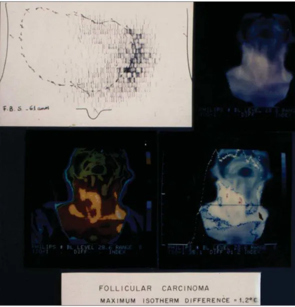
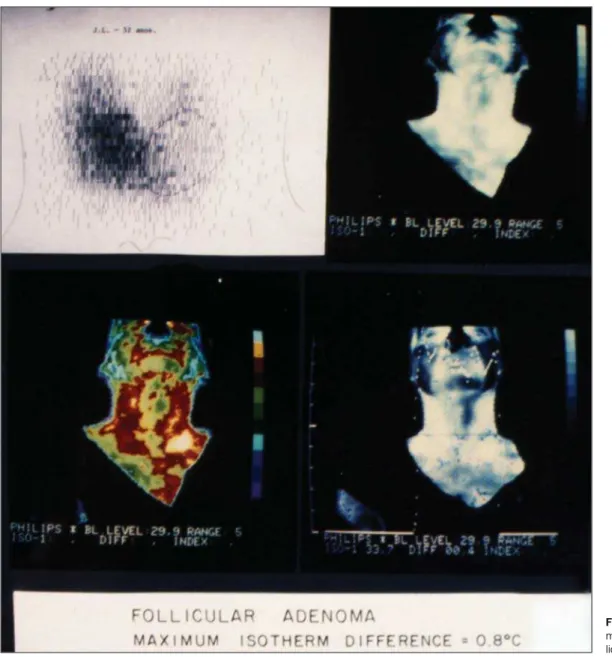
The thermographic study was performed with a digital (dynamic) thermography imaging system according to the technique devised by Mansfield et al.(18) and modified by Alves et al.(19) The study was conducted on a group of 110 patients (98 women and 12 men). Among the women, ages ranged from 18 to 68 years (mean, 50 years; median, 60 years), whereas they ranged from 18 to 78 years (mean, 67 years; median, 58 years) among the men. A total of 124 nodules were analyzed (105 in women and 19 in men) at the maximum isotherm stipulated (0.9°C) and later with FNAB (Figures 1 and 2). All 110 patients were submitted to exci-sion (nodulectomy, in 95 cases, and total thyroidectomy due to malignancy confirmed by intraoperative freezing, in 15).
RESULTS
The power Doppler study revealed 80 lesions catego-rized as Lagalla-Chammas class 2 (77 for women and 3 for men), 971 categorized as Lagalla-Chammas class 3 (918 and 53, respectively), 23 categorized as Lagalla-Chammas class 4, and 4 categorized as Lagalla-Chammas class 5.
patients (26 women and 3 men), there was a suspicion of malignancy (Bethesda category V).
The anatomopathological examination of the lesions evaluated by power Doppler revealed 39 malignant nodules (35 in women and 4 in men), 1 lesion being categorized as Chammas class 2, 32 being categorized as Lagalla-Chammas class 3, and 6 being categorized as Lagalla-Cham-mas class 4. The diagnoses were 26 cases of classical papillif-erous carcinoma, 10 cases of multicenter papillifpapillif-erous car-cinoma, 1 case of the tall-cell variant of papilliferous noma, 1 case of the follicular variant of papilliferous carci-noma, and 1 case of medullary carcinoma.
Thermography of 124 nodules revealed 31 malignant nodules (in 15 women and 6 men) and 93 benign nodules. FNAB of the same nodules revealed that 17 were malignant and 107 were benign. Among the 31 nodules that were found to be malignant by thermography, anatomopathological ex-amination revealed that 18 were classical papilliferous cinoma, 2 were follicular carcinoma, 1 was medullary car-cinoma, 1 was anaplastic carcar-cinoma, 2 were epidermoid carcinoma with metastasis to the thyroid, 5 were
inflamma-tory processes, and 2 were follicular adenomas. One case of papilliferous carcinoma, which had been diagnosed as sus-picious for malignancy by thermography, was found to be benign by FNAB.
Power Doppler and thermography, respectively, showed a sensitivity of 95.16% and 100%, a specificity of 23.52% and 58.06%, a positive predictive value of 96.22% and 87.73%, a negative predictive value of 16.70% and 100%, and an accuracy of 91.83% and 89.51%.
DISCUSSION
The power Doppler study of TN is based on the suspi-cion that abnormal cell proliferation is related to increased vascularization and is based on the modification of normal patterns(13,14,21–23). The thermographic study of TN is based on the fact that the increased cell metabolism due to a be-nign or malignant inflammatory process increases the tem-perature of the nodular region involved(16). It is clear that increased cell metabolism also increases the need for vascu-larization. Therefore, power Doppler and thermography are equivalent for the evaluation of nodular vascularization. Figure 1. Scintigraphy and
Malignancy seems to be indicated by an increased cen-tral TN vascularization when power Doppler is used(14,15) and by a maximum isotherm difference of more than 0.9° (the hottest point inside the nodule compared with the tempera-ture of thyroid tissue around the nodule) when thermography is used(19). However, this increase in central vascularization is not observed in medullary or anaplastic carcinomas or even in some papilliferous carcinomas showing a higher degree of fibrosis(24). Although an increase in cell metabolism oc-curs before angiogenesis and in all types of neoplasias, ther-mography reveals a change in its pattern earlier than does power Doppler, thus being more precise for the detection of malignancy in the TN examined. In conclusion, thermogra-phy was more precise than was power Doppler for the indi-cation of FNAB of TNs suspected of malignancy.
Acknowledgments
The authors wish to acknowledge the invaluable support of the Radiology Department, Biochemistry Laboratory, and
Endocrinology Group of the University of Ribeirão Preto; of the Departments of Gynecology & Obstetrics, Surgery, and Pathology of the University Hospital of Ribeirão Preto; and of the Surgery and Pathology Divisions of the Brazilian Unified Health Care System.
REFERENCES
1. Krohn K, Führer D, Bayer Y, et al. Molecular pathogenesis of eu-thyroid and toxic multinodular goiter. Endoc Rev. 2005;26:504– 24.
2. Tan GH, Gharib H. Thyroid incidentalomas: management ap-proaches to nonpalpable nodules discovered incidentally on thyroid imaging. Ann Intern Med. 1997;126:226–31.
3. Holden A. The role of colour and duplex Doppler ultrasound in the assessment of thyroid nodules. Australas Radiol. 1995;39:343–9. 4. Varverakis E, Neonakis E, Tzardi M, et al. Role of color Doppler ultrasonography in the preoperative management of cold thyroid nodules. Hormones (Athens). 2007;6:44–51.
5. Galofré JC, Lomvardias S, Davies TF. Evaluation and treatment of thyroid nodules: a clinical guide. Mt Sinai J Med. 2008;75:299– 311.
6. Gharib H, Papini E. Thyroid nodules: clinical importance, assess-ment, and treatment. Endocrinol Metab Clin North Am. 2007;36: 707–35,vi.
7. Papini E, Guglielmi R, Bianchini A, et al. Risk of malignancy in nonpalpable thyroid nodules: predictive value of ultrasound and color-Doppler features. J Clin Endocrinol Metab. 2002;87:1941– 6.
8. Pitman MB, Abele J, Ali SZ, et al. Techniques for thyroid FNA: a synopsis of the National Cancer Institute. Thyroid Fine-Needle Aspiration State of the Science Conference. Diagn Cytopathol. 2008;36:407–24.
9. Maia AL, Ward LS, Carvalho GM, et al. Thyroid nodules and dif-ferentiated thyroid cancer. Brazilian consensus. Arq Bras Endocrinol Metab. 2007;51:867–93.
10. Rosário PW, Ward LS, Carvalho GA, et al. Thyroid nodule and differentiated thyroid cancer: update on the Brazilian consensus. Arq Bras Endocrinol Metab. 2013;57:240–64.
11. Mazzaferri EL, Sipos J. Should all patients with subcentimeter thy-roid nodules undergo fine-needle aspiration biopsy and preopera-tive neck ultrasonography to define the extent of tumor invasion? Thyroid. 2008;18:597–602.
12. Cerbone G, Spiezia S, Colao A, et al. Power-Doppler improves the diagnostic accuracy of color Doppler ultrasonography in cold thy-roid nodules: follow-up results. Horm Res. 1999;52:19–24. 13. Lagalla R, Caruso G, Novara V, et al. Flowmetric analysis of
thy-roid diseases: hypothesis on integration with qualitative color-Dop-pler study. Radiol Med. 1993;85:606–10.
14. Chammas MC, Gerhard R, Oliveira IR. Thyroid nodules: evaluation with power Doppler and duplex Doppler ultrasound. Otolaryngol Head Neck Surg. 2005;132:874–82.
15. Faria MAS, Casulari LA. Comparação das classificações dos nódu-los de tireoide ao Doppler colorido descritas por Lagalla e Chamas. Arq Bras Endocrinol Metab. 2009;53:811–7.
16. Samuels BI. Thermography: a valuable tool in the detection of thy-roid disease. Radiology. 1972;102:59–62.
17. Belossi A. Echography and thermography in the pathology of thy-roid tumors. A study of 135 hypofixated nodules. Ann Radiol. 1978;21:409–10.
18. Mansfield CM, Farrell C, Asbell SO. The use of thermography in the detection of metastatic liver disease. Radiology. 1970;95:696–8. 19. Alves MLD, Andrade J, Cherri J, et al. Papel da termografia na seleção de nódulos tireoideanos de indicação cirúrgica. Arq Bras Endocrinol Metab. 1988;32:97–9.
20. Cibas ES, Ali SZ; NCI Thyroid FNA State of the Science Confer-ence. The Bethesda system for reporting thyroid cytopatology. Am J Pathol. 2009;132:658–65.
21. Leblowska UM, Dzieciol J, Lemancewicz D, et al. The influence of the vascularization of the follicular thyroid nodules on the prolif-erative activity of the follicular cells. Folia Morphol (Warz). 2004;63:79–81.
22. Lyshchik A, Moses R, Barnes SL, et al. Quantitative analysis of tu-mor vascularity in benign and malignant solid thyroid nodules. J Ultrasound Med. 2007;26:837–46.