83
Radiol Bras. 2013 Mar/Abr;46(2):83–88
Thyroid arterial embolization as a therapeutic option
for hyperthyroidism: a case series
*
Embolização de artérias tireoidianas como opção terapêutica para o hipertireoidismo: série de casos
Josivan Gomes de Lima1, Alexandre Barbosa Câmara de Souza2, Raphael Pinto de Mendonça3, Lúcia Helena Coelho Nóbrega3, Fernando Antônio de Araujo Moura4, Zankennedy Jales de Queiroz5
Objective: To evaluate the therapeutic response to thyroid arterial embolization for primary hyperthyroidism. Materials and Methods: Five women whose pharmacological treatment with thionamides failed to control Graves’ disease were submitted to embolization of three dominant thyroid arteries (following assessment with ultrasound Doppler and arteriography) and followed-up at the 1st, 8th and 16th weeks after the procedure, with ultrasound Doppler, calcium blood test, thyroid function test and clinical examination. Three of the patients completed 16-week follow-up. Results: None of the patients achieved permanent remission after 8 weeks. Disease recurrence was observed at the 24th week, despite the decrease in thyroid volume (49.5 ± 15.2%) observed at the 16th week. Complications were not observed, but radioiodine therapy was required for three of the patients followed-up. Conclusion: Embolization of three dominant thyroid arteries with polyvinyl alcohol allowed reduction in goiter volume in the three patients who completed the protocol, but was not effective to control hyperthyroidism.
Keywords: Hyperthyroidism; Treatment; Embolization.
Objetivo: Avaliar a resposta da embolização arterial tireoidiana como terapêutica para o hipertireoidismo primário. Materiais e Métodos: Cinco mulheres com falha ao tratamento farmacológico com tionamida foram submetidas a vaso-oclusão em três artérias dominantes tireoidianas (avaliadas por ultrassonografia Doppler e arteriografia) e acom-panhadas até 8 semanas após o procedimento (três acomacom-panhadas até 16 semanas) com ultrassonografia Doppler, calcemia, função tireoidiana e controle clínico. Resultados: Nenhuma alcançou remissão permanente de doença após 8 semanas. Houve recidiva de hipertireoidismo em 24 semanas, mesmo com redução do volume tireoidiano de 49,5 ± 15,2% em 16 semanas. Não encontramos complicações, mas radioiodo foi necessário após 24 semanas em três das pacientes acompanhadas. Conclusão: Nas pacientes que concluíram o protocolo, a vaso-oclusão arterial com polivinil álcool nas três artérias dominantes permitiu redução volumétrica do bócio, entretanto, foi ineficiente em con-trolar o hipertireoidismo.
Unitermos: Hipertireoidismo; Embolização; Tratamento. Abstract
Resumo
* Study developed at Hospital Universitário Onofre Lopes da Universidade Federal do Rio Grande do Norte (UFRN), Natal, RN, Brazil.
1. Specialization in Endocrinology, Professor of Endocrinology at Universidade Federal do Rio Grande do Norte (UFRN), Natal, RN, Brazil.
2. Graduate Student of Medicine at Universidade Federal do Rio Grande do Norte (UFRN), Natal, RN, Brazil.
3. MDs, Endocrinologists, Centro de Endocrinologia de Natal, Natal, RN, Brazil.
4. MD, Interventional Radiologist, Hospital Universitário Ono-fre Lopes da Universidade Federal do Rio Grande do Norte (UFRN), Natal, RN, Brazil.
5. MD, Radiologist, Hospital Universitário Onofre Lopes da Uni-versidade Federal do Rio Grande do Norte (UFRN), Natal, RN, Brazil. Mailing Address: Dr. Josivan Gomes de Lima. Rua Joaquim Fabrício, 233, Petrópolis. Natal, RN, Brazil, 59012-340. E-mail: [email protected].
Lima JG, Souza ABC, Mendonça RP, Nóbrega LHC, Moura FAA, Queiroz ZJ. Thyroid arterial embolization as a therapeutic option for hyperthyroidism: a case series. Radiol Bras. 2013 Mar/Abr;46(2):83–88.
resolution of hyperthyroidism, but it may present complications caused by anesthesia, and fail in the control of the clinical condi-tion (particularly in attempts of partial resec-tion of the gland), besides being an inva-sive procedure involving risks for injuries to the recurrent laryngeal nerve and cervi-cal vessels, and possibility of hypoparathy-roidism. Currently, with improvements in operative techniques and in the knowledge on corticosteroid preparation for surgery, the complication rates have decreased(2).
The radioactive iodine therapy causes actinic lesions which culminate in atrophy of the thyroid tissue. Such an effect, how-ever, presents a variable response, depend-ing upon several factors such as thyroid gland volume, dose delivered, dietary iodine peutic modalities, three are currently
fea-sible, namely, antithyroid drugs, radioac-tive iodine and surgery(2,3).
In spite of the lack of a global consen-sus on the initial therapeutic approach, pharmacological treatment with thiona-mides (methimazole or propylthiouracil) is considered in many countries as the first choice in the attempt to control in patients hyperthyroidism. The main obstacles to their use are represented by remission dif-ficulties and possible side effects resulting from such treatment option.
Thyroidectomy (whether subtotal or total) provides the possibility of definitive INTRODUCTION
Hyperthyroidism, in most cases, is a chronic disease and common cause of goi-ter and exophthalmos(1). Among the
content, previous utilization of iodinated contrast agents and thionamides for con-trol, concomitance or not of utilization of antithyroid drug during iodine administra-tion and gastrointestinal tract integrity(1–3).
Main contraindications include pregnancy and lactation (absolute contraindications) and active Graves’ ophthalmopathy classi-fied as intense (relative contraindication). However, this definitive therapeutic proce-dure is widely utilized nowadays(2–7).
In cases of side effects or contraindica-tions for some specific therapy, the therapy by embolization, as already utilized in other organs (liver, uterus, etc.) may be a new modality for treating hyperthyroidism.
In 2002, Xiao et al.(8) reported
satisfac-tory results with the utilization of a new technique in an attempt to obtain temporary or definitive remission of hyperthyroidism activity: occlusive endovascular emboliza-tion of nourishing arteries of the thyroid tissue. Such study consisted in performing the occlusion of two of the four thyroid arteries in 22 patients with 27-month fol-low-up. Over the whole follow-up period, remission was observed 14 (63%) of the patients, with one patient presenting hy-pothyroidism, and the others requiring supplementary dose of radioactive iodine. In 2007, Zhao et al.(9) reported a remission
rate of 78.6% in patients of another Asian center, over a 12-month follow-up period. In their series, such Asian centers reported a sustained hyperthyroidism, goiter and symptoms management with the men-tioned technique(8,9).
The present study was aimed at evalu-ating the therapeutic response of thyroid ar-teries embolization (TAE) in five Brazilian patients with hyperthyroidism and diffuse goiter after failure of conventional treat-ment.
MATERIALS AND METHODS
The study followed-up five hyperthy-roidism patients who did not achieve re-mission over consecutive 24 months under treatment with thionamides (Table 1). The clinical and laboratory criteria for consid-ering the failure of the pharmacological treatment were suppressed TSH (< 0.4 mUI/ml), free T4 and/or T3 levels above the reference range of normality, as well as
Table 1 Characteristics of the patients at the beginning of the study.
Characteristic
Age (years) Race
Sex Thionamide Volume US (ml) Body weight (kg)
HR (bpm) BP (mmHg)
Patient 1
43 Melanoderm
Female TPZ 64.0 66.7 82 115 × 70
Patient 2
32 Feoderm
Female TPZ 20.5 52.7 90 100 × 70
Patient 3
39 Feoderm
Female TPZ 62.5 51.8 110 160 × 100
Patient 4
25 Feoderm
Female PTU 57.0 54.2 105 110 × 70
Patient 5
58 Feoderm
Female PTU 40.0 46.0 108 120 × 70
US, ultrasonography; HR, heart rate; BP, blood pressure; TPZ, methimazole; PTU, propylthiouracil.
presence of hyperthyroidism symptoms under pharmacotherapy (propylthiouracil or methimazole, with or without beta-blockers). Only patients with Graves’ dis-ease were included (patients with ophthal-mopathy and/or TRAb – thyroid-stimulat-ing hormone receptor antibody – positive). Patients with severe ophthalmopathy (ac-tivity score showing important injury of conjunctiva, corneas, extraocular muscles and/or optic nerve) were excluded. The patients were followed-up from Septem-ber/2005 to December, 2006.
The study project was previously ap-proved by the committee for Ethic in Re-search of the institution. All the patients signed a term of free and informed consent and were allowed to leave the investigation at any moment if they so wished.
Pre-embolization evaluation
Initially, all the patients were submitted to laboratory tests, comprising blood count, renal and hepatic function, whose results should be. Doppler ultrasonography of the thyroid arteries and plain chest radiography were performed before the procedure.
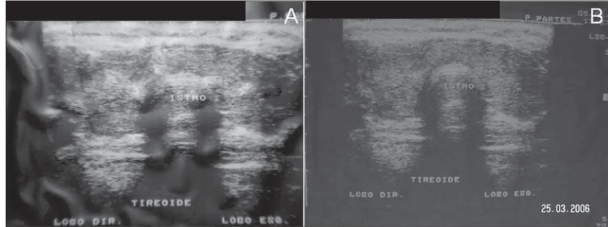
The patients were submitted to volumet-ric measurement and evaluation of vascu-larization (by means of Doppler flowmetry) before and after the embolization proce-dure, for investigation of revascularization (in case it occurred).
Thyroid artery embolization
The patients received sedation with diazepine or thiopental, with heart rate monitoring. Intravenous antibiotic prophy-laxis was performed with a single 2 mg cephalozine dose. Prophylactic hepariniza-tion (5,000 UI intravenously) was per-formed in some patients after arterial
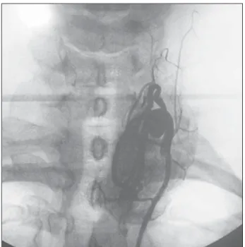
punc-ture in order to avoid hypercoagulability phenomena. The Seldinger technique was utilized for right transfemoral insertion, with series in both superior and inferior and lower thyroid arteries for angiography and embolization(10). Subsequently,
angiog-raphic contrast was utilized to identify the thyroid vascular anatomy coming from the thyrocervical trunk and from the external carotid arteries (Figure 1).
At the beginning of the procedure, he-modynamic angiography evaluated the pa-tency and dominance of the arterial supply to the thyroid in order to perform the arte-rial occlusion of the most calibrous vessels, with embolization of the three largest arter-ies with polyvinyl-alcohol (150–300 µm in diameter), leaving the artery with the smaller diameter totally pervious. At the end of the procedure, a panoramic cervical angiography was performed, with iodi-nated contrast injection in the aortic arch to demonstrate the absence of thyroid blush. The femoral catheter was removed, with manual compression on the puncture site for 15 minutes, followed by bed rest for four to six hours.
Prednisone 20 mg/day was initiated the day after the procedure and maintained for 7 days, and methimazole was reinitiated, at the same pre-procedural dose, 24 hours after the procedure and maintained until the fourth week after the procedure. The beta-blocker was maintained at a personalized dose until euthyroidism was reached. All the procedures were performed in a single unit, by a single interventional radiologist who followed a pre-established protocol.
Post-procedural follow-up
hemo-dynamic changes were performed during the first 48 hours. Thereafter the patients underwent weekly clinical and laboratory evaluations over the first four weeks, and at the 8th, 16th, 20th and 24th weeks (Table 2), with thyroid ultrasonography being per-formed under basal conditions and then at the 1st, 8th, 16th and 24th weeks (Table 3). The laboratory tests were performed by chemiluminescence, with an Immulite 1000 Siemens apparatus, with the following ref-erence levels: TSH, 0.4–4.0 mUI/ml; free T4, 0.8–2.2 ng/dl; total T3, 70–170 ng/dl. The primary objective was analyzing the responses regarding hyperthyroidism con-trol, clinical and laboratorial remission, goi-ter volume decrease and prevention of com-plications in 24 weeks. Persistent hyperthy-roidism would be treated with radioactive iodine, 12 to 15 mCi doses. The secondary objective was evaluating changes in thyroid function (hyper or hypothyroidism), be-sides identifying undesirable collateral ef-fects from the procedure, such as emboliza-tion of a non-target organ, vascular dissec-tion, phlebitic syndrome, aneurysm or pseudoaneurysm and hypoparathyroidism secondary to from parathyroid ischemia.
RESULTS
Five patients were submitted to the pro-cedure, which had a mean duration of 77.4 ± 23.1 minutes. Two patients left the study
protocol (patient 3 on the 4th week and patient 4 on the 12th week). Thus, only three patients underwent the pre-deter-mined period on minimum follow-up (20 weeks) of the project. The initial data of the patients enrolled in the study, such as thy-roid volume at ultrasonography (ml), weight (kg), heart rate (bpm) and arterial pressure (mmHg), are shown on Table 1. Data on weight (kg), arterial pressure
(mmHg), heart rate (bpm), free T4 (ng/dl), T3 (ng/gl), TSH (mUI/ml) and calcium (mg/dl) levels in the patients before embo-lization and after follow-up are shown on Table 2. Size of the thyroid gland, basal volume and treatment progress after one and eight weeks are described on Table 3. Out of the five patients initially evalu-ated, only three completed a minimum postprocedural follow-up of 20 weeks. The whole study sample (five patients) could complete the follow-up only up to the 8th week. Before embolization, one patient (patient 3) presented more severe hyperten-sion, one patient was intolerant to glucose (patient 1), and the others did not present any comorbidity.
After embolization, all the patients pre-sented subacute thyroiditis and, among the patients who completed the follow-up, even for the minimum of 16 weeks, or at the 24th week re-evaluation, none achieved complete hyperthyroidism remission, not-withstanding the significant reduction in the volume of goiter previously observed in all the patients.
Clinical case 1
A 43-year-old, melanoderm, female pa-tient followed-up at the endocrinology clinic for hyperthyroidism secondary to
Table 3 Sonographic volume (ml) and thyroid volumetric decrease (%) in four patients who completed 8 weeks in the study.
Patient 1 Patient 2 Patient 4 Patient 5 Median
US basal (ml)
64.0 20.0 62.5 57.0 59.75
US week 1 (ml)
46.4 17.5 50.0 40.0 43.20
US week 8 (ml)
19.6 14.6 24.7 33.7 22.15
Volumetric decrease (%)
69 27 61 41 51.0
US, ultrasonography.
Table 2 Results at the end of follow-up in patients 1, 2 and 5.
Data
Body weight (kg) BP (mmHg)
HR (bpm) Free T4 (ng/dl)
T3 (ng/dl) TSH (mUI/ml) Calcium (mg/dl)
Basal
52,7 100×70
90 2.93
207 < 0.004
9.7
Final †
53,0 105×70
80 1.7 131 0.06 9.3 Basal
66,7 115×70
82 1.05 170 0.1 10.7
Final*
69,9 110×70
70 2.6 156 0.01 9.5
Basal
51,8 160×100
110 2.70 325 0.068
10.6
Final †
51,5 130×95
100 2.2 259 0.40 9.4
Patient 1 Patient 2 Patient 5
*24 weeks after the procedure. †20 weeks after the procedure. BP, blood pressure; HR, heart rate.
Graves’ disease, undergoing 60 mg/day methimazole therapy. Upon admission, prediabetes was diagnosed.
The patient was submitted to thyroid embolization, with no vascular complica-tion (disseccomplica-tion, non-target organ embo-lization, aneurysms) (procedure shown on Figure 2). During hospital stay, diabetes was diagnosed and insulin therapy was ini-tiated with an adjustment scheme accord-ing to capillary glycemia. Algic cervical syndrome was reported on the first week. No post-procedural infectious complica-tion was observed. A noticeable aesthetic
improvement was observed as a result from the decreased in goiter (Figure 3), as well improvement of the cervical compressive symptoms. After six months, ultrasonogra-phy demonstrated a 16.7 ml gland (Figure 4), with hypervascularization at Doppler analysis, even in previously embolized thy-roid lobes. There were neither reports of visual disorders nor hyperglycemic compli-cation. At the end of the follow-up, the patient was referred to a public nuclear medicine service for radioactive iodine therapy at a dose of 15 mCi, as previously determined by the protocol.
Clinical case 2
A 32-year-old, feoderm, female patient followed-up in the endocrinology clinic for hyperthyroidism secondary to Graves’ dis-ease, and under 60 mg/day methimazole therapy. At the admission for the procedure, the patient complained of insomnia, head-ache and weight loss.
Embolization was performed and, dur-ing the procedure, the thyroid presented signs of anatomic variation, with five thy-roid arteries being identified. Occlusion was performed in the three arteries with larger diameters. The procedure was
suc-Figure 2. Patient 1. A: Pre-embolization image. B: Em-bolization with polynivyl alco-hol particles. C,D: Post-em-bolization image.
C
Figure 3. Patient 1. A: Dif-fuse thyroid goiter with 64 ml, before arterial emboliza-tion. B: Anterior cervical re-gion 8 weeks after emboliza-tion, with 19 ml.
cessful, with no vascular complications (dissection, non-target organ embolization, aneurysms). The patient progressed with-out complaints, and reported improvement of initial symptoms.
There was no report of visual disorders. After follow-up, the patient was referred to a reference public nuclear medicine service for radioactive iodine therapy at a dose of 15 mCi, as previously determined by the protocol.
Clinical case 3
A 39-year-old, feoderm, female patient with hyperthyroidism secondary to Graves’ disease, undergoing 60 mg/day methima-zole therapy, and hypertensive, under 75 mg/day captopril. The patient was admit-ted into the service to undergo the proce-dure, with report of tremors, palpitations and weight loss.
Embolization was performed, with no vascular complication (dissection, non-tar-get organ embolization, aneurysm). After the procedure, the patient experienced a hy-pertensive peak, and was successfully treated with sodium nitroprusside, with no sequel. Appropriate blood pressure control was restored with 150 mg/day captopril.
After the fourth week, follow-up of this patient was lost.
Clinical case 4
A 25-year-old, feoderm, female patient undergoing follow-up in the endocrinology service for hyperthyroidism secondary to Graves’ disease treated with propylthiou-racil 100 mg/day. The patient reported in-somnia, menstrual irregularity, tremors, palpitations and weight loss.
Embolization was performed and, dur-ing the procedure, signs of anatomical variations were found with five thyroid arteries being identified. Occlusion was performed in the three arteries with larger diameters. There was no vascular compli-cation (dissection, non-target organ embo-lization, aneurysm). In the post-operative period, the patient presented a hypertensive peak, and was treated with captopril, re-maining stable and with no other com-plaints. Doppler analysis revealed hyper-vascularization at the 8th week, even in the previously embolized thyroid lobes.
During the follow-up, the patient re-ported partial improvement of her initial complaints and quality of life, but the fol-low-up of this patient was lost after 12 weeks.
Clinical case 5
A 58-year-old, feoderm, female patient referred to the endocrinology service for hyperthyroidism secondary to Graves’ dis-ease, undergoing treatment with 100 mg/ day of propylthiouracil. The patient re-ported insomnia, weight loss, headaches, tremors and palpitations.
Embolization was performed without vascular complications (dissection, non-target organ embolization, aneurysm). In the post-operative period the patient devel-oped subacute thyroiditis which compli-cated with cachexia. Support treatment was instituted, with improvement of the algic symptoms, and the patient reported partial decrease of her initial complaints.
There was no report of visual disorders. After the follow-up period, the patient was referred to a reference public nuclear
medi-cine service for radioactive iodine therapy at a dose of 15 mCi, as previously deter-mined by the protocol.
DISCUSSION
In the present study, after embolization of three thyroid arteries, a volumetric de-crease was observed, besides management of the cervical symptoms caused by goiter in the patients with Graves’ disease whose clinical treatment had failed. However, despite the aesthetic improvement reported by the patients, as well as improvements in cervical compressive symptoms, the serum TSH concentration did not reach the lev-els compatible with euthyroidism at the end of the protocol follow-up period (24 weeks for three patients). Additionally, there was a tendency towards increase in the free T4 levels, characterizing established hyperthy-roidism. The persistence of the disease af-ter the TAE procedure, has deaf-termined the necessity of treatment with radioactive io-dine at the 15 mCi dose to control the hy-perthyroidism as recommended in the study design. No sign of hypervasculari-zation was found at the Doppler analysis, even in the previously embolized thyroid lobes in two of the five patients (patients 1 and 4) who underwent repeated ultrasonog-raphy at the end of the 8th week. A study has demonstrated that the expression of VEGF (vascular endothelial growth factor) is increased until six months after embo-lization, and is decreased after one year, suggesting a decrease of angiogenesis in the long term(11).
Xiao et al.(8) have developed a study
with 22 patients who refused treatment Figure 4. Patient 1. A:
with radioactive iodine, and had severe side effects with thionamides or presented vo-luminous goiter. In such study, 14 patients achieved hyperthyroidism remission and goiter control in 50 months of follow-up. Later, two patients required use of thionamides and six, surgery (without com-plications) because of voluminous goiter persistence. No case of hypothyroidism was reported. Zhao et al.(9) have reported
control of hyperthyroidism in patients with Graves’ disease, with significant decrease in TRAb levels in 37 patients. In another study, those authors have found decrease in the CD4+/CD8+ ratio to normal levels at immunophenotyping(12). Indications on the
elevation of post-immunohistochemical apoptosis parameters demonstrated a ten-dency towards volumetric decrease months after arterial occlusion(13). In a series with
28 patients, Zhao et al. have described eu-thyroidism in 78% of the patients in a 22-month follow-up period(10). On the other
hand, Hiraiwa et al. have reported exacer-bation of ophthalmopathy after TAE pro-cedure(14).
Some investigators have already initi-ated the utilization of TAE with a perspec-tive of tumor reduction in patients with thy-roid anaplastic carcinoma(15) or for tumor
mass reduction, but published data on such utilization are still scarce(15–17).
The present study had a small number of patients, which impairs comparative analysis with other studies. Additionally, the follow-up of two of five patients was lost. All the patients presented Graves’ dis-ease, and even in the absence of TRAb test-ing durtest-ing the follow-up, the noticeable non-remission, even in 24 weeks, already demonstrated an ongoing autoimmune hy-perthyroidism activity. In the present study, the follow-up demonstrated a rapid mean thyroid volumetric reduction of 49.5% of
the thyroid parenchyma within 8 weeks after embolization, with a sustained re-sponse on the following eight weeks. Thus, from a primary observation, arterial embo-lization has offered satisfactory results for reduction of thyroid volume without the need for hyperfunction control of the or-gan. This fact can be corroborated by stud-ies aimed at tumor mass reduction(15,16).
The failure in controlling hyperthyroid-ism may have been a result of the irregu-larity of the utilized polyvinyl alcohol par-ticles which microscopically were roughly “H”-shaped, perhaps favoring the recanali-zation of the thrombi located in the spaces of such “H”, and also by the persistence of the blood flow in the remaining thyroid ar-tery. In a normal functioning organ, smaller occlusive particles make the procedure more selective and effective. However, it should be considered that in a hyperstimu-lated organ, like in the case of Graves’ dis-ease, recanalization or neovascularization after embolization with smaller particles may occur, impairing the hyperthyroidism remission.
Novel protocols with embolization of the four arteries with different sizes of oc-clusive particles, as well as the utilization of calibrated acrylate gel particles are be-ing undertaken, and shall provide new data on such therapeutic modality in the control of hyperthyroidism.
REFERENCES
1. Page S. Hipertireoidismo. In: Lima JG, Nóbrega LHC, Nóbrega MLC. Aulas em endocrinologia. Rio de Janeiro, RJ: Atheneu; 2001. p. 63–74.
2. Lima JG, Nóbrega LHC, Nóbrega MLC, et al. Fa-tores associados com recidiva precoce do hiper-tireoidismo após tratamento com radioiodotera-pia. Arq Bras Endocrinol Metab. 2003;47:701–4. 3. Lopes MHC. Hipertireoidismo. In: Bandeira F, Macedo G, Caldas G, et al. Endocrinologia e dia-betes. Rio de Janeiro, RJ: Medsi; 2003. p. 213– 35.
4. Bonnema SJ, Bennedbaek FN, Veje A, et al. Con-tinuous methimazole therapy and its effect on the cure rate of hyperthyroidism using radioactive iodine: an evaluation by a randomized trial. J Clin Endocrinol Metab. 2006;91:2946–51.
5. Bartalena L, Marcocci C, Bogazzi F, et al. Rela-tion between therapy for hyperthyroidism and the course of Graves’ ophthalmopathy. N Engl J Med. 1998;338:73–8.
6. Kerr L, Vidigal MEL, Rozenkwit D. Pode a gesta-ção influenciar a evolugesta-ção de nódulo tireoidiano maligno? Radiol Bras. 2012;45:65–6.
7. Brandão CDG, Antonucci J, Correa ND, et al. Efeitos da radioiodoterapia nas gerações futuras de mulheres com carcinoma diferenciado de ti-reóide. Radiol Bras. 2004;37:51–5.
8. Xiao H, Zhuang W, Wang S, et al. Arterial embo-lization: a novel approach to thyroid ablative therapy for Graves’ disease. J Clin Endocrinol Metab. 2002;87:3583–9.
9. Zhao W, Gao BL, Tian M, et al. Graves’ disease treated with thyroid arterial embolization. Clin Invest Med. 2009;32:E158–65.
10. Zhao W, Gao BL, Yi GF, et al. Thyroid arterial embolization for the treatment of hyperthyroid-ism in a patient with thyrotoxic crisis. Clin Invest Med. 2009;32:E78–83.
11. Zhao W, Gao BL, Liu ZY, et al. Angiogenic study in Graves’ disease treated with thyroid arterial embolization. Clin Invest Med. 2009;32:E335– 44.
12. Zhao W, Gao BL, Jin CZ, et al. Long-term immu-nological study in Graves’ disease treated with thyroid arterial embolization. J Clin Immunol. 2008;28:456–63.
13. Zhao W, Gao BL, Yi GF, et al. Apoptotic study in Graves disease treated with thyroid arterial em-bolization. Endocr J. 2009;56:201–11. 14. Hiraiwa T, Imagawa A, Yamamoto K, et al.
Ex-acerbation of thyroid associated ophthalmopathy after arterial embolization therapy in a patient with Graves’ disease. Endocrine. 2009;35:302–5. 15. Tazbir J, Dedecjus M, Kaurzel Z, et al. Selective
embolization of thyroid arteries (SETA) as a pal-liative treatment of inoperable anaplastic thyroid carcinoma (ATC). Neuro Endocrinol Lett. 2005; 26:401–6.
16. Dedecjus M, Tazbir J, Kaurzel Z, et al. Selective embolization of thyroid arteries as a preresective and palliative treatment of thyroid cancer. Endocr Relat Cancer. 2007;14:847–52.